Applications are invited from highly motivated individuals who are interested in fundamental mechanisms of neuronal migration and axon guidance. The main focus of our research is to understand the molecular and cellular mechanisms underlying the development of neural circuits using the embryonic spinal cord as a model system (http://www.ucmm.umu.se/english/research/sara-wilson/). The fellowship is funded for two years and is available immediately upon approvals. The laboratory is located at IMB, Umeå University, Sweden. IMB is an interdisciplinary department, which focuses on questions in basic and medical sciences and provides an interactive modern environment with easy access to good core facilities. The working ‘day to day’ language in the laboratory is English.
Requirements: Individuals with a background in developmental biology, neuroscience, molecular and or cell biology or related discipline and with a keen interest in developmental neuroscience are encouraged to apply. The successful candidate will have or about to receive a Ph.D. in a relevant discipline and be proficient in written and spoken English. Technical experience with imaging, vertebrate embryonic model systems – especially mouse handling and genetics, chick or mouse embryo electroporation and /or neurite outgrowth assays, is a big advantage although full training will be given. Experience with molecular/cellular biology will be positively evaluated. The most successful candidatewill have ahigh level of motivation, be creative, organized, rigorous and have the ability to work both independently and within a team.
Please submit your application (reference 2018SW200) by 23rd February 2018 to anita.dreyer-perkiomaki@umu.seby sending the following documents as pdf files:
1) A short cover letter (not more than 1 page) to include a description of your research experience and suitability for the position.
2) Curriculum Vitae including: publication list, technical expertise, names and contact information for three referees.
Informal enquiries may be directed to Dr. Sara Wilson (sara.wilson@umu.se).
Vertebrate brain development is characterised by cell migration, as neurons are often born far from where they need to end up. Migration is regulated by guidance cues and their receptors, but, problematically, many of these molecules are expressed throughout the brain, complicating efforts to spatially and temporally pin down their function. A paper in the latest issue of Development addresses this problem using cell-specific knockouts for Slit and Robo, crucial migration regulators. We caught up with first author Chloé Dominici and her supervisor Alain Chédotal of the Institut de la Vision in Paris to find out more.
Alain and Chloé
Alain, can you give us your scientific biography and the questions your lab is trying to answer?
AC Our lab studies the cellular and molecular mechanisms controlling the development of brain connections, mostly focusing on so-called commissural neurons which interconnect the right and left side of the nervous system. We mainly use genetics and mice but have also started to study other organisms (birds, amphibians, marsupials, humans etc) to address the question of the evolution of midline guidance processes.
I started to study the development of commissural circuits during my PhD in Paris, using various neuroanatomical methods including electron microscopy. For my post-doc, I wanted to learn other techniques such as genetics, molecular biology and biochemistry and joined the lab of Corey Goodman at UC Berkeley who had just discovered the first vertebrate Semaphorin. It was an exciting time as little was known about midline crossing in vertebrates. Within a few years all the main guidance cues, Slits, semaphorins, netrins, ephrins, and their receptors were characterized. After starting my own group, I continued to work on axon guidance, but also neuronal migration, angiogenesis and myelination. In parallel, we have been developing new 3D imaging techniques combining whole-mount immunostaining, tissue clearing and light-sheet microscopy, to facilitate the phenotypic analysis of our mutant mice.
And Chloé, how did you come to be involved with this project?
CD When I first joined Alain’s lab, my project was aiming to understand the role of Slit2 in the development of motorneurons. We initially attempted to do so by using conditional lines and wanted to show, as a control, that we could also remove Slits in the floor plate. After several attempts, I wasn’t able to remove Slits in the motorneurons but the conditional line for the floor plate worked very well. So, we began to analyse these mutants by focusing mainly on the hindbrain as it had been previously reported that Slits were involved in the migration of precerebellar neurons, a model system in the lab.
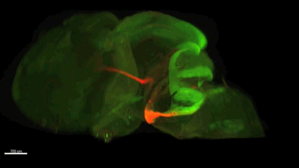
Lightsheet microscopy of the forebrain in different genetic conditions, from Fig 1, Dominici, et al. 2018
What were the limitations of previous genetic studies on the role of Slit and Robo in precerebellar neuron migration, and how did you set out to overcome these?
AC & CD The current model proposes that Slits act as repulsive factors for precerebellar neurons (PCNs), acting through Robo receptors. Accordingly, Slits are found at the floor plate, that most PCNs do not cross, and in the facial nucleus that one class of PCNs, the pontine neurons, avoid during migration. This conclusion was primarily supported by the phenotypic analysis of Slits and Robo knockouts in which many PCNs fail to stop at the midline or migrate over the facial nucleus. However, the existing lines were full knockouts. As Slits and Robos are expressed in many cell types and control the development of many organs, it was possible that some of the PCN migration defects were secondary to anomalies in other systems. To address this question, we have used conditional knockouts to selectively delete Slits and Robos from a limited number of cells. This was particularly challenging for Slits as there are three mouse orthologues with high redundancy. For instance, they are all co-expressed by floor plate.
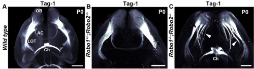
Wild type mouse brain showing Barhl1 in green and Robo3 in red. Movie 1 from Dominici, et al., 2018
Can you give us the key results of the paper in a paragraph?
AC & CD We show that the floor plate-specific deletion of all Slits perturbs midline crossing of spinal cord commissural axons (many accumulate at the midline as expected from lower repulsion). But surprisingly, it does not have any noticeable consequence on the development of precerebellar neurons, which still stop at the midline. Likewise, the simultaneous deletion of Slits from the floor plate and the facial nucleus does not alter pontine neuron migration. Moreover, precerebellar neurons lacking Robo1and Robo2 receptors (the two main mediators of Slit repulsion) migrate normally. Together these results suggest that the migration of precerebellar neurons is independent of Slit/Robo signalling and that other guidance cues control midline crossing in this system. They also show that the mechanisms controlling midline crossing differ between spinal cord and hindbrain commissural neurons.
Do you have any clues as to which other signalling cues might be involved in stopping precerebellar neurons at the midline?
AC & DC Semaphorins are obvious candidates as they are repulsive and control midline crossing in the spinal cord as well as in the cortex. However, this is a large protein family (around 30 members), and in most cases conditional knockouts are not available. Semaphorins are also redundant and single knockout often display minor axon guidance defects. Moreover, none of the semaphorin knockouts that were analysed display PCN migration. Alternatively, Ephrins/Eph receptors are also good candidates and a previous study showed that some PCN neurons cross the midline in EphA 4mutants. However, I would rather support the existence of additional guidance cues, outside the usual suspects. In addition, it is likely that different guidance mechanisms control the migration of the different types of PCNs.
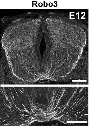
Robo3 in E12 spinal cord sections, from Fig 4, Dominici, et al., 2018
AC & CD Most midline guidance cues (Slits, Netrins, Semaphorins) are expressed in other cell types, including commissural neurons themselves. Therefore, even if the corresponding knockouts have clear axon crossing phenotypes, the direct demonstration of the floor plate as a main source of these factors was lacking. This pushed us to generate conditional knockout lines (Slit2 and Netrin 1) to selectively ablate them from specific cells. In addition, we wanted to use the mice to study the function of these molecules in the postnatal and adult nervous system, which is impossible with the classic knockouts which all die shortly after birth.
The increase in CNS size during vertebrate evolution has put commissural neurons at a larger distance from the floor plate, in particular in the hindbrain, which might explain why they might rely less on floor plate cues, at least initially. This might have been accompanied by a relocation of the main guidance cues to other cell types, closer to commissural neurons.
The imaging in your paper is stunning: can you give us an introduction to technique and how has it changed your view of developing nervous system?
AC & CD The 3DISCO-tissue clearing method was initially developed by Ali Ertürk and Frank Bradke who published it in 2011 in Nature Medicine. The basic principle is to use a series of solvents to solubilize lipids and homogenize the refractive index of the sample (such as a whole embryo, or an adult mouse brain). The method is fast, cheap and works with a wide array of tissues and species. Our addition to this field was to adapt whole-mount immunostaining on top of tissue clearing. Fluorescent dyes are not altered during clearing and samples can be quickly imaged in 3D using a light-sheet microscope. This has significantly facilitated and accelerated the phenotyping of our axon guidance mutants and also diminished the number of samples needed to understand the defects (as they can be oriented in all directions using a 3D Imaging software). We can also be much more precise in the evaluation of the phenotypes of compound mutants. Dissecting the embryo is not needed with this method, therefore you can also easily extend your analysis to organs that are outside the CNS, something that would be too time-consuming using classic histology.
Tracking inferior olive neurons, from Fig 8, Dominici, et al. 2018
When doing the research, did you have any particular result or eureka moment that has stuck with you?
CD When I began to study motorneuron development, I wanted to visualize motor pools in 3 dimensions. This coincided with the emergence of the first tissue clearing techniques and commercial light sheet microscopes. We first used the 3DISCO clearing protocol, but GFP, the reporter used to label axons in our lines, quickly bleached after clearing. I wondered whether we could still visualise GFP using immuno-labelling. I decided to test an in toto immunostaining on a small portion of spinal cord and then cleared it, but I wasn’t really expecting to see anything. To my surprise, it worked! The images were incredible! We then optimised the technique and I could later apply the same technique to visualise migrating precerebellar neurons in 3D.
Not only were we able to visualise the migration of this neuronal population like never before, but we also were able to show that conditional Slits removal had no impact on precerebellar neuron migration.
And what about the flipside: any moments of frustration or despair?
CD Whilst it was a eureka moment, it was also a moment of despair when we realised that conditional removal of Slits in motor neurons showed no clear phenotype. Moreover, the generation of the mouse lines (more than 30) for my project was very difficult. In some mutants we had a one in sixteen chance of getting the right combination of genes. Indeed, the revision of this paper took almost a year due to the difficulty of obtaining additional mutants.
What next for you Chloé?
CD My PhD was quite intense. So I decided to take a year off after defending my PhD in 2016. I travelled and looked for multiple other opportunities. After taking a step back from research, I have realised that it is still something I am incredibly passionate about and would like to pursue. I am currently applying for postdocs.
Where will this work take the Chédotal lab?
AC Our work shows that the midline puzzle is not solved. Commissural neurons are highly diverse: some migrate to the midline, some cross it, others don’t, some project to both sides of the CNS and so on. How could a single unifying model account for this diversity? It has been considered for a long time that midline crossing mechanisms were similar in all bilaterians but this is not the case. We are now trying to identify new regulators of midline crossing using genetic screens, proteomics and transcriptomics. We are also comparing guidance mechanisms in multiple vertebrate species to determine if changes in receptors and ligands played a role in brain evolution, by modifying neuronal connectivity and migration. Some of our preliminary results seem to support this hypothesis.
Finally, let’s move outside the lab – what do you like to do in your spare time?
AC Cooking is one of my favorite hobbies and is the best method I know to rake my mind off science. This said, running an experiment and cooking a dish have a lot in common.
CD Unfortunately during my PhD, I had little time to do much extracurricular activities. But when I managed to squeeze some experiments, I took some time out of Paris to visit my family and friends. This really allowed me to take a step back from the stress of the PhD.
We are looking to appoint a talented post-doctoral researcher to work on an exciting new regenerative medicine project. In this project we will evaluate the effectiveness of using a physiological matrix scaffold created by organ decellularisation to support the differentiation of stem cells into insulin-producing beta cells. This collaborative 3 year project is funded by Diabetes UK and is based in the research groups of Dr Natasha Hill at Kingston University and Dr Aileen King at Kings College London (Guys Campus).
The Person
The project will involve stem cell culture and differentiation, pancreas decellularisation, fluorescence microscopy, flow cytometry, methods to evaluate dynamic insulin release and calcium flux, and in vivo evaluation of beta cell function. Candidates with a relevant PhD degree, ideally with experience in stem cell differentiation or organ decellularisation, and who are interested in developing new treatments for people with diabetes are particularly encouraged to apply.
Further information
Candidates must be able to demonstrate their eligibility to work in the UK in accordance with the Immigration, Asylum and Nationality Act 2006. Where required this may include entry clearance or continued leave to remain under the Points Based Immigration Scheme.
Please also submit a CV and covering letter outlining your interest and suitability for the role.
This is a fixed term contract for 3 years.
To apply click here to go to the Kingston University website.
If, for accessibility reasons, you need to apply in an alternative format, please email jobs@kingston.ac.uk for an application pack or call the HR Shared Services team on 020 8417 3118.
The epic journey of embryogenesis begins with a set of maternal instructions. These instructions are in the form of transcribed mRNA, some even translated into proteins and ready for action. However, many of the critical maternal mRNAs are inactive and must be delivered to the right cell and activated at the right time to encode proteins necessary for development. Regulation of inactive mRNA transcripts is largely dependent on RNA binding proteins (RBPs) (1). However, the mechanisms whereby RBPs reactivate maternal mRNAs in the early embryo are largely unknown. As a doctoral student in Craig Mello’s lab, I was interested in how some RBPs regulate maternal mRNAs responsible for determining the developmental fate of embryonic cells. I wondered how these RBPs turn some cells into muscle and others into neurons, for example.
To pursue this quest, I used the nematode C. elegans, a powerful genetic model to discover genes involved in embryogenesis and to study how they interact together. Mutations in several RBP-encoding genes result in dead C. elegans embryos with cell fate errors, such as germ-cells turning into gut, or neurons becoming muscle (2-4). This further highlights the importance of RBPs in cell fate determination. I was especially fascinated by POS-1, an RBP present in posterior cells of early embryos (5). Depletion of POS-1 results in embryonic death and a complete loss of intestine (gut) (Figure 1, pos-1 mutant). I set out to study the role of POS-1 in the regulation of maternal mRNAs critical for gut fate.
Figure 1. Gut development in wildtype embryo, pos-1 mutant (-, pos-1 loss-of-function), or pos-1 mutants in conjunction with gld-3, gld-2, or neg-1 RNAi silencing. pos-1 mutants are gutless whereas gut is restored upon treatment with either gld-3, gld-2 or neg-1 RNAi. Additional defects that affect the anterior lineages of pos-1 mutants are not restored (compare the anterior ends of embryos to that of wildtype).
To identify the mechanism whereby POS-1 specifies gut fate I conducted an RNAi suppressor screen. I silenced hundreds of genes and asked whether gut fate in POS-1 deficient embryos could be restored. Interestingly, silencing another RBP-encoding gene, gld-3, restored gut in pos-1 mutant embryos (Figure 1, gld-3 RNAi). This result showed that RBPs can positively (POS-1) or negatively (GLD-3) affect gut fate.
Previous studies had shown that GLD-3 recruits the cytoplasmic poly(A) polymerase GLD-2 to mRNAs (6). GLD-2 then adds adenosine (A) nucleotides to mRNA tails to activate their translation to protein. Consistent with GLD-3 and GLD-2 functioning together, silencing gld-2 also restores gut in POS-1 deficient embryos (Figure 1, gld-2 RNAi). This suggested that both GLD-2 and GLD-3, by elongating poly(A) tails, were involved in POS-1 regulation of gut fate specification. Therefore, I asked which maternal transcripts being activated by GLD-2 and GLD-3 were relevant for gut fate?
Among the genes that I discovered as regulators of gut development in pos-1 mutants was the putative transcription factor neg-1(Figure 1, neg-1 RNAi). Remarkably, silencing GLD-2 or GLD-3 decreased the length of neg-1 poly(A) tails, resulting in neg-1 inactivation. This suggested that GLD-2 and GLD-3 could prevent gut formation through neg-1 activation. Pursuing neg-1 was risky, however, since it had no homologs and therefore nothing hinting at its potential function. During a lab meeting, I expressed reluctance to study neg-1 with the excuse that “there is nothing known about it, nothing, and no homologues!”. To which a postdoc retorted, “We spend our careers looking for something new instead of more of the same. You have a shot at something novel and you’re throwing it away!” It was then that I realised we were before true novelty, for it was even beyond my own preconceived impressions of what qualifies as new.
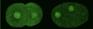
We asked whether POS-1 actually specifies gut fate by inactivating neg-1. We hypothesized that POS-1 inhibits GLD-2/GLD-3 polyadenylation of neg-1, leading to gut fate specification. Consistent with this hypothesis, we found that silencing pos-1 increases the length of neg-1 poly(A) tails and consequently NEG-1 protein levels. Moreover, tagging NEG-1 protein with GFP showed that it was absent in the posterior cells where POS-1 is present after the two-cell stage (Figure 2). Discovering that NEG-1 was an asymmetric protein was the moment the entire project started making sense and the pursuit of a novel gene paid off (See Video below for the Eureka! moment). Most importantly, we also discovered that POS-1 can bind regions in neg-1 mRNAs near the poly(A) tails (Figure 3, Posterior). Taken together, our results showed that POS-1 promotes gut fate in the posterior by preventing GLD-2/GLD-3 activation of neg-1 mRNA (7).
Figure 2. NEG-1:GFP in the two and four-cell stage C. elegans embryo. Note the nuclear localization is asymmetric in the 4-cell stage (two anterior nuclei only).
Considering the dramatic effect of gld-2 or gld-3 knockdown in pos-1 mutants (complete restoration of gut) (see Figure 1), I then asked if cytoplasmic polyadenylation was a general process in the early embryo rather than a neg-1 specific affair. We assayed poly(A) tails of thousands of transcripts following GLD-2 or GLD-3 depletion using a method based on next-generation sequencing (PAT-seq). I found that maternal mRNAs of hundreds of genes are polyadenylated by GLD-2 and GLD-3 and that some, like neg-1, are regulators of cell fate. In the future it would be exciting to explore the roles these GLD-2/GLD-3 activated maternal mRNAs.
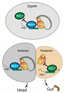
One critical question remained: If POS-1 represses NEG-1, how could both proteins be present in the zygote? I noticed that one of the POS-1 binding sites in neg-1 mRNAs overlapped with a region recognized by another protein called MEX-5. POS-1 and MEX-5 belong to the same group of RNA binding zinc-finger proteins. MEX-5 is also critical for cell fate determination, but unlike POS-1 is present mostly in anterior cells of the early embryo (Figure 3, Anterior) (3). In the zygote, however, MEX-5 and POS-1 are present in the same cell. We found that MEX-5 outcompetes POS-1 in binding to neg-1 mRNA in vitro. Moreover, translation of neg-1 mRNA to protein required MEX-5. This suggested that MEX-5 enables the activation of neg-1 transcripts by blocking POS-1 binding. An intricate circuit was emerging before us: POS-1 represses neg-1 in the posterior to protect gut fate. Conversely, MEX-5 liberates neg-1 from POS-1 repression in the zygote and future anterior cells so that neg-1 can in turn repress gut fate where the head must develop (Figure 3). Morever, the PAT-seq analysis revealed that POS-1 represses mex-5 mRNA as well, consolidating the anterior/posterior divide.
Figure 3. Diagram of one-cell and two-cell stage embryos. In the zygote, MEX-5 blocks POS-1 to allow GLD-2/GLD-3 polyadenylation of neg-1 mRNA, which then is translated to NEG-1 protein (not shown). In the two-cell stage embryo, the posterior blastomere gives rise to the intestinal (gut) lineage, whereas the anterior blastomere gives rise to neuronal, hypodermal and muscle lineages that form the head. POS-1 binds to neg-1 mRNA in the posterior blastomere and prevents GLD-2/GLD-3 mediated polyadenylation.
In the perennial quest to discover the logic of life, the embryo has been the tome of reference. My doctoral research in the Mello lab has contributed to this long tradition of biological studies by linking a family of RNA binding proteins (CCCH zinc-fingers) with cytoplasmic polyadenylation and demonstrating the importance of this connection during embryogenesis. Cytoplasmic polyadenylation is prevalent throughout the tree of life. It is a crucial, for example, to quickly activate mRNAs and produce synaptic proteins in mammals during learning (8). CCCH zinc-fingers are also deeply rooted in life, from single-cell organisms to humans. These RBPs have been shown to mediate the swift removal of mRNAs, for example, in the immune system during acute inflammation (9). My doctoral research has shown that these conserved mRNA regulators collaborate at the commencement of nascent life to decipher the cache of maternal instructions for cell fate and beyond.
The epic journey continues, for the tales of fate do not end here.
References:
L. Weill, E. Belloc, F. A. Bava, R. Mendez, Translational control by changes in poly(A) tail length: recycling mRNAs. Nat Struct Mol Biol 19, 577-585 (2012)
C. C. Mello, B. W. Draper, M. Krause, H. Weintraub, J. R. Priess, The pie-1 and mex-1 genes and maternal control of blastomere identity in early C. elegans embryos. Cell 70, 163-176 (1992)
C. M. Schubert, R. Lin, C. J. de Vries, R. H. Plasterk, J. R. Priess, MEX-5 and MEX-6 function to establish soma/germline asymmetry in early C. elegans embryos. Mol Cell 5, 671-682 (2000)
B. W. Draper, C. C. Mello, B. Bowerman, J. Hardin, J. R. Priess, MEX-3 is a KH domain protein that regulates blastomere identity in early C. elegans embryos. Cell 87, 205-216 (1996)
H. Tabara, R. J. Hill, C. C. Mello, J. R. Priess, Y. Kohara, pos-1 encodes a cytoplasmic zinc-finger protein essential for germline specification in C. elegans. Development 126, 1-11 (1999)
L. Wang, C. R. Eckmann, L. C. Kadyk, M. Wickens, J. Kimble, A regulatory cytoplasmic poly(A) polymerase in Caenorhabditis elegans. Nature 419, 312-316 (2002)
A. Elewa, M. Shirayama, E. Kaymak, P. F. Harrison, D. R. Powell, Z. Du, C. D. Chute, H. Woolf, D. Yi, T. Ishidate, J. Srinivasan, Z. Bao, T. H. Beilharz, S. P. Ryder, C. C. Mello, POS-1 Promotes Endo-mesoderm Development by Inhibiting the Cytoplasmic Polyadenylation of neg-1 mRNA. Developmental cell 34, 108-118 (2015)
M. Ivshina, P. Lasko, J. D. Richter, Cytoplasmic polyadenylation element binding proteins in development, health, and disease. Annual review of cell and developmental biology 30, 393-415 (2014)
S. A. Brooks, P. J. Blackshear, Tristetraprolin (TTP): interactions with mRNA and proteins, and current thoughts on mechanisms of action. Biochimica et biophysica acta 1829, 666-679 (2013)
The Feburary 1st deadline to apply to the Embryology Course at the Marine Biological Laboratory (MBL) is quickly approaching. Maybe you’ve only just learned about the Embryology Course, or perhaps you’ve been meaning to apply to it for years but never pulled the trigger. Either way, I’m writing this blog post to (1) convince you to apply to the course and (2) answer any questions (I imagine) you might have about the experience.
First, some background. The Embryology Course was founded in 1893 at the MBL in the picturesque New England town of Woods Hole. The MBL is a world-renowned institution, and its visiting and resident scientists have made many major scientific contributions over the years, including the discovery of microtubules, kinesin, the squid giant axon, cyclin and GFP. The Embryology Course is one of the outstanding summer courses offered at the MBL. Since its inception, 6 Embryology students and 8 Embryology faculty members have won Nobel Prizes, and many others have become leaders in the field.
I took the Embryology Course in 2017 with 23 other scientists from around the world. It was an amazing and, literally, life-changing experience for me (see later). However, I was hesitant about applying because I wasn’t sure I had the time to take the course, and I didn’t know very much about it. Below I have asked questions that Tessa-circa-January-2017 would have wanted the answers to, and I’ve answered them with my newfound wisdom. I hope you find it useful.
Who can apply for the course? What stage of your career should you be in?
The course is designed for scientists at a range of career stages. The course directors strike a great balance of admitting junior to senior graduate students, postdocs, and a token faculty member (we love you Steve!) In general Embryology students are already in graduate school (or beyond), although we had the pleasure of being joined for a week by undergraduate Evan Brooks, who was selected to participate as a Society for Developmental Biology (SDB) Choose Development! Fellow.
Embryology students come from all over the world, which is one of the forms of diversity we really cherish. Last year we had students from Brazil, Poland, Chile, Australia, the USA, Spain, India, the Czech Republic, France, the U.K., Canada, Germany and Sri Lanka. Not bad!
The 2017 Embryology students re-enacted the scientific event of fertilization during the Woods Hole 4th of July parade, complete with powder paint and a unicorn drummer (AKA course director Rich Schneider). Photo credit: Maren Caruso
Is it okay to have never studied developmental biology in your life? Is it okay to already know quite a lot about developmental biology?
Yes and yes. Students came into our course with very different specialties, which was a great strength. There were experts in computational biology, engineering, evolutionary biology, physics, cell biology, genetics, developmental biology and mathematics. The scientific diversity made our discussions all the more interesting, strengthened the partnerships we formed to tackle biological questions, and was a fantastic way to learn from one another.
Why do people usually take the course?
There are many reasons. For my classmates the motivations included: (i) to learn how to do experimental biology; (ii) to find an exciting lab to do a postdoc in; (iii) to learn more about the field of developmental biology before transitioning into it; (iv) to find the model organism and question to motivate their research for the next 5+ years; (v) to finally meet Ray Keller in person.
How is the course structured?
The course comprises 6 modules (1 per week) that are organized by organism. We had modules that covered the more traditional model systems, such as ‘Zebrafish and Frogs’ and ‘Chicks and Mice’ and others that introduced weird and wonderful beasts, such as ‘Urochordates, Hemichordates, Cephalochordates, and Ctenophores’ and ‘Nematostella, Hydra, Acoels, Planaria, Molluscs, & Annelids’. In total, we estimate we were exposed to almost 100 different animal species! With each module came an outstanding array of faculty who introduced us to their organism, explained their lab’s research, and showed us the tricks of the trade.
What happens on a typical day on the Embryology Course?
I wrote an article for SDB that outlines a typical day on the course. Enjoy!
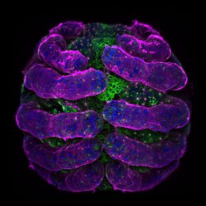
A spider embryo (Parasteatoda tepidariorum) I imaged during the Embryology Course. Blue: DAPI, Green: Acetylated tubulin, Pink: HRP
I’m kind of busy this year. Maybe I’ll apply next year.
That’s not a question…
Most of us find excuses for not applying to the course… “Now’s not a good time – maybe next year”… “Think of all the ground-breaking research I could do in my home lab in 6 weeks”… “I like sleeping”…
It will never be the perfect time to take the course, so just make it work. You will; we did. Anna Czarkwiani wrote a great article about why you should be fearless and apply for the course.
The course is expensive. How do I pay for it?
You should try to find some funding from your home institution, fellowship or advisor, but the course can also provide generous support to help cover the costs (we are very grateful to a number of private and public sponsors including the Company of Biologists and SDB). Money shouldn’t prevent you from applying to the course, and you don’t need to have all the funding when you send in your application.
What is the living and eating situation?
Housing is provided on campus in a room you share with another student at the course. It may sound a little daunting to share a room, but it is easy and fun and another great way to make a close friend. Our meal plan provided 3 meals per day at MBL’s cafeteria, Swope. It may not be the luxurious dining experience of your dreams, but Swope has multiple healthy (and some non-healthy) options, a full salad bar every day, an array of desserts, and lots of coffee. Most importantly, it is quite pleasant to have food prepared for you every day. What a luxury! I welcomed the 6-week period I didn’t have to rummage for food every time I got hungry.
When students experienced palate fatigue, we ate at the restaurants in Woods Hole. In addition, each faculty member was taken to dinner by two students, so on those nights we enjoyed some very fine (and free) dining. There is also a great bakery in town, Pie in the Sky, which is open late, so there was always somewhere to get snacks to keep us going.
Will I get no sleep for 6 weeks? I’m a mess when I don’t sleep.
Wow, me too! As you will read in the other articles about the Embryology Course, sleep is often forsaken in favor of science, socializing or both. That’s one of the magical elements of the course and the MBL: there is so much enthusiasm and energy in the air that people don’t even notice they’re staying up later and later and sleeping less and less. With that said, some people can tolerate extreme sleep deprivation while others can’t. I am one of the people who can’t, so if I got a short night, I would usually follow it with a longer one – we make our own evening schedules so there’s no pressure to stay up really late. Some students took power naps during the day, and others maintained a ‘healthy’ blood caffeine concentration. I settled into the habit of taking an afternoon stroll to Pie in the Sky and getting an afternoon coffee and PBJ bagel to pick me up.
What do you do in your time off? Do you have any time off?
Lectures and labs run from Monday-Saturday, so Sunday is our day off. We usually did some combination of: sleeping, going for bike rides on the beautiful ocean bike path, cycling/driving to Falmouth to visit the French bakery, sleeping, lying on the beach, playing softball and basketball, having parties and barbecues at course directors Dave Sherwood’s or Rich Schneider’s house, swimming in the sea, sleeping, etc. One Sunday we went on a great whale watching trip from Provincetown.
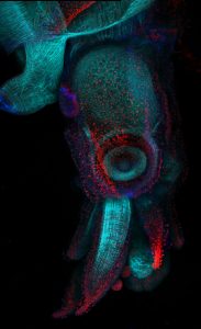
A squid embryo (Doryteuthis pealeii) I imaged during the Embryology Course. Blue: DAPI, Cyan: phalloidin (F-actin) and Red: FMRF (neurons)
What lasting effect has the course had on you?
For me, it has had two major effects: (1) it has given me confidence, and (2) it has changed my life (remember?)
In addition to learning new experimental skills and honing my problem-solving and microscopy prowess, the course gave me the confidence to ask lots of questions during the so-called Sweat Box that follows lecture. I stopped worrying about whether I was asking “good” questions and it was quite liberating. In addition, although the majority of my experiments failed, I also experienced moments of awe and wonder (for instance, watching a tardigrade dump an almighty poop before my eyes) and I had some successes. Since completing the course, a time lapse I made of 9 organisms developing for 24 hours with my classmate Zuzka Vavrušová recently became a winner of the FASEB BioArt Competition. In addition, some of my work has been shared online by Zeiss and SDB, and a recent article on the Harvard Medical School news site used a video I made of the crazy contractions of a fertilized Xenopus egg to illustrate new research by Marc Kirschner’s group. It turns out that everyone thinks embryos are amazing (..duh).
And the course changed my life. I went into it hoping to find an organism and a question to challenge and inspire me for my postdoc and beyond, and I found it. Since the course ended I have interviewed for postdocs on a crazy cephalopod project I would never have conceived before the course, and I have been awarded a Grass Fellowship to begin my cephalopod project at the MBL next summer for 3 months. I am really excited about my scientific future, and I am so inspired by and grateful for the experience I had on the Embryology Course.
Final thoughts?
The Embryology Course is a wonderful, messy, exciting and extremely fun experience that will leave you with friends-for-life, a network of faculty members around the world who might be your colleagues one day, skills in microscopy, embryology, troubleshooting, softball, and a constant source of inspiration and confidence. But to really know what the Embryology Course is like, you’ll have to take it yourself.
A motivated postdoctoral fellow is being recruited to study the role of Hedgehog signaling in craniofacial morphogenesis and birth defects. The project integrates clinically-relevant mouse models, cutting edge microphysiological approaches, and advanced imaging modalities. The applicant should have a Ph.D. and a strong background in developmental biology. Preference will be given to candidates with experience with mouse embryo manipulation and bioinformatics. Anticipated start date is June 1st, 2018. More about the research and mission of the Lipinski laboratory can be found at: https://www.vetmed.wisc.edu/lab/lipinski/
The University of Wisconsin is a great place to postdoc. UW-Madison is among the top five public institutions in research expenditures, second among public institutions in the nation for research and development, and has more NIH-funded postdoctoral training positions available than any other institution. The UW campus is the heart of Madison, which consistently ranks among the top 5 best US cities for young professionals to live, work, and raise a family. More about postdoctoral opportunities and support at UW can be found at: https://uwpa.wisc.edu/ and https://postdoc.wisc.edu/.
Please email a CV, statement of research interests and contact information for at least three referees to:
The impact of developmental biology on society is particularly acute when it comes to reproduction – research informs efforts to assist reproduction and understand what happens when pregnancy goes wrong. Recent developments in stem cells, culturing conditions, gene editing and sequencing are also revealing aspects of human embryonic development previously hidden from us. Here at Development, we are committed to promoting human developmental biology, through a dedicated section in the journal, regular meetings (you can book your place on our September ‘From stem cells to human development’ meeting now) and special issues (our upcoming one is accepting submissions until March 1st). As well as promoting the science, we like to encourage discussion of its impacts on society, and have published front section articles on various aspects of embryo ethics.
A collection of Development Spotlight articles focused on the human embryo
Given all of this I was very excited to attend the Progress Educational Trust (PET) meeting ‘Crossing Frontiers: Moving the Boundaries of Human Reproduction’, held in Camden, London, last month. The PET was established in 1992 with roots in an earlier campaign for the continuation of human embryo research, and through events and publications aims to ‘inform debate on assisted conception and genetics’. The day conference brought together representatives from research, law, policy, ethics and science communication, and an audience of medical students, patient group representatives, genome biologists, journalists, academics and members of the public with an interest in the topic.
Below are some of my highlights of what turned out to be an engaging and fascinating day. If you were at the meeting I would love to hear your thoughts on the debate, and for you to clarify any mistakes or misrepresentations I have (inevitably) made.
Making gametes in and out of the body
Allan Pacey and Richard Anderson kicked off proceedings by discussing the cells that kick it all off to begin with: sperm and eggs. Human reproduction can be challenging, and much of the challenge lies in making viable gametes (‘Being a good egg ain’t easy’, as Anderson put it). We’re also in a society where, as Anderson pointed out, the birth rate in women under 20 is exceeded by that of women over 40. Pair this with the dramatic reduction in fertility of eggs used for IVF as women get older, and we have a problem. (Unless, as a member of the audience later pointed out, one does not want to have children; she even asked whether as a society, given the planet’s population pressures, we should be doing more to encourage women not to have children.) Thousands of women are freezing their eggs, but this procedure comes with its own issues, not least the limited supply of eggs, and of course has its own costs.
One way these challenges could be met is to achieve gametogenesis in vitro: make gametes from stem cells. Azim Surani described how little we know about human gametogenesis and the incredible journey the gamete precursors, primordial germ cells, make in the embryo, but also how research performed on human embryos and animal models was changing things. Truly ‘closing the gap’ between the soma and the germline would have powerful implications: just imagine, a participant in one of the panel discussions said, making thousands of viable eggs from a single skin sample from a female donor over the age of forty, or fifty, or sixty; it would be quite a game changer. (I was a little dumbfounded by the possibility, discussed later over a coffee with someone who wanted to invest in the technology, of generating eggs from men, though I really don’t know where the science is in that regard – any insights would be appreciated in the comments ?)
Definitions, definitions
Technological advances can call into question existing terminologies as well as forcing us to come up with new ones. Sue Avery described how this is nothing new: the advent of IVF in the late seventies had not only transformed fertility but also revolutionised our understanding of human development. Before then, we really didn’t have much of an idea of what an early human embryo looked like, and seeing the process of embryogenesis under a microscope raised the issue of how and when exactly we defined an embryo. Magdalena Zernicka-Goetz then introduced the more recent creation of ‘embryo-like structures’ from human stem cells, made possible by tinkering with culturing conditions and inspired by lessons from the embryo. This give a glimpse into the black box of implantation, the point at which the body plan is established, and which is a crucial nexus for pregnancy failure.
It is remarkable to think how these experiments (and earlier ones using mouse embryonic stem cells) subvert biology, in the sense of not requiring fertilisation to generate something that looks like an embryo. Hence the inevitable question, explored by Robin Lovell-Badge, of when ‘embryo-like’ becomes ‘embryo’, and of the moral and legal status of these structures. Lovell-Badge introduced ‘synthetic human entities with embryo-like features’ (SHEEFs), a term coined by John Aach, George Church and colleagues that encompasses ‘embryo-like structures’ as well as stem-cell derived organoids and gastruloids (embryos derived from fertilised eggs, whether through sex or assisted reproduction, are, in this parlance, rather jarringly termed ‘non-synthetic’).
The ethical questions raised by SHEEFs often include reference to the 14 day rule – the fortnight limit for the culturing of human embryos in vitro, around the time that the primitive streak appears. So should similar limits exist for SHEEFs? It makes little sense, it would seem, to give a defined time limit to a process that does not involve a day 0 (i.e. fertilisation). According to the 14 day rule, gastrulation marks the point at which you should not pass, but, Lovell-Badge asked, what if synthetic biology allowed us to bypass it all together? (I think the idea was to 3D print tissues to make something that looked or like a neural tube for instance without it ever having passed through gastrulation.) And then there are the inevitable questions of sentience: if a SHEEF develops the neural structures for pain recognition, but not those for consciousness, should we care?
For Lovell-Badge, we shouldn’t get too far ahead of ourselves – he pointed out that in terms of self-organisation/self-assembly, we’ve had teratomas in the lab for years, and they can often make incredibly complex structures like eyes or teeth. SHEEFs could thus have moral equivalence with teratomas. Furthermore, since they are derived from stem cells, SHEEFs currently hold the same status as any stem-cell derived structure (at least the UK, as far as I could tell), and so might not require a legalistic overhaul. It all brought me back to a session in last year’s ISSCR meeting in Boston (I wrote about it here), where in a session on the ethics of organoids Melissa Little argued that organoids were just extensions of stem cell differentiation protocols and did not require a radical rethink of stem cell guidelines, and Jeurgen Knoeblich argued that ethical debates about consciousness in cortical organoids were based on perceived rather than real risk.
Stepping back from visions of Gattaca
In the ‘What next for genome editing?’ section, Andy Greenfield began with a plea to avoid adopting tedious tropes when we discuss ethics – people immediately bring up Orwell, Huxley, Gattaca, designer babies and eugenics, when the more interesting and pressing ethical questions concern what researchers are developing right now. Not only are these more interesting, they are also positive, in terms of promoting how research is helping human health. I liked this positive, contemporary angle: it’s not that we shouldn’t have discussions about future advances, it’s that they can become a bit like a session in the Black Mirror writing room, where imagination outpaces reality, and we fail to deal with what is feasible today.
Many of the ethical concerns in the meeting hinged around the differences between gene editing of the soma and of the germline. Greenfield made the point that we already perform germline therapy, in the form of preimplantation genetic diagnosis (PGD), which imposes non-random transmission of genetic material and leads to a change in future generations. Much ethical discussion of potential future gene editing and the human population could thus be refocused on PGD. Philippa Taylor was stridently against any form of germline editing – you can fix the patient (somatic editing is in line with medicine as she sees it), but the patient’s offspring have no say in the issue, and as they in a sense represent the human race, that is another issue entirely. Here, Greenfield’s point seemed pertinent: we already prevent the birth of individuals whose genotypes are deemed unfavourable, so there is not much ethical distance between PGD and a safe and efficacious form of gene editing, which, he stressed, we donot yet have. So, for those who oppose germline editing, the question might be turned on its head – do you support PGD, a practise which has become routine? Unless I missed it, I’m not sure opponents of germline editing addressed this question in the session.
****
Update 19/01: I heard back from Philippa Taylor on Twitter:
“To answer q about my view-yes we’ve always opposed PGD for ethical reasons & I said so in panel discussion”
So I did miss it – apologies to Philippa, and thanks for the clarification. I should further clarify here that not everyone at the event who opposed germline editing did so for the same reason (many would be in favour of PGD, I imagine). It wasn’t a simple case of two sides.
*****
Greenfield proposed that not only should we continue gene editing, we also need to continue literal editing, in terms of proper science communication as a prerequisite for a mature, informed debate about the science. Sandy Starr agreed – not only is there a societal duty to do so, but human embryo research is dependent on fertility patients as donors of ‘non-synthetic’ embryos, and so is dependent on their continued support. Starr described how, with help from patient groups and practitioners, the PET generated a report on the basic understanding of genome editing. Starr argued getting your terms and definitions right is crucial. For instance, he insists on ‘genome editing’ rather than terms like ‘gene engineering’, as it best describes what is going on (I have undoubtedly strayed from his terminology in this article!). Practitioners and science communicators should be able to describe technologies like CRISPR clearly, but also to emphasise that not all editing is CRISPR-based, and indeed that CRISPR is not the end of the story. But how to define a genome in the first place? It seems a conceptually simple and complex at the same time – there are, proposed someone from the floor (I think it might have been Philip Ball), more than 7 billion human genomes, so it is not one steady state. Another audience member pointed out that one place to increase scientific literacy was in the classroom, but unfortunately the ethical implications of science tend to be taught separately from the science itself.
Where next?
The final session took quite a broad look at the future of reproduction. Anna Smajdor explored how artificial gametes like those discussed by Azim Surani earlier in the day had the potential to ‘democratise’ reproduction. Biological boundaries were, with technological advances, dissolving in front of our eyes. (Again, currently, while artificial gametes might be realistic, we do not have artificial wombs, so would still need women involved in the process as far as I can tell.) Echoing an above-mentioned audience member, Smajdor wondered whether we focus too much on the positives of reproduction, and argued (in what she later admitted was a bit of a ‘rhetorical flourish’) that sexual reproduction is incompatible with Western liberal values, citing the inverse correlation between increasing indices of female emancipation, for instance education and career opportunities, and reduced child rearing.
Guido Pennings then argued that wide scale (in terms of the genome) and widely adopted germline editing was inevitable, to the demise of PGD. He imagined a future where embryos were screened in a more statistical manner over a range of genetic indicators and altered accordingly, and where parents would be faced not with the ability of creating an ideal embryo, but a good enough one. This drew some critique from the audience – the more we understand genomes, the more complex we understand them to be, and the more we appreciate that so many traits are multifactorial. Thus the dream of a checklist of genetic changes to be edited was not realistic. We are also not anywhere near demonstrating safe and efficacious germline editing in humans, and presumably will not be for some time, so PGD is not going anywhere for now.
This discussion reminded me of the reaction to the Mitalipov paper last year – multiple commentators pointed that, since PGD does very well in selecting against disease-carrying embryos, why would you edit those embryos you would normally discard after PGD? So should we be putting more effort into PGD rather than germline editing?
~
There really were so many interesting ideas floating about, arguments and counter arguments, ideas I winced at initially but later gave me cause for thought, and those I wholeheartedly supported until another came up to question my assumptions. As Andy Greenfield said, some of the most exciting debates concern what we are doing right now, and it must be a very exciting time for anyone involved in human development and reproduction.
The first Development issue of the year is now complete! Here are the highlights:
RESEARCH HIGHLIGHTS:
Micro-lenses focus on cataract development
Cataracts have many potential risk factors but the molecular mechanisms underlying their development are unclear. Aggregates of lens epithelial cells (LECs) derived from human pluripotent stem cells (hPSCs) are a potentially powerful in vitro tool to tackle this problem, but existing protocols have a number of shortcomings, including the aggregates’ inability to focus light. Michael O’Connor and colleagues describe an efficient system for the derivation of LECs from hPSCs, and the subsequent creation of light-focusing ‘micro-lenses’ (dev155838). The cell surface marker ROR1 allows for sorting and purification of LECs, which are then cultured as spherical aggregates. Over the course of around three weeks, the aggregates develop the ability to focus light, associated with the expression of crystalline genes and anatomical maturation to mimic lens morphology in vivo. The micro-lenses promise to be clinically relevant, as shown by an analysis of Vx-770, an emerging cystic fibrosis drug that has an as-yet-unclear association with cataract formation. The authors find that culturing with high concentrations of Vx-770 reduces the light-focusing ability of micro-lenses. hPSC-derived micro-lenses therefore provide a powerful in vitro model for research into lens disorders, their risk factors and their molecular underpinnings.
Plant miRNAs: the root of interspecies variation
Plant roots have evolved a variety of anatomical patterns. For example, it is known that the number of root cortical layers varies amongst plant species. But what are the genetic mechanisms that underlie such morphological differences? Raffaele Dello Ioio and colleagues investigate this by analysing root development in Arabidopsis thaliana and Cardamine hirsuta, a genetically tractable close relative of Arabidopsis (dev153858). They first show that, unlike Arabidopsis (which has a single cortical layer), Cardamine has two cortical layers that arise during embryogenesis from a tissue with mixed cortical/endodermal (CEM) identity. They further reveal that Arabidopsis mutants in which the miRNA biogenesis machinery is perturbed also exhibit an additional cortical layer, likely owing to ectopic expression of the HD-ZIPIII transcription factor PHABULOSA (PHB), which is known to be regulated by the microRNA miR165/6. Following this, the authors show that HD-ZIPIII factors are required in the Cardamine CEM tissue for double cortex formation and that the activity domain of miR165/6 differs between the two species; the absence of miR165/6 in Cardamine CEM tissue allows a broader expression domain of PHB, resulting in the development of an extra cortical layer. Together, these findings highlight that variations in miRNA distribution can lead to differences in plant morphology.
All agog for zebrafish Nanog
The Nanog transcription factor is a core regulator of pluripotency in early mammalian embryos, but its role in other vertebrates – which may offer more tractable models for how it works in embryogenesis – has remained unclear. In zebrafish, Nanog morphants fail to complete gastrulation. While a requirement in extra-embryonic tissue development was suggested to underlie this effect, an additional role in zygotic genome activation in the embryo proper has also been proposed. Now, in this issue, two groups report the generation of zebrafish nanog mutants using TALENs, and describe their effects on embryonic and extra-embryonic tissues during development.
James Gagnon, Kamal Obbad and Alexander Schier show that maternal-zygotic (MZ) nanog mutants arrest at gastrulation, phenocopying the morphants, and that Nanog’s function is provided maternally (dev147793). MZnanog mutants fail to normally form the yolk syncytial layer (YSL), an extra-embryonic tissue that attaches the embryo to the yolk, and fail to express key YSL genes while also showing reduced expression of a subset of early zygotic genes within the embryo. Supporting the argument of a predominantly extra-embryonic functional requirement, wild-type blastoderms fail to undergo epiboly when transplanted onto MZnanog mutant yolk cells, while mutant blastoderms transplanted to wild-type yolk cells undergo epiboly. Finally, MZnanog mutant cells proliferate and differentiate into derivatives of all germ layers when transplanted into wild-type hosts, and are detectable in the adult using CRISPR lineage tracing.
Daria Onichtchouk and colleagues show that MZnanog mutants are arrested in gastrulation movements, and exhibit an increase in cell death (dev155366). Gastrulation arrest in the mutants is accompanied by a failure to properly organise the microtubule and actin cytoskeletons in the yolk. In the absence of Nanog, maternal RNAs fail to degrade; this failure can be rescued by mir-430. The authors show that injection of mxtx2 or miR-430 mRNA into the 1-cell stage embryo can rescue the failure of epiboly, but not cell survival, in the MZnanog mutants. While MZnanog mutant cells can differentiate when transplanted into the wild-type embryos, viability is substantially reduced compared with co-injected wild-type cells, suggesting an autonomous role for Nanog in embryonic cell survival.
Taken together, these papers extend our understanding of Nanog’s relative roles in embryonic and extra-embryonic tissues in the fish, and provide a platform for further comparison with other vertebrate models.
PLUS:
And one last thing
In his Editorial, Olivier Pourquié takes stock of his nine years as Editor in Chief of Development, highlighting the key changes that have been implemented by the journal during this period and reflecting on how the field has changed over this time. Olivier also discusses what’s in store for the journal this year, before he steps down as Editor in Chief in September.
An interview with Cliff Tabin
Cliff Tabin is George Jacob and Jacqueline Hazel Leder Professor and Chairman of the Department of Genetics at Harvard Medical School. His lab aims to understand the genetic control of morphogenesis during embryonic development and its change over evolutionary time. We met Cliff at the Pan-American Society for Evolutionary Developmental Biology’s second biennial meeting, held in August 2017, and heard about how he got into development, how a long-standing interest in the limb has been complemented by ventures into new models, and why he thinks we are in a golden age for evo-devo. Read the Spotlight article here.
Towards stem cell based therapies for Parkinson’s disease
Treating neurodegenerative diseases with cell transplantation has been within reach since the first pioneering clinical trials in which dopamine neuron progenitors from the fetal brain were transplanted to individuals with Parkinson’s disease. However, the use of fetal tissue is problematic in terms of low availability and high variability, and it is also associated with ethical concerns that vary between countries. For decades, the field has therefore investigated new scalable source of therapeutic cells from stem cells or via reprogramming. Here, Malin Parmar, discusses how it is now possible to generate authentic midbrain dopaminergic neurons from pluripotent stem cells and how clinical trials using such cells are rapidly approaching.
Mechanisms of erythrocyte development and regeneration: implications for regenerative medicine and beyond
Hemoglobin-expressing erythrocytes (red blood cells) act as fundamental metabolic regulators by providing oxygen to cells and tissues throughout the body. Whereas the vital requirement for oxygen to support metabolically active cells and tissues is well established, almost nothing is known regarding how erythrocyte development and function impact regeneration. Furthermore, many questions remain unanswered relating to how insults to hematopoietic stem/progenitor cells and erythrocytes can trigger a massive regenerative process termed ‘stress erythropoiesis’ to produce billions of erythrocytes. In their Review, Emery Bresnick and colleagues discuss the cellular and molecular mechanisms governing erythrocyte development and regeneration, and highlight the potential links between these events and other regenerative processes.
mTOR signaling in stem and progenitor cells
The mammalian target of rapamycin (mTOR) senses nutrients and growth factors to coordinate cell growth, metabolism and autophagy. Extensive research has mapped the signaling pathways regulated by mTOR that are involved in human diseases, such as cancer, and in diabetes and ageing. Recently, however, new studies have demonstrated important roles for mTOR in promoting the differentiation of adult stem cells, driving the growth and proliferation of stem and progenitor cells, and dictating the differentiation program of multipotent stem cell populations. In their Review, Jenna Jewell and co-workers review these advances, providing an overview of mTOR signaling and its role in murine and human stem and progenitor cells.
Two postdoctoral positions are open at the Institute of Developmental Biology (IBDM) in Marseille (France) to visualize the dynamics of adhesion complexes and probe the cellular and tissue-level mechanics of developing embryos. Successful candidates will develop a project in the context of an interdisciplinary collaboration between the groups of Thomas Lecuit, a biologist, and Pierre-François Lenne, a physicist, and with theoretical expertise of Raphaël Clément, from the Lenne group.
DESCRIPTION OF PROJECTS
The overarching goal is to understand how tissue scale morphogenesis emerges from subcellular mechanics and dynamics. The projects will tackle two aspects related to this goal. To understand mechanics, the first candidate will develop and use biophysical approaches to probe cellular to multicellular mechanics, including optical and magnetic manipulation. These measurements should shed light on how cells respond to mechanical stress and how they are mechanically coupled. These measurements will be combined with genetic and molecular perturbations, and will feed theoretical models.
To unravel the dynamics of adhesion complexes, the second candidate will develop/implement lattice light sheet microscopy to determine how forces organize and regulate adhesion complexes.
EXPECTED PROFILE OF THE CANDIDATES
Candidates should have at least one of the following skills:
Biophysical instrumentation, microscopy design, design and implementation
Cell and/or tissue mechanics
Computational image analysis, algorithm development
Candidates should have a strong interest for collaborative research and experience in an interdisciplinary environment will be much appreciated. English is the working language.
The two postdocs will benefit from an interdisciplinary environment with expertise in cell developmental biology, biophysics and imaging (IBDM, Labex Inform , CENTURI).
Applicants should send a CV, names of two referees, and a short outline of their research interests to P.-F. Lenne and T. Lecuit.
A post-doctoral position is available in the research group of Dr Anthony Gavalas. The group investigates the role of signaling pathways and metabolism in the late stages of endocrine pancreas development, the application of novel signals for the conversion of human pluripotent stem cells into functional beta cells and the function of adult pancreas stem cells. A combination of directed pluripotent stem cell differentiation, genomics, in vivo genetic analyses and ex vivo experiments using explants and organoids is being used. (https://www.digs-bb.de/research/research-groups/anthony-gavalas/).
The post-doctoral fellow is primarily required for a project that focuses on the manipulation of signaling pathways for the efficient conversion of human pluripotent stem cells into fully functional beta cells. However, depending on prior experience, projects on the metabolic regulation of endocrine pancreas development and newly identified adult pancreas stem cells may also be available.
The successful candidate will have a Ph.D. degree in Biology or related disciplines, a good publication record and extensive experience in cell culture and differentiation of human pluripotent stem cells. Experience with CRISPR-Cas9 mediated genome engineering will be an asset.
The lab is located in the Center for Regenerative Therapies Dresden (CRTD) with full access to state of the art core facilities for Deep Sequencing, including single cell RNA Seq, Genome Engineering, Imaging and FACS analysis.
The salary will be according to the TV-L scale commensurate with experience and qualifications. The contract will be initially for two years with the possibility for renewal. Applicants are requested to send their CVs along with names and emails of at least two referees to Dr Anthony Gavalas (Anthony.Gavalas@tu-dresden.de), before March 20, 2018.
 (No Ratings Yet)
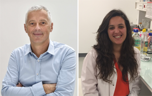
 (3 votes)
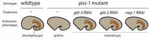


 In his
  Cliff Tabin is George Jacob and Jacqueline Hazel Leder Professor and Chairman of the Department of Genetics at Harvard Medical School. His lab aims to understand the genetic control of morphogenesis during embryonic development and its change over evolutionary time. We met Cliff at the Pan-American Society for Evolutionary Developmental Biology’s second biennial meeting, held in August 2017, and heard about how he got into development, how a long-standing interest in the limb has been complemented by ventures into new models, and why he thinks we are in a golden age for evo-devo. Read the Spotlight article
  Treating neurodegenerative diseases with cell transplantation has been within reach since the first pioneering clinical trials in which dopamine neuron progenitors from the fetal brain were transplanted to individuals with Parkinson’s disease. However, the use of fetal tissue is problematic in terms of low availability and high variability, and it is also associated with ethical concerns that vary between countries. For decades, the field has therefore investigated new scalable source of therapeutic cells from stem cells or via reprogramming.
  Hemoglobin-expressing erythrocytes (red blood cells) act as fundamental metabolic regulators by providing oxygen to cells and tissues throughout the body. Whereas the vital requirement for oxygen to support metabolically active cells and tissues is well established, almost nothing is known regarding how erythrocyte development and function impact regeneration. Furthermore, many questions remain unanswered relating to how insults to hematopoietic stem/progenitor cells and erythrocytes can trigger a massive regenerative process termed ‘stress erythropoiesis’ to produce billions of erythrocytes. In their
  The mammalian target of rapamycin (mTOR) senses nutrients and growth factors to coordinate cell growth, metabolism and autophagy. Extensive research has mapped the signaling pathways regulated by mTOR that are involved in human diseases, such as cancer, and in diabetes and ageing. Recently, however, new studies have demonstrated important roles for mTOR in promoting the differentiation of adult stem cells, driving the growth and proliferation of stem and progenitor cells, and dictating the differentiation program of multipotent stem cell populations. In their