Department of Clinical and Experimental Medicine, Linkoping University, Sweden.
How cells are generated in proper numbers in order to form specific structures in an organism has been a subject of interest for many years. This issue is particularly intriguing in in the central nervous system (CNS), where the number of neurons of each type plays a crucial role in neural circuitry and cognitive capabilities in different species across evolution1. An intriguing feature of the CNS is that different regions have different cell numbers, and the number of neurons along the anterior-posterior (A-P) axis seems to be organized in a wedge-like appearance: the anterior part contains more neurons than the posterior part. This overall feature of CNS organization holds true for many vertebrate and invertebrate species. So how is the generation of proper number of neurons determined in the different regions of the central nervous system? This was, and still is, one of the questions around when I joined Stefan Thor’s lab in Linkoping, and the one which we try to shed light on in this post.
The total number of cells generated in a certain region is determined by several factors: the initial number of progenitors, the number of divisions that those progenitors go through, the proliferative behavior of the daughter cells produced by progenitors, and programmed cell death (PCD) present within the lineage. Drosophila CNS is subdivided into the brain, and the ventral nerve cord (VNC), equivalent to the mammalian and spinal cord respectively. Recent studies in the Drosophila VNC have provided an essential framework for addressing how the proper number of cells are generated in each segment. First, the recent meticulous mapping of the number of neural progenitors or neuroblasts (NBs) in each of the 13 segments of the VNC 2–6; second, our previous discovery of different daughter proliferation modes in NB lineages, where NBs initially bud off daughters that divide once, to generate two neurons/glia, denoted Type I, and subsequently switch to generating daughters that do not divide, instead directly differentiating, denoted Type 0 7; third, the identification of mutants that lack PCD in embryonic stages8. These findings gave us the opportunity to make a global and systematic study of proliferation in the VNC.
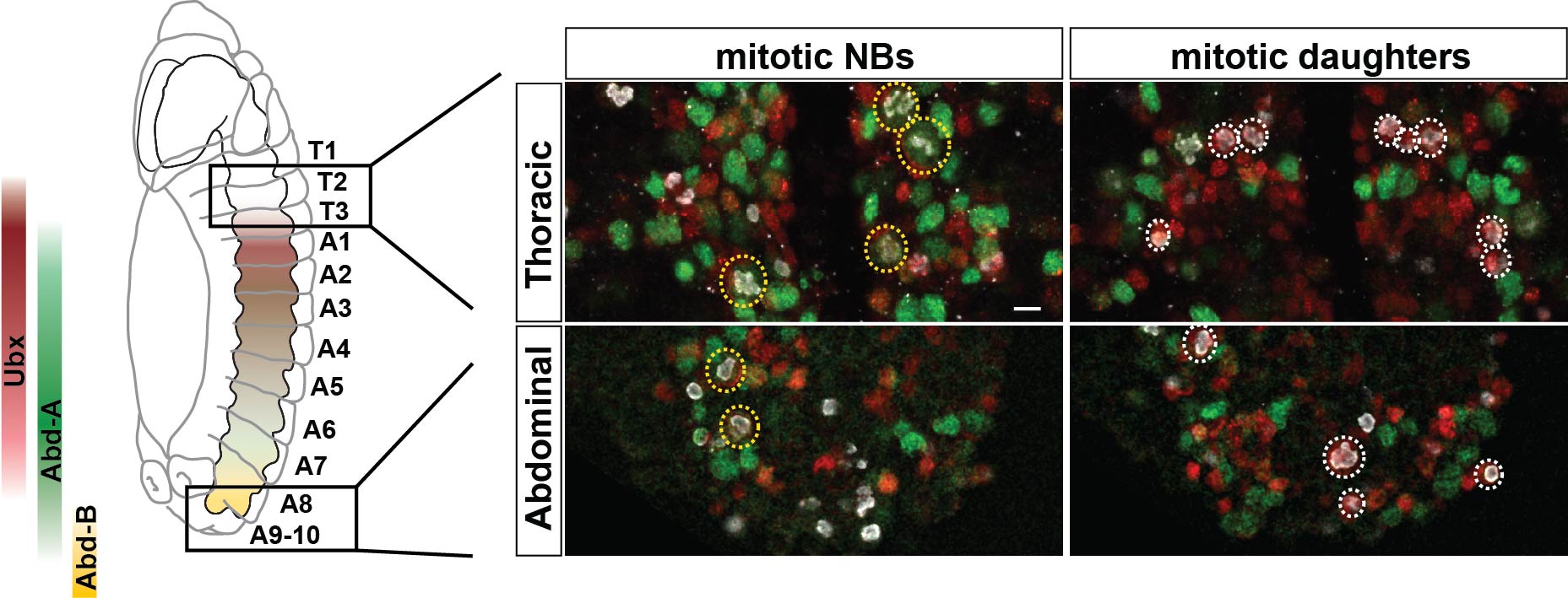
With this set up, our first challenge was to establish just how different the anterior segments are from the posterior ones, and that was one of the most exciting parts in the project. The 13 segments of the VNC are generated by a similar number of progenitors, about 64 per segment, in each segment. Our cell number analysis at the end of neurogenesis revealed that these 64 NBs generate different numbers of cells in different segments, with more cells in anterior segments than posterior ones. Based upon this finding we postulated two main processes that could contribute to this wedge-like appearance: removal of excess cells, by PCD, in posterior segments, and/or increased proliferation in anterior segments. Looking at PCD mutants, the A-P differences in numbers were still noticeable, suggesting PCD could not explain the difference. In contrast, we observed increased proliferation, of both NBs and daughters, in anterior segments, which pointed to proliferation as the main contributor to the A-P differences. To further address this notion, we analyzed proliferation in specific NB lineages, serially presented in all VNC segments. Whereas in anterior segments, NBs undergo more rounds of division and display a late or absent Type I->0 daughter proliferation switch, in the posterior segments NBs undergo fewer rounds of divisions and display an earlier Type I->0 switch. The combination of cell number analysis with global and single-lineage proliferation analysis allowed us to extract an average lineage behavior for thoracic segments versus posterior segments.
Anterior-posterior differences in proliferation behavior. Anterior thoracic segment show a higher number of dividing neuroblasts (NBs) and dividing daughter cells than the posterior abdominal ones.
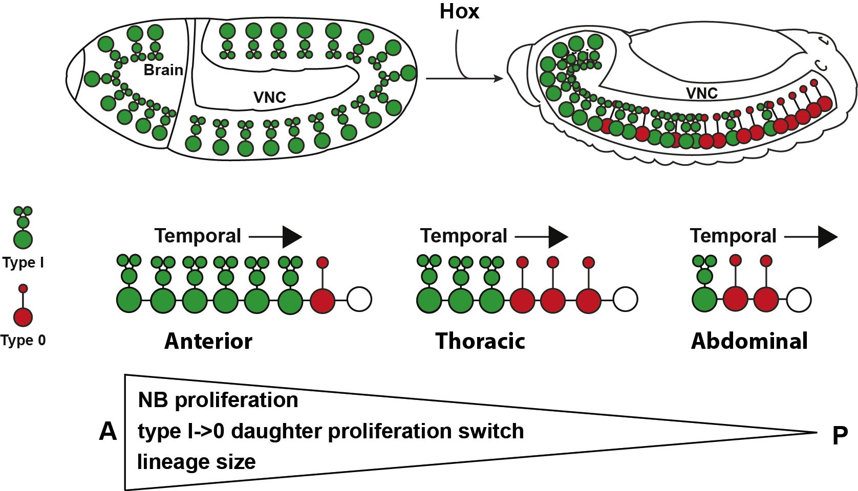
The next goal was to begin identifying some of the players controlling this gradient in proliferation behavior. The A-P axis differences obviously pointed to homeotic domains, and our previous work has identified the Hox gene Antennapedia (Antp) as one of the main triggers of the Type I->0 daughter proliferation switch in the thoracic segments7. Therefore, we analyzed the role of the more posteriorly acting Hox genes of the Bithorax-Complex (BX-C); Ultrabithorax (Ubx), abdominal-A (abd-A), and Abdominal-B (Abd-B). Strikingly, we indeed observed that BX-C genes were important for the earlier Type I->0 daughter proliferation switch and NB proliferation exit observed in posterior segments. However, the triggering of the Type I->0 switch during later stages of lineage progression implied that the action of BX-C was gated in a temporal manner. Our previous studies of Antp had revealed that its expression in thoracic NBs commences during later stages. Studying this, we found that although Hox genes are indeed expressed in an A-P manner during early embryonic stages, early NBs display little if any BX-C protein expression. As NBs lineage progression proceeds, we observed onset of Hox protein expression, establishing the characteristic overlapping domains along the A-P axis, and this late onset in NBs then promotes the Type I->0 daughter proliferation switch. Based upon these findings we propose a model where at early stages NBs progress in a Hox-free “ground state”, allowing for high proliferative modes, whereas at later stages, the onset of Hox expression in NBs promotes the Type I->0 daughter proliferation switch, and eventually NBs stop dividing. This action of Hox genes establishes a gradient in NB lineage size, and results in the A-P wedge appearance.
Hox-Mediated Proliferation Control along the A-P Axis in the VNC
The hypothesis of the ground state is not new in Drosophila, and has been proposed by Fernando Casares and Richard Mann for appendages9. This idea proposes a “default” program, that we believe applies also to NB proliferation behavior, in thoracic segments, which is modified along the A-P axis by the same cues that determine the body plan. This program allows for homologous NBs in different segments to modify their proliferation behavior to generate the proper number of cells needed for that specific segment.
Since Hox genes are evolutionary conserved, establishing the body plan of invertebrates and vertebrates equally, it is reasonable to envision that they may have a conserved function in proliferation control in the nervous system. Indeed, several studies have identified Hoxc6, Hoxb9 and Hoxb13 related to suppression of proliferation in the vertebrate CNS10,11. Most intriguingly, while our current study was focused on the VNC, the most obvious wedge-like appearance in nervous systems is brain size when compared to nerve cord. Given that we have shown that Hox expression suppresses neural progenitor and daughter proliferation, and that the brain is a Hox free region, it seems reasonable to think that Hox genes could be one of the motors of the evolutionary expansion of the brain.
We are now very enthusiastic about the open possibilities of what is happening in the brain. The work is now focused on addressing how proliferation is controlled in the brain, and to what extent could alternate neural progenitor and daughter cell proliferation modes be a common feature, using similar or homologous tools, to establish A-P differences across different species.
We are looking for a motivated, ambitious and collegial person interested in joining us as a postdoc to pursue projects centered around host-pathogen interaction in human stem cell derived macrophages. Prior exprience with stem cells or high throughput microscopy is required. Please contact Eva directly with your CV or with queries if you are interested.
eva.frickel@crick.ac.uk
frickellab.com
This project will dive into regulation of host immunity to infection or infectious stimuli using macrophages derived from human stem cells. More emphasis will be put on deciphering molecular signaling or cellular response pathways than the actual stimuli/infection under study. This will require efficient gene editing approaches for stem cell derived macrophages (CRISPR, siRNA) and the ability to work with high throughput analysis (image based) as well as handling large datasets (RNASeq, proteomic or metabolomic analysis). Thus I believe a person with an interest in using human stem cells to study intracellular mechanisms is best suited for this post.
Please consider visiting us in Sheffield for this exciting symposium this September (register at lifecourse.group.shef.ac.uk). The symposium is being organised by Marysia Placzek, Stephen Renshaw and David Strutt on behalf of the Bateson Centre at the University of Sheffield (https://www.sheffield.ac.uk/bateson). There is an array of outstanding external speakers already confirmed and the opportunity to present your own work (abstract deadline is 15th June). Sign up by 28th July! We look forward to welcoming you to Sheffield!
An NIH R01-funded postdoctoral position is available immediately in the laboratory of Dr. Beth Roman in the Department of Human Genetics at the University of Pittsburgh. This position provides an outstanding opportunity for cross-disciplinary training at the interface of physics and human health, requiring close collaboration with the laboratories of Dr. Lance Davidson (Bioengineering) and Dr. Andrew Hinck (Structural Biology). We are looking for a productive, highly motivated scientist to dissect the interaction between bone morphogenetic protein (BMP)/ALK1 signaling and mechanical force in controlling directed endothelial cell migration, using zebrafish and endothelial cell culture models. Our ultimate goal is to understand how ALK1 signaling disruption in the genetic vascular disorder, hereditary hemorrhagic telangiectasia (HHT), causes arteriovenous malformations (AVMs). AVMs are direct connections between arteries and veins that can lead to hemorrhage and stroke.
The University of Pittsburgh is a collaborative, collegial environment for biomedical research. Our laboratory is part of a large, interactive zebrafish community, and we are affiliated with the Heart, Lung, and Blood Vascular Medicine Institute (VMI), which houses numerous laboratories focused on basic and translational cardiovascular research. Our laboratory is also integral to the UPMC/Pitt HHT Center of Excellence, affording the opportunity to translate research findings to a clinical care setting.
Qualified candidates must have a PhD in biology, bioengineering, or other relevant field, a strong publication record, and excellent oral and written English communication skills. Expertise in fluid mechanics and endothelial cell culture is preferred. Previous experience with zebrafish, dynamic live-cell imaging, confocal microscopy, and image analysis is desired. Interested candidates should send a brief cover letter (maximum 1-page) summarizing scientific accomplishments and outlining motivation for this position, a CV, and full contact information for three professional references to Dr. Beth Roman at romanb@pitt.edu.
For more information, please see
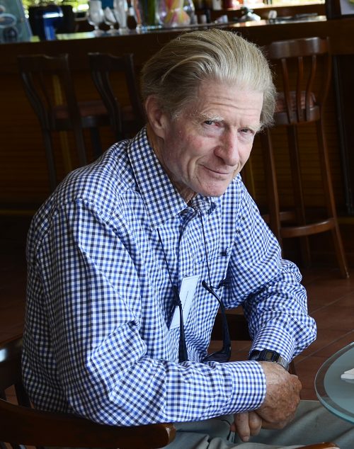
On a bright, cold morning at the beginning of March, I went back to the institute I once worked in to interview the man after whom the place was named. Greeting me at the entrance, John Gurdon apologised for being a little late and asked if it was alright to delay the interview for five minutes while he finished up some lab work. I followed him up the stairs to his lab and through the lab to his office, where he took a plate out of an incubator and placed it between old microscopes and pipettes on a bench that ran the length of one side of the room. Sunlight streamed in and the frog embryos bobbed around as John replaced the media – it was something to do with Halo tagging, apparently – and put the plate back in the incubator, before pulling a chair out to start the interview. As anyone who has met him will not be surprised to hear, he was engaging, humble and lucid in his recollection of experiments started half a century ago and which his lab continues today.
John Gurdon is a Distinguished Group Leader in the Wellcome Trust/Cancer Research UK Gurdon Institute and Professor Emeritus in the Department of Zoology at the University of Cambridge. In 2012, he was awarded the Nobel Prize in Physiology or Medicine jointly with Shinya Yamanaka for work on the reprogramming of mature cells to pluripotency, and his lab continues to investigate the molecular mechanisms of nuclear reprogramming by oocytes and eggs. We met John in his Cambridge office to discuss his career and hear his thoughts on the past, present and future of reprogramming.
Your first paper was published in 1954 and concerned not embryology but entomology. How did that come about?
Well, that early paper was published in the Entomologist’s Monthly Magazine. Throughout my early life, I really was interested in insects, and used to collect butterflies and moths. When I was an undergraduate I liked to take time off and go out to Wytham Woods near Oxford to see what I could find. So I went out one cold spring day and there were no butterflies about, nor any moths, but, out of nowhere, there was a fly – I caught it, put it in my bottle, and had a look at it. The first thing that was obvious was that it wasn’t a fly, it was a hymenopteran, but when I tried to identify it I simply could not work out what it was. I don’t like to be defeated, so I went to the Hope Department in Oxford and they didn’t know what it was either, and then to the Natural History Museum, where a curator told me that, amazingly enough, this was a species never recorded in England before! This was intensely irritating to the Entomology Department in Oxford because the professor at the time had a major ecological study of all the insects in those woods, and here was a student who had just caught the first thing he could find, and picked up a new species. So I wrote a couple of paragraphs announcing the discovery, and that’s how I came to have that paper.
And did you keep up your interest in insects?
Not really in a proper scientific way, though I keep thinking I’d like to go back to that, mainly because the colour patterns of lepidopterans are so remarkable. We really know almost nothing about how colour patterns are formed – in any species. You won’t have a gene that puts a spot on one wing, it’s a more complicated process, including diffusion of molecules. I keep thinking that when I actually retire I’ll take that up, but I’ve yet to get to that point!
Half a century ago you started your nuclear transfer experiments, and today your lab is still publishing on it. Why do you think such a conceptually simple experiment has had such a remarkably long shelf life?
When I was doing those early nuclear transfer experiments – and I am permanently grateful to my supervisor, Michael Fischberg, for putting me onto that work – the question at the time was whether all cells in the body have the same genes. One way to determine this was to take a nucleus from one kind of cell, put it into the egg, and see if it can develop. This experiment was conceived as far back as the late 19th century: there’s a paper by a man named Rauber who describes an experiment of putting a toad nucleus into a frog egg, and simply says he didn’t get a result, so it’s not clear whether he did the experiment or not!
Anyway, in the 1950s Briggs and King, two Americans, developed the technique of transplanting the nucleus, and Fischberg decided we should try this in Xenopus. There were several very troublesome technical difficulties which we eventually overcame – as much by luck as skill – and the end result was that you can get essentially normal development by taking the nucleus of a specialised cell, in this case an intestinal cell, and transplanting it to an enucleated egg. That clearly said that the same genes are present in all different kinds of cells.
And then there was this gap of 50 years before Yamanaka developed the induced pluripotent stem cell technique, which really opened the field to useful clinical potential. The frog experiments (and much subsequent work, including the generation of Dolly the sheep in the 1990s) said you can reverse or rejuvenate a specialised nucleus right back to the beginning again, but clinical translation became a realistic possibility in humans only when Yamanaka showed you did not need to obtain human eggs or embryos to make stem cells. This idea that you could derive new cells of one kind starting with adult cells of a completely different kind obviously chimed with our work from half a century beforehand but, interestingly enough, this was absolutely not evident when these early experiments were done. ‘Reprogramming’ wasn’t even the aim of the experiments. I imagine I would not get support for carrying on these nuclear transfer experiments today were it not for their relevance to reprogramming in humans.
So then the question is how does this process work? What underlies the egg’s ability to rejuvenate a nucleus? We were always interested in that question, but it became increasingly interesting with Yamanaka’s experiments. And I would point out that people still don’t really know why the Yamanaka procedure works – even after ten years, they don’t really understand the mechanism. So we take the view, and it is true, that the egg does a rather better job of reversing differentiation compared with overexpressing transcription factors, and therefore think that if you knew what all the egg components are, and knew how to make them exchange with the somatic ones, you wouldn’t need the Yamanaka factors. That is why we are actively pursuing the mechanism of reprogramming by the egg, using the same procedure aswas done 60 years ago, but with awhole lot of new ways of investigating it. To me, this exemplifies the interesting principle that work which was done at one time can have a subsequent, much greater relevance in the light of later advances.
“We are actively pursuing the mechanism of reprogramming by the egg, using the same procedure as was done 60 years ago, but with a whole lot of new ways of investigating it”
And what is your current understanding of the molecular mechanisms of reprogramming by the egg?
It’s almost certainly due to a high concentration in the egg of chromatin components, particularly histones. There are numerous variants of histones, in terms of how they are modified, and quite a lot of our recent work has been describing the histone changes that are imposed by the egg on an incoming nucleus. This chromatin change is perhaps the first key stage – there’s a particular histone variant present in eggs which is very important, and it’s likely that the replacement of adult chromatin components by ones present in the egg is ultimately what helps to cause the change.
There are two aspects to this problem. One is how does the egg use its components to replace those of the somatic nucleus, and so rejuvenate it? The second is why doesn’t reprogramming work perfectly? I like to illustrate it like this: there’s a battle between the egg, trying to turn everything back into an embryonic state, and the somatic nucleus, which is designed to be exactly the opposite – it’s meant not to change. Most of our cells don’t change, and it’s quite important that cells are extraordinarily stable. So the egg tries to overrule the nucleus, and the nucleus tries to resist it; those are the two complementary parts of our research project at the moment.
To complement this, we’re also looking at the changes that occur to a sperm nucleus that make it so remarkably receptive to reprogramming; ultimately, we’d like to convert the somatic nucleus into the same condition as the sperm, and then reprogramming should work very well.
While I think most readers will be familiar with your reprogramming experiments, I’d like to discuss some of your other work. In a series of papers in the 1970s you studied the translation of injected RNA in frog oocytes: can you tell us a bit about this work?
The experiment that appealed to me enormously at the time, and still does, is to inject messenger RNA (mRNA) into eggs. I was doing this work when people, notably Hubert Chantrenne in Belgium, had first isolated mRNA. I was a good friend of a wonderful man named Jean Brachet, and told him that what I’d really like to do is to transplant not nuclei but mRNA into eggs.
Jean Brachet and Hubert Chantrenne, from Jean Brachet and his school by H Alexendre, Int. J. Dev. Biol, 1992.
He gave me an introduction to Chantrenne, who was making rabbit globin RNA and gave us some, thanks to Brachet. The stuff was known to be extremely RNase sensitive, so you almost had to take a bath in chromic acid before you touched anything! Now had I proposed that experiment as a grant it would have been rejected because the egg was known to be full of ribonucleases: to put sensitive mRNA into a ribonucleic environment would make no sense. Nevertheless, it worked, and astonishingly well – the globin message went into eggs, and by the time the eggs had turned into tadpoles there was still rabbit globin being made. Almost certainly the reason for the success is that microinjection doesn’t open up the lysosomes, where the ribonucleases are partitioned. So there’s another interesting principle: when someone tells you something won’t work, it’s much better to try it than to take their word for it. And mRNA injection has turned out to be a very useful approach for all sorts of questions. These RNA experiments were really a derivative of the technological results of nuclear transfer – if it works for nuclei, what else can you transfer? Eddy de Robertis and I even had a paper calling the Xenopus egg a living test tube.
You were also interested in the process of induction, and identified a ‘community effect’ in the induction of the Xenopus mesoderm. What is the basis of this effect?
For many decades people had been transplanting tissue – take a piece of tissue and graft it onto another host. But the tissue is obviously composed of many cells, which may not all be the same, and for me it was always desirable to do a single-cell transplant. And so I did a lot of those, moving single progenitor cells from one part of the embryo to another, but I could never get it to work – the cells always died. There must have been some reason why you can successfully transplant multiple cells but not single cells. That led me to perform injections of smaller and smaller numbers of cells. It turned out that transplanted cells release secreted molecules – signalling proteins for instance – that are necessary for them to do anything in the host. A single cell has difficulty doing much with what it secretes – the concentration is too low – but multiple cells will build up a high enough concentration to actually work. This ‘community effect’ is somewhat analogous to the quorum sensing identified in bacteria.
What is your perspective on where developmental biology as a field is today? What are the gaps in our understanding, and what do we need to do to fill the gaps?
My own view of development is that one has to try to narrow things down to single entities, whether it’s a cell or a nucleus or a molecule, and I’m often ridiculed because I always ask people what concentration their molecule is at, and they’ll say that it doesn’t matter.
I’d say that concentration and time are the two critical things in development. You need to know the concentration, and you need to know how long it has to be there to make a difference – because for cells, a particular concentration of a molecule for a few seconds may not be the same as that concentration for 10 minutes. So I would take the view that what we really lack in developmental biology at the moment is any ability to determine the concentration of proteins, analogous to the measurement of nucleic acids using PCR.
In my own experience, I got involved in experiments on a protein called Activin, a TGF-β molecule. Rather amazingly – and I still like this experiment – you can take blastula cells, completely dissociate them in suspension, and then add Activin at a known concentration for a known time. Then you wash the cells and let them reaggregate and ask how they differentiate. It turned out that the outcome – whether these cells made ectoderm, mesoderm or endoderm – depended not only on the amount of Activin but also on the time you bathed the cells in it. It was an interesting principle that concentration and time can have completely different effects depending on which one you alter, and by how much.
But to really understand amazing phenomena like this in vivo, knowing the concentration of proteins is really going to be important, and I think we completely lack that at the moment. In the future we will gradually be working with single cells, known concentrations, known amounts of time, and then we can get to an understanding of what’s going on in these differentiation events.
“Concentration and time are the two critical things in development”
Your work will probably be most clinically influential in the field of cell replacement – what do you think of the current challenges and prospects?
I think the prospects for cell replacement are very good, but scientific progress might be hindered by other things. The example I often use is of age-related macular degeneration, where the photoreceptors die and so you go blind. These photoreceptors are supported by retinal pigmented epithelial cells, and researchers in London and elsewhere can use the Yamanaka procedure to make thin layers of the epithelial cells, and then insert them into the eye by a process that is no more complicated than lens replacement. Whenever I talk about this, people come up to me and ask when they can get it done. The answer is that they are not allowed to, and the reason in my opinion goes back ultimately to legal issues. If something goes wrong, the lawyers will fight for huge amounts of compensation. If you do the procedure one hundred times, and it goes wrong once – ninety-nine people will gain tremendously in not going blind, but one will get such a massive financial award that the medical profession will shy away from it. I think this is a real challenge to the field – the resistance of the medical profession because of potential legal and financial consequences.
You’ve previously talked about the importance of the guidance your PhD supervisor, Michael Fischberg, and many of your mentees have talked of you as a great mentor. What is the Gurdon style of leadership?
Well, I would be highly self-critical here – I don’t sit down with everyone for an hour a week to go through their results, I just wait until I see them over coffee and ask how things are going. So I must be a terribly bad mentor in the sense of not really doing a regular and methodical check of things. But I do like to think that people will get something just from ordinary conversation. Someone like Doug Melton was a really fantastic colleague, but that was all through his own ability – I can’t think what he got from me! I simply try to persuade people coming in to my lab to work on a worthwhile project, and then let them enjoy it.
I should just comment that Michael Fischberg really was a remarkable and generous mentor. He put me on to this nuclear transfer work, telling me that I should try anything I wanted to, and was extremely supportive. The very first paper on nuclear transfer – he didn’t do the experiments, but he was an author on it, and quite rightly so. But after that, almost to my embarrassment, he said ‘you take the endoderm cells, I will take the rest’. And so he wasn’t an author on the further papers – it was remarkably generous, really.
I had planned to ask if you are still connected to the lab bench, but I got my answer when I arrived to your office today as you were changing the media for a batch of Xenopus eggs. Is it important to you to maintain this connection?
I’ve always maintained my lab work, even when I was doing other things as well, and still teach nuclear transfer to my colleagues. This connection to the bench of course is not realistic for everyone, but I like to think that by doing it you sometimes discover things that might not be obvious. There’s no point in me using PCR machines or that sort of thing, and one of my colleagues at the moment is running a western blot for me. But the lab work I am doing now is more dependent on trying to find ways of getting these cells to do what I want them to do – and this is something I know well.
Has the Nobel Prize changed your life appreciably?
Well yes, in the sense that I get a ludicrous amount of invitations, which is running now at about 200 per year. You can’t begin to handle that – I travel less than I used to, and I am rather selective about what I accept. I get a lot of invitations not for my scientific contribution but rather for my school report, in which my biology master wrote that I would have no chance of succeeding as a scientist, and which is framed above my desk. That story obviously made a big impression too.
There’s also the recognition of the public. Very soon after the Nobel award was made known I happened to be in South Korea. Walking along the street, someone stopped me and asked if I was Dr Gurdon, and told me my photograph was in the paper. It was quite remarkable really, the coverage that the award got. It’s also obviously nice for people to appreciate my work, and Yamanaka’s, and that people were talking about reprogramming.
Is there anything that Development readers would be surprised to find out about you?
I take the view that it is important to keep reasonably fit and healthy. I’ve always kept an interest in various sporting activities, most notably skiing, skating and squash, which were my major activities, though I have turned in recent years to tennis from squash.
But I suppose what might surprise readers is that I am a complete non-intellectual. I just don’t read books, I hate reading, and I don’t go to the theatre either. If I’m asked why I don’t enjoy reading, I’ll say that it takes a long time, it’s much easier to talk to someone who has read the book and ask for the bottom line! I’m not interested in fiction, it’s just not for me. So I am really the ultimate non-intellectual.
Check out Development’s collection of print interviews here, and our video interview collection here
A growing aging population means that age-dependent neurodegenerative disorders such as Alzheimer’s disease, Parkinson’s disease, Huntington’s disease and amyotrophic lateral sclerosis are affecting increasing numbers of people, making research into these diseases more important than ever. As a crucial resource for researchers and to showcase the vital research being done in the field, Disease Models & Mechanisms (DMM) is launching a new special collection – Neurodegeneration: from models to mechanisms to therapies.
A special issue of DMM is being published to launch the collection, guest edited by Aaron Gitler (Stanford University) and James Shorter (the Perelman School of Medicine at the University of Pennsylvania), both renowned specialists in protein-misfolding neurodegenerative disorders. Our guest editors open the issue with an Editorial outlining historical models in neurodegeneration research, and the potential for future breakthroughs using cutting-edge iPSC and 3D brain organoid models. They also discuss new therapies such as the recent successful clinical trials for antisense oligonucleotide therapy for spinal muscular atrophy, outline articles in the special issue and highlight key neurodegeneration articles published previously in DMM.
Following the Editorial, DMM Founding Editor Huda Zoghbi describes in an interview how the cases she saw as a clinical neurologist inspired her to move into the lab. A long-time advocate of using model systems in human disease research and epitomising the dedication and passion of the researchers in the field, Huda shares why it has taken her 25 years to understand the biology of neurological disorders well enough to start developing therapeutics. Another highlight of the special issue is a poster by Edward Lee and colleagues that provides a detailed visual realisation of how impaired RNA metabolism can underlie the pathophysiology of neurodegeneration, as well as discussing potential therapeutic interventions to target these processes.
Amyotrophic lateral sclerosis (ALS) is covered extensively in this special issue, beginning with a review by Wim Robberecht and colleagues, which provides a comprehensive discussion of model systems used to study ALS, from cell-based systems to fruit flies to rodents. A research article from Javier Fernández-Ruiz and colleagues describes a new spontaneous canine degenerative myelopathy model for ALS that offers several advantages over traditional models relying on transgenic overexpression. Further research articles discuss the molecular basis for weight loss in neurodegenerative disorders with a focus on tyrosine receptor kinase 3 (Kim et al., 2017), the role of autophagy dysregulation in ALS (Wald-Altman et al., 2017), and a new Ranbp2 knockout mouse model for studying the impairments in nucleocytoplasmic transport that can underlie neurodegenerative diseases (Cho et al., 2017).
A second review article presents a discussion by Gabriela Caraveo and colleagues on the current evidence for dysregulation of calcium-dependent pathways in Parkinson’s disease (PD), and its implications in drug development. Research articles focusing on PD describe how an analysis of blood samples from a large Turkish family revealed complexin-1 as potential new blood biomarker in early PD risk screening (Lahut et al., 2017), and how genetic mutations linked to PD lead to stage-specific deregulation of the nucleolus (Evsyukov et al., 2017).
Less well-known neurodegenerative disorders also feature in this special issue, including an in vitro cellular model exploring Miller Fisher syndrome (Rodella et al., 2017), and a study into the role of the IKBKAP gene in hereditary sensory and autonomic neuropathies (HSANs) of the peripheral nervous system (Chaverra et al., 2017).
No compilation of current neurodegeneration research would be complete without taking a moment to reflect on the extraordinary contributions to the field by Susan Lindquist, who passed away last year. Her legacy is clear in the number of articles in DMM authored by, citing or mentioning Susan, including a poster co-authored with Julie Valastyan that provides a snapshot of protein-level mechanisms implicated in neurodegenerative and related disorders, and a moving tribute by Vivian Siegel. This special collection is dedicated to her memory.
The primary aim of DMM is to promote human health by inspiring collaboration between basic and clinical researchers in translational science. All our articles are free to read, and we are proud to launch this new special collection on neurodegeneration as part of our ongoing focus on the use of model systems to better understand, diagnose and treat human disease. Look out for new additions to this collection in upcoming issues of the journal – you can sign up for email alerts here.
Here are the highlights from the current issue of Development:
SEGGA: new software for analysing cell shape and dynamics
Changes in epithelial cell shape and organisation are essential for tissue morphogenesis, regeneration and repair. In recent years, advances in microscopy have made it possible to capture these changes in living animals, but the quantitative analysis of changes in cell shape, behaviour and polarity in large cell populations remains a significant challenge. Here, on p. 1725, Jennifer Zallen and colleagues describe new image analysis software that allows automated image processing, image segmentation, cell tracking, data analysis and data visualization. Using this software, which they term SEGGA (for image SEGmentation, Graphical visualization and Analysis), the team analysed cell behaviours during convergent extension in the Drosophila embryo. Their analyses reveal that cell intercalation is a key mechanism that drives convergent extension and that planar polarity is rapidly established, prior to the onset of elongation, and is dynamically remodelled as cells intercalate. The researchers also demonstrate the general utility of this software by analysing images of epithelial cells from other tissues and organisms. This software, which is freely available and can run on Mac, Windows and Linux operating systems, promises to be a valuable tool for the community.
CDC42 shapes up the epicardium
The epicardium – a single layer of cells that covers the heart – contributes to multiple cardiac lineages and is essential for heart development and regeneration. During development, the epicardium arises from a transient structure known as the pro-epicardium (PE): pro-epicardial cells (PECs) dissociate from the PE, translocate across the pericardial cavity and attach to the heart surface. The molecular and cellular mechanisms that control this process have thus far been elusive but, now, Mingfu Wu and co-workers reveal four different mechanisms of PEC translocation to myocardium and a key role for the small GTPase CDC42 during epicardial development in mice (p. 1635). They first report that the conditional knockout of Cdc42 in PECs results in epicardial defects, with PECs failing to form villous protrusions and floating cysts and failing to translocate to the myocardium. The authors further demonstrate that Cdc42 null PECs exhibit loss of polarity and impaired cell dynamics. Finally, they reveal that CDC42 also regulates the trafficking of FGFR1 in PECs and hence is likely to be required for FGF2-mediated signalling activity in these cells. Overall, these findings provide new insights into the mechanism by which the epicardium forms and highlight a pivotal role for CDC42 in epicardial development.
Controlling somatic cell NUMB-ers during gonadogenesis
During gonadogenesis in mice, cells within the coelomic endothelium (CE) proliferate to give rise to additional CE cells as well as somatic cells of the gonads. But how is this cell fate decision – CE versus gonadal – determined? In this issue, Blanche Capel and colleagues show that NUMB, a known antagonist of Notch signalling, plays a central role (p. 1607). They show that, during early gonadogenesis in mice, NUMB is asymmetrically localised to the basolateral domain of CE cells, which also exhibit high levels of Notch signalling. Importantly, the authors report that the temporal deletion of Numb results in gonadal defects; mutant gonads contain patches of undifferentiated cells, reduced numbers of differentiated somatic cells and, curiously, reduced numbers of germ cells. The polarity of CE cells in mutant gonads is also disrupted. Finally, the researchers demonstrate that blocking Notch signalling (using the g-secretase inhibitor DAPT) can rescue the somatic cell defects. Based on their findings, the authors propose that asymmetric divisions in the CE give rise to one daughter that remains in the CE, and one daughter that inherits NUMB and the competence to differentiate.
PLUS:
An interview with John Gurdon
John Gurdon is a Distinguished Group Leader in the Wellcome Trust/Cancer Research UK Gurdon Institute and Professor Emeritus in the Department of Zoology at the University of Cambridge. In 2012, he was awarded the Nobel Prize in Physiology or Medicine jointly with Shinya Yamanaka for work on the reprogramming of mature cells to pluripotency. We met John in his Cambridge office to discuss his career and hear his thoughts on the past, present and future of reprogramming. Read the Spotlight article on p. 1581
Human organomics: a fresh approach to understanding human development using single-cell transcriptomics
Innovative methods designed to recapitulate human organogenesis from pluripotent stem cells provide a means to explore human developmental biology. New technologies to sequence and analyze single-cell transcriptomes can deconstruct these ‘organoids’ into constituent parts, and reconstruct lineage trajectories during cell differentiation. In their Spotlight article, J. Gray Camp and Barbara Treutlein summarize the different approaches to performing single-cell transcriptomics on organoids, and discuss the opportunities and challenges of applying these techniques to generate organ-level, mechanistic models of human development and disease.
Development of the hypothalamus: conservation, modification and innovation
Yuanyuan Xie andRichard Dorsky review the factors involved in the induction, patterning and neuronal differentiation of the hypothalamus, highlighting recent evidence that illustrates how changes in Wnt/β-catenin signaling during development may lead to species-specific form and function of this important brain structure. Read the Review article on p. 1588.
Color is a key aspect of graphic design, but for many years was not relevant for scientific figures that were largely black and white. Falling prices for color print and electronic publishing changed this dramatically and scientists now frequently produce multi-colored figures. Using color functionally is not always straightforward but few rules exist: do not combine red and green!
Already in 1939 Willard Brinton advised his readers to not use red letters on a green background as they become invisible to color-blind people (and are hideous for the rest of us!). [his great book on data visualization is available for free here]. A century later, when browsing through figures in scientific periodical, this message has not reached everyone.
Red/Green line chart
Color-blind friendly
Direct data labels abolish color need
In charts, it is very straightforward to avoid mixing red and green. If you want to use red, combine it with blue or cyan, if you want to use green, combine it with magenta or orange. That way also color blind people can distinguish the data points. A side note: try starting a chart in black and white, and only add color if absolutely essential.
Overlapping data is problematic
Color-blind friendly
Alternative without using color
In laser-microscopy green and red fluorophores are widely used, often in combination. But: Simply because a wavelength of your fluorophore is 488nm this does not mean you have to use green for its display! The camera output doesn’t have color anyway, so you are at liberty to choose a suitable lookup table. Why not be color-blind friendly and choose colors visible to your entire audience. Options that still preserve a little information on the wavelength are green/magenta or cyan/red.
Red/green confocal image
Color-blind friendly version
Again, consider if two black and white images instead of a composite color. In fact, the contrast is usually higher in greyscale which benefits the display of structure details and subtle intensity differences.
More detail visible…
…in black/white images
*Rm62 RNA in Drosophila egg chambers part of my postdoc project, find more subcellular RNAs on the Dresden Ovary Table.
In this post, I would like to introduce to “The Node” our website, reviewercredits.com, launched last July by myself and my friend Robert Fruscio. We are both physicians (I’m in critical care, he is in Gynecological oncology) and several times we chatted about peer review: we always realized how this activity is poorly recognized, despite its invaluable role. As scientists we felt, often, hard to devote time and energies to something which has no metrics or repository or publicity. Of course, we are not so small-minded to deny that, as part of sciectific community we are all devoted to this “common effort”. At the same time it is tempting to spend an afternoon writing your own paper or preparing lessons or analyzing data (where you are more likely to get an “immediate” reward) rather than peer reviewing someone else’s paper. This is becoming more and more relevant, since scientist are increasingly “metered” in terms of productivity.
Scientist and researchers can subscribe to ReviewerCredits.com and get recognition for all the reviews they perform. ReviewerCredits keeps a history of all the reviews performed, assigns an Index and, in the future, will give tangible rewards!
Researchers, after subscribing to the website, can fill a claim for each peer review performed in the previous twelve months. We verify that the review has actually been performed by asking a confirmation from the journal. This step is essential in the process to create a solid and reliable history of reviews performed and is a feature unique to ReviewerCredits.com. Upon confirmation from the Editor, the review is added to the personal account of the reviewer.
We decided to create our own metric, the Reviewer Index, to appraise and quantify the work of reviewers. The reviewers are able to accumulate points towards their Reviewer Index. For each review performed, the reviewer will receive one point. The reliability of this Index is guaranteed by the fact that each single review is verified and certified by the Journals. A high Reviewer Index reflects a very active reviewer. This Index reflects and measures the value created by all the hard work performed with peer reviewing.
Furthermore, subscribers on their profile will be able to keep track of the reviews performed for any Journal, and will be able to download a pdf file with the list of all approved reviews.
Reviewers can also earn credits and redeemed these credits for tangible rewards. One review certified by the journal earns 10 credits for the reviewer (20 if the Journal has an account at reviewercredits.com). Credits can also be earned by inviting colleagues to join ReviewerCredits.
Our vision is to make credits convertible in real, tangible rewards. We would like to offer the possibility to choose between several alternatives, including, for example, discount on publishing fees for articles, free subscription to Journals, gift cards, small reseach grants, or others. The amount of this kind of reward will depend on you, on your activity, and on your willingness to change things that are, apparently, unchangeable.
So far, about 2400 scientists joined our community. This number is still very small, if compared to the total number of the scientist active in the field of peer review, but we need the word to spread, to grow more and more!
At the same time we aim to provide a service for journals, too.
Journal’s editors often struggle to find experts willing to review articles submitted or waste time chasing reviewers whose peer reviews are overdue.
There is the lack of any mechanism by which reviewers can be rewarded for their effort. Reviewercredits wants to become this mechanism to encourage and motivate reviewers easing the job on editors, journals and publishers. Reviewercredits wants to reward reviewer and help them create value for themselves by assigning a reviewer Index for the work performed.
Journals can actively participate in this new direction by opening a profile on our website. Registered journals provide twice the credits for each peer review. This will likely increase the involvement and efficiency of peer-reviewers for these journal.
We met, personally, by email and skype so many people, many liked the idea, others did not, but we always tried to get all the good suggestions to improve ourselves. We received many emails, some nice, some odd, some aggressive. Sometimes there is a natural difference toward new ideas, but we hope that the members of this community will like reviewercredits.com and decide to join us, so that, more and more, the efforts of peer reviewers get the reward which they deserve!
A postdoctoral position is available in the Cisse laboratory in the department of neurological surgery, the Brain and Mind Research institute and the Children Brain Tumor Project (CBTP) at Weill Cornell Medicine in New York city.
Our research interest is on the transcriptional control of cell fate in the immune system (e.g. Cissé et al., Cell 2008) and current projects will focus on the transcriptional regulation of brain tumor-associated immune cell functions using mouse models and human brain tumor samples.
We are looking for highly qualified and motivated candidates with strong backgrounds in immunology, developmental biology, molecular biology and/or genetics for a 2-3 year post-doctoral position. Experience with computational biology and bioinformatic tools would be advantageous for the position.
Please submit your CV and a cover letter outlining your research interests, career goals and the names of three referees with contact information to Babacar Cisse at the following address: CisseLab@med.cornell.edu
 (1 votes)
 (No Ratings Yet)
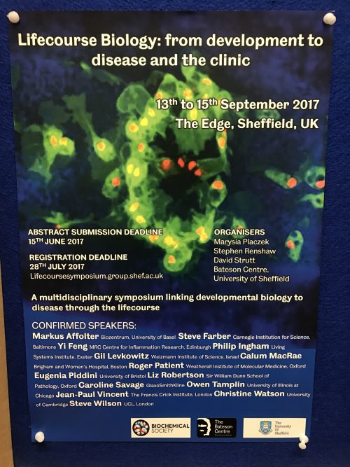


 John Gurdon is a Distinguished Group Leader in the Wellcome Trust/Cancer Research UK Gurdon Institute and Professor Emeritus in the Department of Zoology at the University of Cambridge. In 2012, he was awarded the Nobel Prize in Physiology or Medicine jointly with Shinya Yamanaka for work on the reprogramming of mature cells to pluripotency. We met John in his Cambridge office to discuss his career and hear his thoughts on the past, present and future of reprogramming. Read the Spotlight article on p.
  Innovative methods designed to recapitulate human organogenesis from pluripotent stem cells provide a means to explore human developmental biology. New technologies to sequence and analyze single-cell transcriptomes can deconstruct these ‘organoids’ into constituent parts, and reconstruct lineage trajectories during cell differentiation. In their