‘Deciphering the link between adhesion and pluripotency,”
University of Copenhagen, Denmark
The Danish Stem Cell Center (http://danstem.ku.dk/) is seeking one or two postdoctoral fellow(s) to join the Brickman Lab/DanStem & Michael Lund Nielsen Lab/Centre for Protein Research (CPR)
The post-doctoral positions will initially be supported by a grant from the Lundbeck Foundation and seek to probe the link between pluripotency and cell-cell adhesion. They will follow up recent observations on that focus the gene regulatory network (GRN) sustaining pluripotency on adhesion. The first project, based in the Brickman lab, will follow up the means by which the GRN regulates adhesion and the second, in the Nielsen lab, will focus on deciphering the nature of the adherns junction complex in pluripotent vs differentiating cells
Project description
Embryonic Stem Cells (ESCs) are genetically normal, immortal cell lines with the capacity to become any cell type in the future organism. This project will explore the role of cell-cell adhesion supporting a pluripotent state, both in vivo and in vitro. We recently found that the evolutionarily conserved gene rergulatory network (GRN) downstream of one of the central pluripotency regulators, Oct4, was primarily concerned with regulating cell-cell adhesion (Livigni et al Curr Biol. 2013). Moreover, we found that force expression of E-cadherin could partially block differentiation in response to reduced Oct4 levels in both ESCs and embryos. These projecst will follow up these observations on at a transcriptional and post-transcriptional level, using a combination of genetic models and mass spectrophotometry.
Qualifications
Candidates should have experience in embryonic stem cells, developmental biology (mouse and Xenopus) or mass spectrophotometry
Must be eligible for and prepared to write fellowship applications with in 12 months of being in the laboratory
Experience with bio-informatics would be considered an advantage
Terms of salary, work, and employment
The employment is for 3 years and is scheduled to start October 1st or upon agreement. Place of work: DanStem and CPR, University of Copenhagen, Blegdamsvej 3B, Copenhagen. Terms of employment are in accordance with the collective agreement between the Danish Government and the Danish Confederation of Professional Associations
Curriculum vitae incl. education, experience, previous employments, language skills and other relevant skills
Copy of diplomas/degree certificate(s)
Other information for consideration, e.g. list of publications (if any), letters of recommendationThe University of Copenhagen welcomes applications from all qualified candidates regardless of personal backgroundHow to apply:
Research assistant position for subsequent appointment as PhD fellow
to the project Deciphering the link between adhesion and pluripotency
The Danish Stem Cell Center (http://danstem.ku.dk) at Faculty of Health & Medical Sciences, University of Copenhagen seeks a Research assistant subsequent appointed as PhD fellow
Job description
Embryonic Stem Cells (ESCs) are genetically normal, immortal cell lines with the capacity to become any cell type in the future organism. This project will explore the role of cell-cell adhesion supporting a pluripotent state, both in vivo and in vitro. We recently found that the evolutionarily conserved gene regulatory network (GRN) downstream of one of the central pluripotency regulators, Oct4, was primarily concerned with regulating cell-cell adhesion (Livigni et al Curr Biol. 2013). Moreover, we found that forced expression of E-cadherin could partially block differentiation in response to reduced Oct4 levels in both ESCs and embryos. This project will follow up these observations. We seek to understand how Oct4 regulates cell-cell adhesion, both at a transcriptional and post-transcriptional level. The project will explore how Oct4 regulates E-cadherin dynamics and how Oct4 targets regulate signaling.
Qualifications
Master’s degree in biology, biochemistry, medicine or human biology, or similar, and a general understanding of developmental and/or stem cell biology
Strong motivation and very good scientific skills are essential
Publications and practical experience are an advantage
Good communication skills oral and written
Terms of salary, work, and employment
The position as research assistant is for 1 year and as PhD fellow for 3 years. Start October 1st 2016 or upon agreement.
The employment as a PhD student is conditioned upon a positive assessment of the candidate´s research performance and enrolment in the Graduate School at the Faculty of Health and Medical Sciences. The PhD study must be completed in accordance with the ministerial orders from the Ministry of Education on the PhD degree and the University´s rules on achieving the degree.
Work place: DanStem, University of Copenhagen, Blegdamsvej 3B, Copenhagen. Terms of employment accord to the agreement between the Ministry of Finance and The Danish Confederation of Professional Associations on Academics in the State
Emergence of functional polarity in a tubular epithelium
Applications are invited for a PhD project in the laboratory of Dr. Barry Denholm in the School of Biomedical Sciences at Edinburgh University, UK.
This exciting scholarship scheme provides a unique framework for postgraduate research students to undertake research training and development, together with experience of teaching and mentoring in an international context. Each scholarship covers the UK/EU rate of tuition fee as well as a stipend of £14,296 per annum. Subject to satisfactory progress, the scholarships are awarded for 4 years.
May was most notable for the departure of the Node’s brilliant Community Manager, Cat Vicente. Cat said her goodbyes here, but we’d like to take this opportunity to thank her again for her great work over the last 3 years.
And Cat still found time to keep posting right up to the end – both with the latest in our Forgotten Classics series, and her reflections on the inspiration behind Eric Satie’s piano composition ‘Desiccated Embryos‘.
In Cat’s absence, the community has been fantastic at keeping the Node full of content – we’ve had a particularly busy month on the jobs page, and lots of other really interesting content. Thanks to all of you for posting!
Erin and Matthias were at the Inaugural Sainsbury Lab Symposium on Induced Plant Development. They tell us all about the latest advances in this fascinating field.
The latest Company of Biologists’ Workshop “Metabolism in Development and Disease” brought together a diverse group of researchers, including Carlos and Patricia – who give us their reflections on the meeting.
Development is hosting a meeting this September, focussing on the use of stem cell technologies to understand human development. Find out more here, and register soon to secure your place!
Also on the Node
The recent Kumamoto earthquakes devastated the Institute of Molecular Embryology and Genetics. Find out about their recovery efforts.
Nestor got involved with the recent ‘Science Saturday‘ event with The Rockefeller University’s outreach team.
And the SDB is offering the John Doctor Education Prize for the best video explaining ‘Induction’ to a lay audience. Get your thinking caps on and enter for the chance to win $1000!
We hope you enjoy these and all the other posts featured on the Node in the past month. June sees the arrival of our new Community Manager – find out more in a couple of weeks!
Disease Models & Mechanisms invites you to submit original research for consideration for an upcoming Special Collection ‘Neurodegeneration: From Models to Mechanisms to Therapies‘. This ongoing collection will focus on mechanistic insights into neurodegenerative diseases using model systems, with an emphasis on key translational advances made in recent years. This collection will be launched with a dedicated issue that will benefit from high visibility to basic researchers through to clinicians interested in understanding and treating neurodegenerative diseases.
High visibility and impact (2014 Impact Factor 5)
Open Access (CC-BY licence) and PMC deposition
Rapid peer review and publication
Indexed in Medline, ISI and Scopus
Not-for-profit publisher
Here are the highlights from the current issue of Development:
A new paradigm for Wnt/β-catenin signalling
The Wnt/β-catenin signalling pathway is a key pathway involved in a myriad of developmental processes, from body axis patterning to cell migration and fate specification. The role of the Wnt pathway is to regulate transcription of specific target genes, and it has long been thought that the driving event for this is the recruitment of β-catenin to specific gene loci on the chromatin. Now, however, on p. 1914, Stefan Hoppler and colleagues provide in vivo evidence that, rather than β-catenin recruitment, it is instead the context-specific events that occur subsequent to β-catenin binding that enable gene-specific regulation. The authors use ChIP-seq to show that β-catenin is recruited to many genomic loci in Xenopus early embryonic development, while RNA-seq reveals that many of these β-catenin-bound loci are not transcriptionally regulated by Wnt signalling in this context. Instead, the transcriptional response depends on the presence or absence of additional mechanisms, for example FGF and BMP signalling. This important study advances the current understanding and interpretation of how Wnt/β-catenin signalling operates to regulate gene-specific transcription in different developmental contexts.
Oocyte maturation: cAMP and PKA called into question
The production of a mature vertebrate egg is a lengthy process in which the developing oocyte undergoes meiotic arrest followed by a long incubation period, before finally resuming meiosis in preparation for ovulation. The prevailing dogma in the field has been that, in Xenopus, meiotic arrest is released through a drop in cyclic adenosine monophosphate (cAMP) levels and protein kinase A (PKA) activity, which occurs following exposure to progesterone. In this issue (p. 1926), Khaled Machaca and colleagues provide evidence that challenges this dogma, as they demonstrate that no change is detectable in cAMP levels and PKA activity as meiotic arrest is released in the Xenopus oocyte. The authors use in vivo reporters to detect cAMP and PKA levels in real time in single cells, and show that there is no correlation between the rate of meiotic resumption and levels of cAMP or PKA inhibition. Furthermore, the authors develop conditions in which meiotic release is indeed possible in the presence of high levels of cAMP. These surprising results provide a new model for the release of meiotic arrest in the Xenopus oocyte and suggest that this phenomenon occurs through a positive signal downstream of the progesterone receptor that overcomes cAMP/PKA inhibition of meiosis resumption.
Uncovering the mechanisms of Müller glia activation
Radial glia cells in the central nervous system serve as an important source of progenitor cells for generating a range of neural cell types. Retinal regeneration in vertebrates relies on a specialised type of radial glia, called Müller glia, which are normally quiescent but can be stimulated to undergo proliferation and differentiation in order to generate new neurons. In this issue (p. 1859 and p. 1874), two studies shed light on the molecular mechanisms that regulate Müller glia activation and proliferation in response to injury and in different vertebrate species.
In the first study, Andy Fischer and colleagues investigate the role of mTor signalling in the formation of Müller glia-derived progenitor cells (MGPCs) in the chick retina. The authors use NMDA to induce a cytotoxic response, and observe that mTor signalling is transiently activated upon activation of Müller glia cells. Inhibition of mTor signalling in vivo prevents the proliferation of the Müller glia cells and blocks the regenerative response. Using a range of inhibitors and readouts, the authors show that mTor signalling is required for the proliferation of MGPCs. The authors further show that mTor signalling is activated in response to insulin, IGF1 and FGF2, and that this response is most likely independent of the MAPK pathway.
In the second study, Joachim Wittbrodt and colleagues look at the role of a single factor, Atoh7, in directing Müller glia cells to proliferate and differentiate in the absence of an injury. The authors use a fluorescent transcriptional reporter of atoh7 to demonstrate atoh7 expression in proliferating Müller glia cells after retinal injury in medaka. The authors then use an inducible system to activate expression of atoh7 in vivo in the Müller glia cells, and find that this is sufficient to drive the cells to re-enter the cell cycle and undergo proliferation. Forced expression of atoh7 in these cells activates Notch signalling, and indeed the authors show that overexpression of the Notch intracellular domain can recapitulate the effects seen by atoh7 overexpression. Importantly, not only did atoh7 overexpression in Müller glia lead to cell cycle re-entry and proliferation, but the authors also observed the formation of neurogenic clusters and subsequent de novo neurogenesis following atoh7 overexpression in these cells. Together, these two studies bring together novel and exciting findings regarding the regulation of Müller glia proliferation following injury and the subsequent regenerative response.
PLUS…
Cell fate control by pioneer transcription factors
Recent studies have shown that pioneer factors are crucial for cellular reprogramming and that they are implicated in the marked changes in gene regulatory networks that occur in various cancers. Here, Makiko Iwafuchi-Doi and Kenneth Zaret provide an overview of the contexts in which pioneer factors function, how they can target silent genes, and their limitations at regions of heterochromatin. See the Development at a Glance poster article on p. 1833
Circular RNAs: analysis, expression and potential functions
In early 2012, circular RNA (circRNA) was shown to be a transcriptional product in thousands of human and mouse genes and in hundreds of cases constituted the dominant RNA isoform. Subsequent studies revealed that the expression of circRNAs is developmentally regulated, tissue and cell-type specific, and shared across the eukaryotic tree of life, suggesting important functions for these molecules. Here, Steven Barrett and Julia Salzman describe major advances in the field of circRNA biology, focusing on the regulation of and functional roles played by these molecules. See the Primer on p. 1838
The regulation and plasticity of root hair patterning and morphogenesis
Root hairs are highly specialized cells found in the epidermis of plant roots that play a key role in providing the plant with water and mineral nutrients. Many studies have shown that the fate of root epidermal cells, which differentiate into either root hair or non-hair cells, is determined by a complex interplay of intrinsic and extrinsic cues. Here, Wolfgang Schmidt and colleagues review these studies and discuss recent evidence suggesting that environmental information can be integrated at multiple points in the root hair morphogenetic pathway and affects multifaceted processes at the chromatin, transcriptional and post-transcriptional levels. See the Review on p. 1848
Earlier this month, two papers were published (from the Brivanlou and Zernicka-Goetz labs) that reported in vitro systems to study development of the human embryo through implantation stages. These experiments have kept human embryos developing for longer than any previous work, and close in on the 14-day limit imposed by many governmental and regulatory bodies. 14 days were set as a limit (at a time when it was technically impossible to keep embryos alive this long) because this is the stage at which the primitive streak emerges – the time at which embryos can no longer split or fuse – and has been considered by some to be the stage at which a ‘morally significant individual’ comes into being (see this discussion in Nature). But now that we can culture human embryos up to this point, is this 14-day rule still appropriate? Would it be ethically ‘wrong’ to try and study human gastrulation in culture, or do the potential advances in our understanding of human development (along with their possible therapeutic implications) outweigh any ethical concerns involved? This month we are asking:
Should the 14 day limit on human embryo culture remain in place, or should it be extended or even dropped?
Share your thoughts by leaving a comment below! You can comment anonymously if you prefer. We are also collating answers on social media via this Storify. And if you have any ideas for future questions please drop us an email!
Mammalian development and tissue homeostasis are complex and dynamic biological processes. Focusing on Wnt signal transduction, our research aims to understand the genetic and epigenetic mechanisms that control gene activation in the mammary gland. We are now recruiting a postdoctoral fellow that fits one of the following profiles:
You are a ‘dry-lab’ candidate with extensive and proven expertise in complex bioinformatics analyses and an interest in analyzing large datasets, who can complement our wet-lab skills.
You are a ‘wet-lab’ candidate with proven expertise in embryonic stem cell targeting and the generation of genetically engineered mouse models.
You will be part of a young and growing research team that uses a combination of in silico, in vitro and in vivo approaches to under- stand normal (stem) cell behavior, with the ultimate goal of trans-lating principles from developmental and stem cell biology to regenerative medicine and breast cancer research. We are embedded within the section of Molecular Cytology at the Swammerdam Institute for Life Sciences and collaborate with other groups in the institute, but also with other labs in the Netherlands (e.g. at the Netherlands Cancer Institute) and abroad. We have strong ties to the field of mammary gland biology (active member of the ENBDC), as well as to the Wnt-signaling community.
Details of the job description (including tasks and requirements) and the appointment are available at the University website, which is also where you can submit you application.
In September, Development will be holding the second of our ‘From stem cells to human development‘ meetings – following on from the hugely successful first event in Autumn 2014 (see here and here). This year’s meeting promises to be equally exciting, with a fantastic line-up of speakers (check out the preliminary program) and a great venue just outside Boston, USA.
This should be a great event for anyone interested in how we can apply stem cell technologies to understand the development of our own species, spanning a broad range of topics from pluripotency and early human embryo patterning through to applying the lessons we’ve learned from development for therapeutic purposes.
Registration closes at the end of next month, but with a limited number of spaces available, we’d encourage anyone interested to apply as soon as possible to secure your place!
We look forward to seeing you at Southbridge in September!
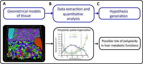
A major challenge in cell and developmental biology is to understand the mechanisms whereby cells interact with each other to form the variety of complex tissue forms present in organisms. This requires visualizing and analysing different cellular processes across multiple scale levels -from the subcellular to the tissue, i.e. generating cell and tissue models at different levels of complexity. The different cells as well as their essential sub-cellular components can be observed by fluorescence microscopy and reconstructed by means of image processing techniques. Then, three-dimensional (3D) digital representations of tissue can be generated using the structural information extracted from the microscopy images. A set of 3D digital representations of tissue at multiple scale levels constitutes a geometrical model of the tissue. Such a geometrical model provides the means of extracting detailed morphological and structural information of the tissue as well as its spatial variability. Based on such information, new hypotheses about tissue organization can be generated (Figure 1).
In our recent study published in eLife, we developed a software platform for the reconstruction and quantitative analysis of tissue architecture in 3D (Morales-Navarrete et al., 2015). We combined newly developed and established image analysis algorithms in a pipeline that allows for a flexible workflow to reconstruct and analyse different structures forming the tissue with high accuracy. It is implemented as part of the stand-alone freely available software MotionTracking (http://motiontracking.mpi-cbg.de; the name is historical as it was first developed for the tracking of endosomes in cultured cells (Rink et al., 2005)) and addresses several unmet computational needs, such as 1) high accuracy of 3D geometric reconstruction, 2) image processing of large volumes of tissue, 3) fast image analysis amenable to middle-throughput, 4) it can be run on a regular PC, 5) it is adaptable to various tissues and imaging conditions, 6) it provides a flexible tool for tissue reconstruction without need of further programming or scripting and 7) it allows quantitative morphometric and spatial analysis at cellular and tissue level.
As proof of principle, the pipeline was applied to the analysis of liver tissue of adult mice. The resulting geometrical model provided a complete description of different morphological parameters across multiple scales from tissue to subcellular level (Figure 1A and http://dx.doi.org/10.7554/eLife.11214.003, http://dx.doi.org/10.7554/eLife.11214.012, http://dx.doi.org/10.7554/eLife.11214.022) and uncovered a significant amount of new biological information. For example, we could map the distribution of different populations of hepatocytes with different number of nuclei and DNA content within the mouse liver lobule. It has been reported that hepatocytes are remarkably heterogeneous in terms of ploidy but whether they are randomly distributed or follow a specific spatial organization remains controversial (Gentric and Desdouets, 2014). Our quantitative and spatial analysis of the geometrical model revealed an unexpected spatial distribution of hepatocytes with distinct DNA content. Whereas hepatocytes with low ploidy were enriched in the peri-central and peri-portal regions, high ploidy hepatocytes were preferentially located in the middle region (Figure 1B and http://dx.doi.org/10.7554/eLife.11214.027). This is the first time that polyploidy is found to be zonated and follow a specific pattern.
Such an unexpected zonation suggests the existence of mechanisms underlying the functional specialization of hepatocytes during tissue homeostasis. One of the most remarkable features of liver tissue is the zonation of metabolic functions (hepatocytes located in the vicinity of the portal vein have different metabolic activities than hepatocytes located near the central vein). Our findings therefore suggest a possible role of polyploidy in liver metabolic functions (Figure 1C). Moreover, low ploidy hepatocytes (enriched in the peri-central and peri-portal regions) may correspond to the cells recently implicated in hepatocyte renewal (Wang et al., 2015, Font-Burgada et al., 2015). Altogether, these results show how tissue geometrical models can be used to unravel principles of tissue architecture but also generate hypotheses about tissue structure and function.
We expect our pipeline to be very useful to the biology community since it can be used for several applications from basic (understanding of how cells form tissues) to clinical research (diagnosis of diseases by 3D digital histology).
Figure 1: Using tissue geometrical models to derive principles of tissue organization and function. A) Geometrical model of liver tissue generated from confocal microscopy images. The main components of liver tissue architecture are central vein (cyan), portal vein (orange), bile canaliculi (green), sinusoids (magenta) and hepatocytes (random colours). B) Measurements of hepatocytes volume, number of nuclei per cell and DNA content were extracted, and the spatial analysis of the localization of hepatocytes with different ploidy (estimated from the volume, number of nuclei and DNA content per cell) revealed zonation patterns within the lobule. C) These zonation patterns showed a correlation with the metabolic zones in liver, which lead to the generation of the hypothesis of the existence of a possible correlation between cell polyploidy and liver metabolic functions.
Software resources:
All resources for our platform, including user manuals, examples and the latest version of the software, can be found on http://motiontracking.mpi-cbg.de/get/.
FONT-BURGADA, J., SHALAPOUR, S., RAMASWAMY, S., HSUEH, B., ROSSELL, D., UMEMURA, A., TANIGUCHI, K., NAKAGAWA, H., VALASEK, M. A., YE, L., KOPP, J. L., SANDER, M., CARTER, H., DEISSEROTH, K., VERMA, I. M. & KARIN, M. 2015. Hybrid Periportal Hepatocytes Regenerate the Injured Liver without Giving Rise to Cancer. Cell, 162, 766-79.
GENTRIC, G. & DESDOUETS, C. 2014. Polyploidization in liver tissue. Am J Pathol, 184, 322-31.
RINK, J., GHIGO, E., KALAIDZIDIS, Y. & ZERIAL, M. 2005. Rab conversion as a mechanism of progression from early to late endosomes. Cell, 122, 735-49.
WANG, B., ZHAO, L., FISH, M., LOGAN, C. Y. & NUSSE, R. 2015. Self-renewing diploid Axin2(+) cells fuel homeostatic renewal of the liver. Nature, 524, 180-5.
 (No Ratings Yet)
 May was most notable for the departure of the Node’s brilliant Community Manager, Cat Vicente. Cat said her goodbyes
  Elizabeth posted on
 Nestor got involved with the recent ‘
 (1 votes)


 In the first study, Andy Fischer and colleagues investigate the role of mTor signalling in the formation of Müller glia-derived progenitor cells (MGPCs) in the chick retina. The authors use NMDA to induce a cytotoxic response, and observe that mTor signalling is transiently activated upon activation of Müller glia cells. Inhibition of mTor signalling in vivo prevents the proliferation of the Müller glia cells and blocks the regenerative response. Using a range of inhibitors and readouts, the authors show that mTor signalling is required for the proliferation of MGPCs. The authors further show that mTor signalling is activated in response to insulin, IGF1 and FGF2, and that this response is most likely independent of the MAPK pathway.
 In the second study, Joachim Wittbrodt and colleagues look at the role of a single factor, Atoh7, in directing Müller glia cells to proliferate and differentiate in the absence of an injury. The authors use a fluorescent transcriptional reporter of atoh7 to demonstrate atoh7 expression in proliferating Müller glia cells after retinal injury in medaka. The authors then use an inducible system to activate expression of atoh7 in vivo in the Müller glia cells, and find that this is sufficient to drive the cells to re-enter the cell cycle and undergo proliferation. Forced expression of atoh7 in these cells activates Notch signalling, and indeed the authors show that overexpression of the Notch intracellular domain can recapitulate the effects seen by atoh7 overexpression. Importantly, not only did atoh7 overexpression in Müller glia lead to cell cycle re-entry and proliferation, but the authors also observed the formation of neurogenic clusters and subsequent de novo neurogenesis following atoh7 overexpression in these cells. Together, these two studies bring together novel and exciting findings regarding the regulation of Müller glia proliferation following injury and the subsequent regenerative response.
 In early 2012, circular RNA (circRNA) was shown to be a transcriptional product in thousands of human and mouse genes and in hundreds of cases constituted the dominant RNA isoform. Subsequent studies revealed that the expression of circRNAs is developmentally regulated, tissue and cell-type specific, and shared across the eukaryotic tree of life, suggesting important functions for these molecules. Here,
  Root hairs are highly specialized cells found in the epidermis of plant roots that play a key role in providing the plant with water and mineral nutrients. Many studies have shown that the fate of root epidermal cells, which differentiate into either root hair or non-hair cells, is determined by a complex interplay of intrinsic and extrinsic cues. Here, Wolfgang Schmidt and colleagues review these studies and discuss recent evidence suggesting that environmental information can be integrated at multiple points in the root hair morphogenetic pathway and affects multifaceted processes at the chromatin, transcriptional and post-transcriptional levels. See the Review on p.