The community site for and by
developmental and stem cell biologists

New insights into sex differences in Drosophila development and physiology

Posted by , on 27 May 2016

Bruno Hudry and Elizabeth J. Rideout

Male and female fruit flies differ in many aspects of development and physiology. For example, males and females differ in abdominal pigmentation, sex comb formation, courtship behaviours, the development and wiring of the central nervous system, internal reproductive organs, and genital morphology. Many elegant studies on the genes that regulate sex differences in these traits have led to the prevailing model of sex determination in Drosophila where the number of sex chromosomes determines male or female sexual identity1,2.

Briefly, the presence of two X chromosomes in female cells causes the production of an X-derived protein called Sex-lethal3. Sxl is a splicing factor, and introduces a splice into the pre-mRNA of an autosomal gene called transformer (tra) that allows a functional protein to be produced4-6. Like Sxl, Tra is a splicing factor, and together with its co-factor transformer2 (tra2), binds to the pre-mRNA of two genes: doublesex (dsx) and fruitless (fru)7-10. Tra-dependent splicing of dsx pre-mRNA causes the production of an mRNA encoding the female-specific isoform of Dsx protein (DsxF). In contrast, Tra binding to fru pre-mRNA introduces a stop codon into transcripts derived from the P1 promoter, and no Fru P1 proteins are produced in females. In males, with one X chromosome, Sxl is not induced, and consequently no functional Tra protein is produced. Lack of Tra means that dsx and fru P1 pre-mRNA undergo default splicing, and the male-specific isoforms of each protein are made (DsxM and FruM).

This sex determination cascade explains most aspects of sexual development and reproduction. However, male and female flies also differ in body size, stress responses, immunity, physiology, and lifespan11-14. Yet the mechanisms underlying sex differences in these additional traits remain unclear. The focus of this blog post is to highlight the findings from our two recent papers on sex differences in Drosophila body size (Rideout et al. 201515) and adult intestinal stem cell homeostasis (Hudry et al. 201616). We will review how these findings advance our understanding of the genetic control of sexual identity in flies, and the use of Drosophila as a model to study the molecular mechanisms underlying sexual dimorphism in body size and gut physiology.

Figure 1 – Drosophila Somatic Sex Determination Pathway. In flies, the number of sex chromosomes (X) determines sex. XX animals develop as females, whereas XY animals develop as males. Two X chromosomes causes the production of an X-derived protein called Sex-lethal (Sxl). Sxl introduces a splice into transformer (tra) pre-mRNA that allows a functional protein to be produced. Many aspects of sexual identity are controlled by Tra and its co-factor transformer2 (tra2) via regulation of doublesex (dsx) and fruitless (fru) pre-mRNA. However, our two recent papers, Rideout et al. (2015) and Hudry et al. (2016), identify at least two additional branches downstream of tra in the regulation of sexual identity: one that is tra2-dependent but dsx-/fru-independent, and another that is independent of tra2 and dsx/fru. These findings reveal additional complexity in the Drosophila sex determination pathway.

 Sex determination: different tissues, different rules

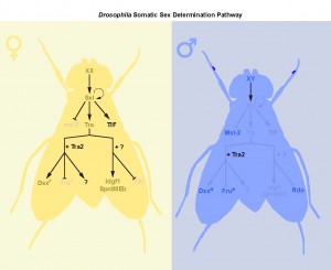
Studies on courtship behaviour, abdominal pigmentation, and sex comb formation show a clear role for the canonical Sxltradsx/fru splicing cascade in the creation of sex differences3,17. However, our findings, together with data from other studies18,

demonstrates that the genetic control of sexual identity involves additional downstream effector pathways (Fig. 1). These pathways operate in different tissues, and at different stages of the life cycle, suggesting that the process of sex determination in flies is more complex, and includes more genes, than the central core of Sxl/tra/dsx/fru (Fig. 2).

Beyond dsx and fru: new downstream targets of tra

Although there is growing evidence to support a tra-independent branch of the sex determination pathway downstream of Sxl, whether an additional branch exists downstream of tra is unclear. Previous studies on dissatisfaction (dsf), a gene with effects on sex determination in males and females, suggested a tra-dependent, dsx-independent branch of the sex determination pathway19. Yet the authors were not able to rule out the possibility that dsf may be target of fru20. Further, there was no evidence supporting sex differences in dsf expression, transcript levels, or splicing. Our two papers now provide concrete evidence of the existence of a tra-dependent, but dsx-/fru-independent, branch of the sex determination pathway in two different contexts: larval body growth, and adult intestinal stem cell (ISC) homeostasis.

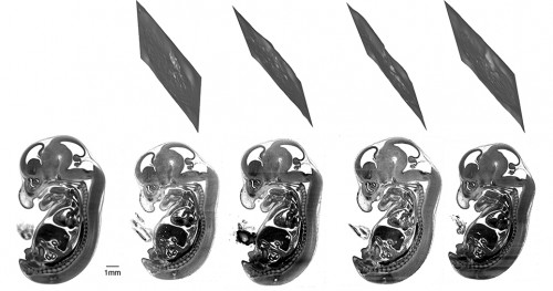
In Rideout et al. (2015) we show that tra promotes the increased body size of female flies. Loss of Tra in female larvae decreases body size, whereas ectopic expression of Tra in male larvae increases body size. Specifically, we found that Tra functions in the larval fat body to control overall body size in a non-cell-autonomous manner, and that these effects on body size were reproduced in larvae with fat-specific loss of Tra2. Surprisingly, body size was unaffected in dsx mutants, females ectopically expressing FruM, and in males lacking FruM. Thus tra/tra2, acting independently of fru and dsx, control male-female differences in body size.

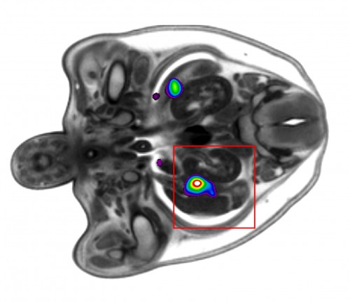
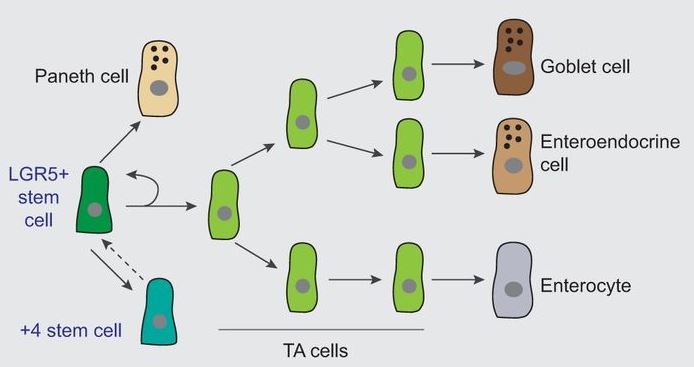
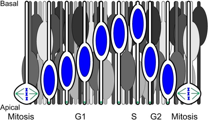
Similarly, in Hudry et al. (2016), we show that tra promotes higher ISC proliferation rate in females. This sex difference in adult stem cell divisions controls sex differences in organ size, plasticity during reproduction, and response to tumorigenic insult. Using a series of rescue, and gain- and loss-of-function experiments with dsxF transgenes, dsx/fru mutants, and RNAi transgenes, we ruled out both direct and indirect actions of dsx and fru on ISC proliferation.

Figure 2 – Sex Determination in Drosophila: Different Tissues, Different Rules. The classical sex determination pathway of Sxl/tra/dsx/fru operates in many tissues, including the adult TN1 neurons (middle panel). Interestingly, the classical pathway operates in parallel to the tra-/tra2-dependent, dsx-/fru-independent pathway in the larval fat body. In contrast, in the adult intestinal stem cells, dsx and fru are not present. Thus sexual identity is governed by multiple branches of the sex determination pathway that act in a highly context-dependent manner.

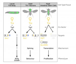
Together, our studies provide strong evidence that Tra’s effects on body size and ISC proliferation are independent of both dsx and fru (Fig. 2). Interestingly, though our findings converge on the existence of a fru-/dsx-independent branch of the sex determination pathway downstream of tra, we found important differences in the molecular mechanisms of tra’s effects on larval growth and ISC proliferation. For example, in larvae, the dsx-/fru-independent effects of tra on body size require tra’s partner tra2. In contrast, tra2 is not required for tra’s effects on ISC proliferation in the adult gut. How tra activity acquires tissue- and cell-type-specificity is not yet understood and emerges as an important new question.

One possibility is that tra acts via co-operation with tissue-specific partners. For example, one study found that among 217 Drosophila genes with Entrez Gene identifiers related to RNA-binding proteins (including splicing regulators), 83 produce multiple transcripts21. The temporal and spatial regulation of the production of these different isoforms is far from being elucidated. However, among these 217 RNA-binding proteins, 35 display very specific expression pattern in adult tissues. Some of these alternative splice variants, or tissue-specific RNA binding proteins, could be context-specific regulators of tra-mediated splicing.

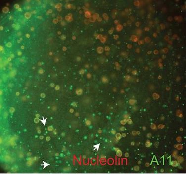
Alternatively, tra might influence ISC proliferation and body size via regulation of other aspects of RNA metabolism. Tra is a member of the SR family of proteins, which play diverse roles in RNA metabolism, including effects on transcript stability, mRNA translation, and transcriptional regulation of gene expression22. A non-splicing function for Tra in ISCs is supported by lack of Tra/Tra2 binding sites near tra-regulated splice junctions in adult ISCs. On the other hand, a genome-wide search successfully identified 18 candidate targets with multiple Tra/Tra2 sites, including tra’s known targets dsx, fru, and Sxl. Indeed, the three functional targets of tra identified in adult ISCs are regulated at the level of mRNA expression rather than splicing, suggesting one explanation for why tra2 is not required for adult ISC proliferation.

Drosophila as a model for sex differences in development and physiology

Sex differences in body size, stress responses and physiology are common to many animals other than flies, including mammals. Decades of research have examined the important contribution that sex hormones make to sex differences in traits such as adiposity and stem cell biology. For example, estrogen in female mammals causes a smaller body size, limits visceral fat accumulation, and increases haematopoietic stem cell self-renewal in females in both homeostatic conditions and during pregnancy23-25.

However, when sex hormones are absent, data from new mouse models shows that chromosomal sex (e.g. XX vs XY) dictates male-female differences in body size and adiposity. For example, in the Four Core Genotypes mouse model (FCG), which allows the effects of sex chromosomes to be directly assessed independently of sex hormones, gonadectomized XX animals achieve a larger body weight than XY animals26. This mirrors the situation in flies, where XX females are larger than XY males. Thus, in the absence of sex hormones, the trends in body size between mammals and flies are remarkably similar.

At present, it remains unclear how sex chromosomes affect body size and adiposity in mice. Flies may therefore provide important insight into the molecular mechanisms underlying these sex hormone-independent effects on body size and adiposity. For example, an important finding from Rideout et al. (2015) is that females achieve a larger body size at least in part due to increased secretion of Drosophila insulin-like peptide 2. Intriguingly, XX FCG mice have higher fasting levels of insulin26. Since the insulin pathway is an important regulator of body weight and adiposity, similar pathways may be involved in mediating the effects of sex on these traits in both flies and mice.

Over the past two decades, Drosophila has provided vital information on the molecular and physiological mechanisms responsible for determining growth during development and final body size31. Drosophila ISCs have more recently emerged as a good model to investigate adult somatic stem cell functions and regulation in vivo32,33. Recent studies using the fly model have identified many conserved intrinsic factors and extrinsic niche signals that drive the proliferation, self-renewal, and differentiation of ISCs34. In addition, a recent study demonstrated that the sex of the gut mediates the sex-biased responses of males and females to lifespan extension by dietary restriction35. We therefore suggest that flies present an excellent model to dissect the molecular mechanisms underlying sex differences in diverse aspects of development and physiology.

References

  1. Bridges, C.B. Triploid Intersexes in Drosophila Melanogaster. Science 54, 252-4 (1921).
  2. Erickson, J.W. & Quintero, J.J. Indirect effects of ploidy suggest X chromosome dose, not the X:A ratio, signals sex in Drosophila. PLoS Biol 5, e332 (2007).
  3. Cline, T.W. & Meyer, B.J. Vive la difference: males vs females in flies vs worms. Annu Rev Genet 30, 637-702 (1996).
  4. Butler, B., Pirrotta, V., Irminger-Finger, I. & Nothiger, R. The sex-determining gene tra of Drosophila: molecular cloning and transformation studies. EMBO J 5, 3607-13 (1986).
  5. Boggs, R.T., Gregor, P., Idriss, S., Belote, J.M. & McKeown, M. Regulation of sexual differentiation in D. melanogaster via alternative splicing of RNA from the transformer gene. Cell 50, 739-47 (1987).
  6. Inoue, K., Hoshijima, K., Sakamoto, H. & Shimura, Y. Binding of the Drosophila sex-lethal gene product to the alternative splice site of transformer primary transcript. Nature 344, 461-3 (1990).
  7. Ryner, L.C. et al. Control of male sexual behavior and sexual orientation in Drosophila by the fruitless gene. Cell 87, 1079-89 (1996).
  8. Heinrichs, V., Ryner, L.C. & Baker, B.S. Regulation of sex-specific selection of fruitless 5′ splice sites by transformer and transformer-2. Mol Cell Biol 18, 450-8 (1998).
  9. Inoue, K., Hoshijima, K., Higuchi, I., Sakamoto, H. & Shimura, Y. Binding of the Drosophila transformer and transformer-2 proteins to the regulatory elements of doublesex primary transcript for sex-specific RNA processing. Proc Natl Acad Sci U S A 89, 8092-6 (1992).
  10. Hoshijima, K., Inoue, K., Higuchi, I., Sakamoto, H. & Shimura, Y. Control of doublesex alternative splicing by transformer and transformer-2 in Drosophila. Science 252, 833-6 (1991).
  11. Alpatov, W.W. Phenotypical variation in body and cell size of Drosophila melanogaster. Biological Bulletin 58, 85-103 (1930).
  12. Taylor, K. & Kimbrell, D.A. Host immune response and differential survival of the sexes in Drosophila. Fly (Austin) 1, 197-204 (2007).
  13. Neckameyer, W.S. & Matsuo, H. Distinct neural circuits reflect sex, sexual maturity, and reproductive status in response to stress in Drosophila melanogaster. Neuroscience 156, 841-56 (2008).
  14. Magwere, T., Chapman, T. & Partridge, L. Sex differences in the effect of dietary restriction on life span and mortality rates in female and male Drosophila melanogaster. J Gerontol A Biol Sci Med Sci 59, 3-9 (2004).
  15. Rideout, E.J., Narsaiya, M.S. & Grewal, S.S. The Sex Determination Gene transformer Regulates Male-Female Differences in Drosophila Body Size. PLoS Genet 11, e1005683 (2015).
  16. Hudry, B., Khadayate, S. & Miguel-Aliaga, I. The sexual identity of adult intestinal stem cells controls organ size and plasticity. Nature 530, 344-8 (2016).
  17. Billeter, J.C., Rideout, E.J., Dornan, A.J. & Goodwin, S.F. Control of male sexual behavior in Drosophila by the sex determination pathway. Curr Biol 16, R766-76 (2006).
  18. Evans, D.S. & Cline, T.W. Drosophila switch gene Sex-lethal can bypass its switch-gene target transformer to regulate aspects of female behavior. Proc Natl Acad Sci U S A 110, E4474-81 (2013).
  19. Finley, K.D. et al. Dissatisfaction encodes a tailless-like nuclear receptor expressed in a subset of CNS neurons controlling Drosophila sexual behavior. Neuron 21, 1363-74 (1998).
  20. Shirangi, T.R. & McKeown, M. Sex in flies: what ‘body–mind’ dichotomy? Dev Biol 306, 10-9 (2007).
  21. Hartmann, B. et al. Distinct regulatory programs establish widespread sex-specific alternative splicing in Drosophila melanogaster. RNA 17, 453-68 (2011).
  22. Shepard, P.J. & Hertel, K.J. The SR protein family. Genome Biol 10, 242 (2009).
  23. Gambacciani, M. et al. Body weight, body fat distribution, and hormonal replacement therapy in early postmenopausal women. J Clin Endocrinol Metab 82, 414-7 (1997).
  24. Grove, K.L., Fried, S.K., Greenberg, A.S., Xiao, X.Q. & Clegg, D.J. A microarray analysis of sexual dimorphism of adipose tissues in high-fat-diet-induced obese mice. Int J Obes (Lond) 34, 989-1000 (2010).
  25. Nakada, D. et al. Oestrogen increases haematopoietic stem-cell self-renewal in females and during pregnancy. Nature 505, 555-8 (2014).
  26. Chen, X. et al. The number of x chromosomes causes sex differences in adiposity in mice. PLoS Genet 8, e1002709 (2012).
  27. Markle, J.G. et al. Sex differences in the gut microbiome drive hormone-dependent regulation of autoimmunity. Science 339, 1084-8 (2013).
  28. Steegenga, W.T. et al. Sexually dimorphic characteristics of the small intestine and colon of prepubescent C57BL/6 mice. Biol Sex Differ 5, 11 (2014).
  29. Cook, M.B. et al. Sex disparities in cancer incidence by period and age. Cancer Epidemiol Biomarkers Prev 18, 1174-82 (2009).
  30. El Ouaamari, A. et al. SerpinB1 Promotes Pancreatic beta Cell Proliferation. Cell Metab 23, 194-205 (2016).
  31. Edgar, B.A. How flies get their size: genetics meets physiology. Nat Rev Genet 7, 907-16 (2006).
  32. Micchelli, C.A. & Perrimon, N. Evidence that stem cells reside in the adult Drosophila midgut epithelium. Nature 439, 475-9 (2006).
  33. Ohlstein, B. & Spradling, A. The adult Drosophila posterior midgut is maintained by pluripotent stem cells. Nature 439, 470-4 (2006).
  34. Li, H. & Jasper, H. Gastrointestinal stem cells in health and disease: from flies to humans. Dis Model Mech 9, 487-99 (2016).
  35. Regan, J.C. et al. Sex difference in pathology of the ageing gut mediates the greater response of female lifespan to dietary restriction. Elife 5, (2016).
Thumbs up (4 votes)
Loading...

Tags: , , , , , , ,
Categories: Research

Navigate the archive

Use our Advanced Search tool to search and filter posts by date, category, tags and authors.

From our sister journals – May 2016

Posted by , on 26 May 2016

Here is some developmental biology related content from other journals published by The Company of Biologists.


CoB_DisModMech_AW_RGB


Gastrointestinal stem cells in health and disease

DMM_1This Review from Li and Jasper highlights recent findings on intestinal stem cell (ISC) diversity in the GI tract of Drosophila, focusing on the role of ISCs in healthy and diseased conditions, and drawing parallels with vertebrate GI stem cells. Read the paper here [OPEN ACCESS].


TET-1 in neural tube closure

Lozanoff and colleagues propose an epigenetic mechanism establishing the regulation of genes that are crucial for neural tube closure. This mechanism could be a novel target for resolving such birth defects and associated disorders. Read the paper here [OPEN ACCESS].


DMM_2Genetic variants of zebrafish muscle myosin

Pack and co-workers report two newly identified Myh11 gene missense mutations discovered in a zebrafish enhancer-suppressor mutagenesis screen. The mutations disrupt myosin regulation and ATPase activity in a graded fashion, and this correlated with their effects on intestinal and vascular physiology. Read the paper here [OPEN ACCESS].


Journal typography


JCS_1Nuclear migration events in development 

Nuclear migration is a central part of many cell and developmental processes. Bone and Starr review the large number of different molecular mechanisms that exist to move nuclei to specific, intracellular locations. Read the paper here.


TrkA mediates Sema3A signallingFigure 5

Goshima and colleagues show that, by interacting with PlexA4, TrkA plays a crucial role in redirecting local Sema3A signaling to retrograde axonal transport, thereby regulating dendritic GluA2 localization and patterning. Read the paper here.


Epiboly expands the mammalian epidermis

Formstone and co-workers find that the nascent mammalian epidermis spreads to enclose the embryo trunk through a process akin to epiboly, which has important implications for human birth defects of the abdominal wall. Read the paper here [OPEN ACCESS].


Journal typography


Tadpole eyes swivel and cross during metamorphosis
Combes and colleagues analyse adaptive neural plasticity of spinal locomotor-extraocular motor circuit coupling that enables Xenopus frogs to continuously generate effective retinal image-stabilizing eye movements throughout the metamorphic transition from fish-like tadpole to quadrupedal adult. Read the paper here.


JEB_1Extra arms destabilise sea urchin larvae

Chan and co-workers show that pre-competent, 6-armed larval urchins swim faster and are less stable in experimental turbulent flow than younger 4-armed larvae, suggesting a potential age/morphology-driven differential transport mechanism in ambient flow conditions. Read the paper here [OPEN ACCESS].


CoB_BioOpen_AW


BiO_1Amyloid assembley in Xenopus oogenesis

Hayes and Weeks find that non-membrane-bound nuclear particles in Xenopus oocytes responsible for RNA transcription, modification and processing contain proteins assembled into amyloids as part of normal development. Read the paper here [OPEN ACCESS].


Thumbs up (1 votes)
Loading...

Tags: , , , , ,
Categories: Research

Fueling embryology and developing metabolism

Posted by , on 25 May 2016

By Carlos Carmona-Fontaine and Patricia Nunes


Most scientific conferences have become large gatherings where little exchange of ideas can actually occur. There are way too many people to start socializing with folks you don’t know and there are so many talks that the idea of sitting through one on a topic on which you are not an expert sounds suicidal. At the other extreme was The Company of Biologists’ “Metabolism in Development and Disease” Workshop. Within a Downtown Abbey-like XVI century house, a few dozen scientists, including early career ones like us, met to discuss the implications of cell metabolism in embryonic development, immune response and disease – particularly cancer.

It was excellent. And not only because of the impressive Wiston House and its surroundings or the great list of invited speakers. It was excellent because this was a true instance where people from disparate fields met, exchanged ideas and learnt from each other. We were no exception -as our backgrounds testify. Patricia is a biochemist that has always worked on metabolism while Carlos is a developmental biologist by training. As early career scientists, it was incredibly exciting to meet and discuss with more seasoned researchers in an open and intimate environment. The pastoral fields that surrounded Wiston House and the only bar of the neighborhood, conveniently located in the same house, also helped to foster friendship and possible collaborations with fellow junior and senior biologists alike.

The Wiston House
Wiston House. Photo: Carlos Carmona-Fontaine

The talks were excellent too. There will be an upcoming meeting report to be published in Development (by Aurelio Teleman) that will summarize some of the scientific content of the meeting, so we will focus more on our personal take on the lessons from the meeting. Molecular biologists have pushed for an understanding of cellular functions based on signal transduction and codes – a visionary idea. For example, the conceptualization of the Central Dogma powered by people like Francis Crick and Sydney Brenner was a milestone in the history of molecular biology. It allowed us to simplify the complexity of cellular activities and think them as signaling problems. This concept helped us greatly to advance our understanding of how cells function and interact with each other. This is similar to how graphical interfaces and operating systems have eased the use of computers, as we do not need to deal with abstract programs and complicated coding anymore. There is a downside to this approach however: we could forget how computers actually work. Similarly, we sometimes forget that “cell markers” are enzymes, or nutrient transporters or pH and oxygen sensors. Transcription factors are critical for the development of the embryo (or a tumor) but they are that way because they ultimately encode for functional proteins that direct specific chemical reactions – such as RNA synthesis – in the cell. Why don’t we think where the methyl or acetyl groups are coming from when we talk about histone modifications and the “epigenetic code”? We risk biasing our understanding of cell biology as a collection of codes and signals, rather than actual chemical reactions.

Caption: The painting “Metabolism” by Edvard Munch depicts Adam and Eve, a metaphor for the initiators of human’s development. https://commons.wikimedia.org
The painting “Metabolism” by Edvard Munch depicts Adam and Eve, a metaphor for the initiators of human’s development. Photo: https://commons.wikimedia.org


The field of cell metabolism is inherently linked with a more biochemical understanding of biological functions. For the last two decades, this field has been reinvigorated by advances in molecular biology and modern genetics.
This synergy has been adopted by a vast number of scientists working in immunology, cancer and many other research fields. Although modern metabolism has also begun to permeate developmental biology, we think that this workshop was absolutely unique and marks the milestone of a hopefully long-lasting interaction between embryology and metabolism. In this meeting, it was clear that the field of development reciprocated with its emphasis on spatial compartmentalization and temporal dynamics of cell metabolism. For example, it was exciting to learn about the efforts to develop and improve machines that allow the detection of metabolites and stable isotopes with a 50nm resolution! We also discussed how nutrients can mediate cell-cell interactions and allow for mutualistic or competitive cell relationships. There is still a long road ahead and these research fields need to cooperate to develop new and better tools to probe complex metabolic systems. For example, promising technologies such as optogenetics will clearly help us to “shine light” onto as yet unknown metabolic networks within and between cells. Although regulation of metabolic networks in certain diseases may sound futuristic, it may well be a crucial step to develop successful personalized therapies – though as we said it’s a long road!

detail of “Embryology” by Magdalena Abakanowicz is an abstract large installation where shapes that can resemble eggs or early embryos that have achieved different sizes. One could imagine that their differential growth is due to differences in their metabolism. Foto: Carlos Carmona-Fontaine
Detail of “Embryology” by Magdalena Abakanowicz is an abstract large installation where shapes that can resemble eggs or early embryos that have achieved different sizes. One could imagine that their differential growth is due to differences in their metabolism. Photo: Carlos Carmona-Fontaine


At the end of the workshop we left with a feeling that cell signals are a key way to understand cell biology, but we cannot forget that ultimately life is about growing and growth requires a exquisite regulation of its basic components: metabolites.

We thank Ben Steventon and Katherine Brown for comments on the text.

Thumbs up (5 votes)
Loading...

Tags: , , ,
Categories: Discussion, Events

2 technical and 1 post-doctoral posts in cell biology, molecular endocrinology and skeletal biology, Imperial College London

Posted by , on 24 May 2016

Closing Date: 15 March 2021

Professors Graham Williams and Duncan Bassett have 3 full-time posts (5 years each) available. One technical post in skeletal biology is funded by the Horizon 2020 grant: Resetting the THYRoid axis for prevention of AGE-related diseases and co-morbidities. One post-doc and one technician post in molecular and cellular biology are funded by the Wellcome Trust: Cellular thyroid hormone availability: regulation of development and tissue repair, and pathogenesis of degenerative disease.


Please see our website for this exciting opportunity to join our cutting-edge research program based at Imperial College London.

http://boneandcartilage.com/opportunities.html


Thumbs up (No Ratings Yet)
Loading...

Categories: Jobs, News

Apply for a grant to study knockout mice

Posted by , on 23 May 2016

The MRC has now launched its first, cross-Board Call for Pump Priming Awards for preliminary research using IMPC knockout mice (http://www.mrc.ac.uk/funding/browse/pump-priming-awards-for-preliminary-research-using-impc-knockout-mice/).

The aim of this initiative is to capitalise on MRC’s (and others’) investments in the International Mouse Phenotyping Consortium (IMPC) (https://www.har.mrc.ac.uk/research/large-scale-functional-genomics/international-mouse-phenotyping-consortium-impc) and make best use of the resources, data and tools available. The expectation is to provide small, flexible awards to allow the take-up and use of the existing phenotyped and banked knockout lines and resources to support UK research.  It is expected that this will catalyse more ambitious studies in the future.

Applications are invited from UK-based research groups to receive, via the MRC’s Mary Lyon Centre (http://www.har.mrc.ac.uk/about/mary-lyon-centre), knockout mice produced by the IMPC, and undertake early biological investigations that build on existing research expertise and underpin longer-term research goals.

This Call will run in May 2016 and 2017, with 10-25 awards made in September 2016 and 2017.  Awards will be of 1-2 years’ duration and 20-40k in value.

The deadline for this year’s applications is 4pm on 8th July 2016.

Thumbs up (1 votes)
Loading...

Tags: , ,
Categories: Funding

Reflecting on the student & postdoc events at the BSCB/BSDB Spring Meeting

Posted by , on 23 May 2016

BSCB-BSDBLast year, the BSDB carried out a student and postdoc survey (see here), and the results clearly highlighted the need for more dedicated program items for young researchers on BSDB conferences. In response, the BSDB student rep Alex Ashcroft, the BSCB postdoc rep Alexis Barr, and the BSDB postdoc rep  Michelle Ware took initiative and organised a serious of highly successful events at the 2016 BSCB/BSDB joint Spring Meeting. Read below what happened.


Career workshop

From the BSDB student/postdoc survey results last year, it was evident that most people wanted to find out more about ‘alternative’ careers other than those on the traditional route of academia. With more PhDs being awarded and few top level jobs there is a need to provide more information as to what else can you do with your PhD. For this reason, we chose to focus this year’s careers session on alternative careers to academia. The highly attended session took the format of roundtable discussions and covered a plethora of topics including but not limited to, consulting, publishing, academic fellowships and engaging with the media. We would like to thank all the table leaders who provided stimulating discussions. This event wouldn’t have been possible without you!

Obtaining a lectureship/fellowship

  • Claudia Barros (Peninsula School of Medicine, Plymouth University)
  • Paul Conduit (Henry Dale Fellow, Department of Zoology, University of Cambridge)
  • James Wakefield (University of Exeter)

Careers outside of academia

  • Katherine Brown (Editor, Development)
  • Anne Wiblin (Research Collaborations Manager, Abcam)
  • Caroline Grant (Senior Manager, Accenture)
  • Valentina Sasselli (Associate Publisher, Cell Biology)

Science Communication

  • Andreas Prokop (University of Manchester)
  • Catarina Vicente (Community Manager, The Node)

From the feedback, we realise how valuable it is for young scientists to talk to other scientists who have trained as cell or developmental biologists and go on to have successful ‘alternative’ careers.  For future workshops, we intend to build on this theme and invite an even more diverse selection of speakers.

Some selected comments from the participants:

  • ‘Open and honest speakers, Enough time to discuss and explore career prerequisites, responsibilities and prospects’
  • ‘Great organisation and table choices, Thank you! I feel quite optimistic now!’
  • ‘Table leaders were friendly, easy to talk to and answered all questions’
  • ‘Learning about career paths, Variety of careers amongst speakers’

Click here for a more in depth summary of the workshop.

student-postdocs-1

Science Breakfasts

This was the first year that we ran science breakfasts, whose goal was to facilitate informal discussions between junior researchers and scientists at the top of their field. A small number of students and postdocs got to participate in this event, discussing everything from research, careers and life in general with Abigail Tucker, Ottoline Leyser, Jordan Raff, Lidia Vasilieva and Thomas Surrey – who we are really grateful for giving up their time.

Student social

student-postdocs-2
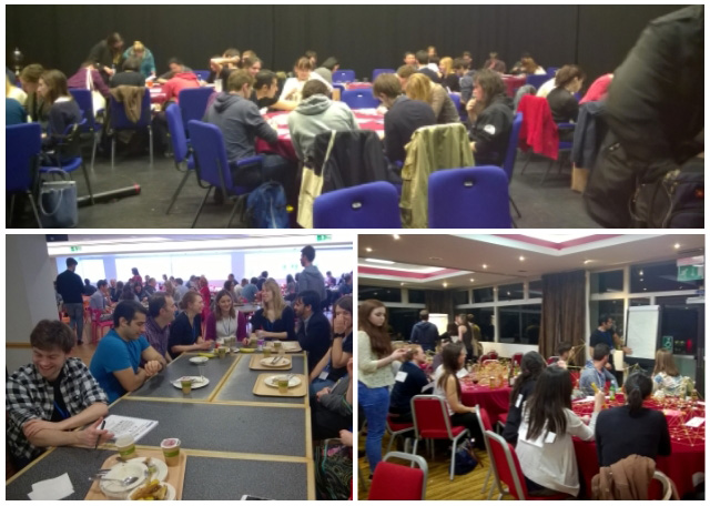
This year the reps decided it would be fun to do something more interactive for the student/postdoc social. We randomly attached a name to the back of every guest, where each name was one-half of a famous pair, such as Romeo and Juliet. Each guest could not see their own name but could see the names of other guests. Using yes-no questions, each guest had to work out who they were and then find the other half of their pair. Each pair were given the task to build the tallest possible tower using marshmallows and spaghetti. The pairs were then grouped into teams of ten which competed against each other in a round of science pictionary.

Thank-you to all the sponsors who donated gifts. For the pair that found their pair first won a bound notebook each from Horizon and sweets. Class Learning provided a voucher for two books up to £100, which was awarded to the winners of spaghetti towers (Erik Clark, Gautham Dey and their winning tower pictured here).

Chocolates and sweets from the BSDB committee were awarded to the winning teams after the Pictionary round. Molecular biology of the Cell (Garland Science) was given to the winner of the best image (Rachna Narayanan with a drawing of WALL-E).

Student Symposium

This year the graduate student symposium was moved to the middle of the meeting, resulting in excellent attendance. This was a truly excellent event – the speakers covered a diverse range of topics in an engaging manner. Some talks even got mentioned in the twitterverse!

The format was also altered so that there were six fifteen minute presentations and six five minute presentations. All the speakers did an excellent job – particular mention must be made for everyone who managed to describe their complex research in just five minutes!

We hope to see many of you next year. If you have any comments or ideas please get in touch with Alex (students@bsdb.org), Alexis (Alexis.Barr@icr.ac.uk) or Michelle (postdocs@bsdb.org); especially if you have ideas for games to play in the student social, know someone who would be a great table leader for the careers workshop or if there is someone with whom you would really like to have breakfast.

Thumbs up (1 votes)
Loading...

Tags: ,
Categories: Events, News, Societies

A mouse embryo atlas using HREM data

Posted by , on 23 May 2016

For the first time, a reference mouse embryo atlas has been created using HREM image data. For other embryo imaging methods such as micro-CT, a reference embryo atlas has previously been shown to be the basis of automated phenotyping (Wong et al, 2014). This new work (a collaboration between the DMDD programme and the Mouse Imaging Centre in Toronto) is a proof of principle for automated phenotyping of HREM data, which can now be tested in more detail.


COMPARING LIKE WITH LIKE

In order to phenotype an embryo, we need to compare it to another embryo that we consider to be normal. With histology this can be very difficult, as it’s virtually impossible to take exactly the same slice through two embryos.

So this leads to the question ‘have I really observed a phenotype, or am I simply looking at a different part of the embryo?’

The ideal scenario for a phenotyper is to have exactly equivalent slices of anatomy to compare. They can then do a direct visual comparison, or an algorithm can be used to do a statistical comparison, therefore automating the process to some degree.


A REFERENCE EMBRYO ATLAS

Using a technique previously employed with micro-CT data (Wong et al, 2012), the team have created a reference embryo atlas from HREM data. The atlas can be used to find these equivalent sections of anatomy.

21 wild-type embryo image stacks were merged together to give an unbiassed ‘average embryo’. Every possible pair of embryos was compared in terms of displacement. The embryos were then combined together so that the displacement for all embryos to join the atlas was a minimum. This technique avoids giving undue weighting to any one embryo in the average.

As HREM data is so large, the resolution was scaled back first to make the computation feasible.

For each individual embryo, a different deformation field is needed to bring the image into alignment with the average. The inverse of this field can then be used to propagate back from any plane in the reference embryo to a section of the original input embryo.

This means it’s possible to find equivalent embryo sections to compare, ensuring that any differences observed are much more likely to be genuine phenotypes.

A merged reference embryo and sections of interest.
Bottom left, the merged reference embryo where the plane of interest is defined. Bottom right, the equivalent data from four individual mouse embryos. Top, the crumpled sections show the effective ‘cut’ plane through the original HREM data that is homologous to the plane of interest in the reference embryo. (Image recreated with the permission of Elsevier).

It’s important to note that these sections are never planes but crinkly slices through the embryo. This means that the sections couldn’t have been identified by manually scrolling through the image stacks.


AUTOMATED PHENOTYPING

Automated selection of equivalent embryo sections is the starting point for automated phenotyping. Given the time-consuming nature of phenotyping embryos, and the large task of phenotyping many mouse lines, automated phenotyping is an attractive prospect to many. It can act as a guide – a primary screen to highlight areas of interest to manual phenotypers.

The next step, then, is to compare other HREM embryo data with this reference atlas.

The results of automated primary screens are typically presented as a heatmap (Wong et al, 2014), highlighting areas of interest. The authors showed a proof of principle for an automated primary screen of HREM data by separating the embryos into male and female groups. After selecting equivalent sections through the gonads to compare between the two groups, they were able to automatically distinguish statistically significant differences in this region.

Statistically significant differences between male and femal embryos.
The coloured regions show statistically significant differences between males and females in the region of the gonads.

This is an exciting result for the future of automated phenotyping of HREM mouse embryo data, which will be tested much more extensively over the coming months.


This post originally appeared on the DMDD blog, Annotations (blog.dmdd.org.uk).

DMDD is a systematic study of embryonic-lethal mouse gene knockouts, and provides a database of high-resolution image and phenotype data – a valuable resource for both developmental biologists and clinicians. Visit the website to find out more: dmdd.org.uk


READ THE FULL PAPER

R. Mark Henkelman1, Miriam Friedel1, Jason P. Lerch1, Robert Wilson2, Tim Mohun2 (2016)
Comparing homologous microscopic sections from multiple embryos using HREM
Developmental Biology [Epub ahead of print], DOI: 10.1016/j.ydbio.2016.05.011

1 Mouse Imaging Centre (MICe), Hospital for Sick Children, University of Toronto, Toronto, Canada
2 The Francis Crick Institute Mill Hill Laboratory, London, UK


REFERENCES

M. D. Wong1, Y. Maezawa2, J. P. Lerch1, R. M. Henkelman1 (2014)
Automated pipeline for anatomical phenotyping of mouse embryos using micro-CT
Development, DOI: 10.1242/dev.107722

1 Mouse Imaging Centre (MICe), Hospital for Sick Children, University of Toronto, Toronto, Canada
2 The Lunenfeld-Tanenbaum Research Institute, Mount Sinai Hospital, Toronto, Canada


M. D. Wong1,2, A. E. Dorr1,3, J. R. Walls4, J. P. Lerch1,2, R. M. Henkelman1,2 (2012)
A novel 3D mouse embryo atlas based on micro-CT
Development, DOI: 10.1242/dev.082016

1 Department of Medical Biophysics, University of Toronto, Toronto, Canada
2 Mouse Imaging Centre (MICe), Hospital for Sick Children, University of Toronto, Toronto, Canada
3 Sunnybrook Hospital, Toronto, Canada
4 Regeneron Pharmaceutecals, Tarrytown, USA

Thumbs up (No Ratings Yet)
Loading...

Tags:
Categories: News

Post-doc position on evolution of non-embryonic developments in urochordates

Posted by , on 22 May 2016

Closing Date: 15 March 2021

A 15 months postdoctoral position is available at the Observatoire Océanologique de Villefranche-sur-Mer -LBDV (UPMC – Sorbonne Universités). We are seeking a highly motivated and passionate researcher to join a 5-years international project, DEVODIVERSITY, funded by the French Agence Nationale de la Recherché (ANR) and the São Paulo Research Foundation (FAPESP).


By using molecular and cell biology, NGS transcriptomic, genomic, and ecological approaches, a multidisciplinary consortium led by two teams, the Tiozzo Lab at the Villefranche-sur-Mer Developmental Biology Laboratory in France (CNRS-UPMC), and the Brown Lab at the Istituto de Biociências in Brazil (USP), will study the evolution of regeneration, asexual reproduction, and clonality in several species of ascidians (Urochordata), and examine how ecological factors affect distribution ranges, evolution of life cycles and developmental strategies. DEVODIVERSITY has the following main aims:


  1. To resolve the phylogenetic relationships and evolutionary transitions between strict sexual reproduction to budding and high regenerative abilities among Styelidae (Ascidiacea).
  2. To provide a coupled morphological and ecological understanding of asexual propagation (budding). We will generate detailed anatomical and developmental descriptions of budding processes, and explore if and which environmental conditions are associated with the use of particular budding modes.
  3. To compare gene pathways involved in stem cell functions or trans/de-differentiation of budding and regenerative processes by the generation and in silico analyses of transcriptomic data.
  4. To launch a comparative genomic approach study in order to better understand the evolution of a major life history transition in marine chordates, in particular the evolutionary transition from sexual to asexual propagation.


The candidate will be mainly based at the LBDV (Villefranche sur Mer, France) and may have the opportunity to do laboratory and fieldwork in both countries.


It is ESSENTIAL:


  • A doctoral degree in animal biology, evolutionary biology or related disciplines.
  • Demonstrated experience in molecular biology, organismal biology and at least bases of bioinformatics and biostatistics.
  • Enthusiasm and genuine commitment to research work.
  • Demonstrated ability to undertake independent research, flexibility and problem solving skills.
  • Excellent communication skills in English spoken/written.
  • Ability to collaboratively work on a team.


Expertise in phylogenomics and transcriptomics is highly desirable but not mandatory. The LBDV has an international working environment, speaking French is not required but it would be a plus.


The applicant should contact directly Dr. Stefano Tiozzo (tiozzo@obs-vlfr.fr) providing a letter of interest, a CV and the contact of three potential referees. Please, add “DD_postdoc” on the subject of your email. Applications review will continue until the position is filled. Starting data could be any time from September 2016.


Contacts

Stefano Tiozzo (tiozzo@obs-vlfr.fr): http://biodev.obs-vlfr.fr/~tiozzo/tiozzo-lab.

LBDV: http://biodev.obs-vlfr.fr/en/index.html

Thumbs up (No Ratings Yet)
Loading...

Categories: Jobs

Carthew & Mani Labs Postdoc Search

Posted by , on 20 May 2016

Closing Date: 15 March 2021

The Carthew and Mani Labs are looking to hire a joint postdoc with familiarity in live-imaging and quantitative analysis. Building on the expertise of both labs, the goal of the project is to construct a live imaging platform for eye development with a view to explore the coupled dynamics of cell-cell signaling, mechanical forces, and gene regulation using state-of-the-art techniques. The postdoc will benefit from the highly collaborative and interdisciplinary environment of the two labs that already share many common interests and students. Both investigators are committed to supporting the development of soft skills, in addition to the experimental and quantitative skills that are required for success in science. Competitive salary, computing resources, and travel money will be provided.

Interested? Contact us at madhav.mani@northwestern.edu

Mani Lab Website: madhavmani.com

Carthew Lab Website: http://groups.molbiosci.northwestern.edu/carthewlab

Thumbs up (No Ratings Yet)
Loading...

Tags: , , , , ,
Categories: Jobs

Madrid Adventure to Study Autophagy in Melanoma Stem Cells

Posted by , on 20 May 2016

I am hugely grateful to the Journal of Cell Science and the Company of Biologists for awarding me a traveling fellowship to visit the laboratory of Dr Guillermo Velasco at Complutense University in Madrid, Spain. During this visit I gained a greater understating of the skills and techniques used to manipulate autophagy regulatory pathways within melanoma stem-like cells, as well as knowledge of autophagy regulation within cancer.

Cutaneous malignant melanoma is the most aggressive form of skin cancer and despite the recent introduction of targeted therapeutics to treat the disease, relapse is common and there remains no effective cure for metastatic melanoma. Therefore, an improved understanding of the underlying biological mechanisms mediating development and progression of melanoma is desperately required. A potential therapeutic approach may be to target melanoma stem-like cells that sustain tumour growth and contribute to drug resistance. Stem-like progenitor cells from other cancers have been shown to use autophagy, the principle lysosomal mediated mechanism for the breakdown and recycling of damaged proteins, for survival. Therefore, as autophagy is also known to be important for melanoma growth, the current project aims to test the hypothesis that melanoma subpopulations that display a stem-like phenotype use autophagy for tumour maintenance, invasion and survival.

The trip to Dr Velasco’s lab allowed me to learn specific practical techniques for the identification and characterisation of cancer stem cells, as well as gain knowledge of autophagy regulation from experts in the field. During my time in Madrid I met a number of very interesting people who I hope will remain close collaborators in the future. Also, the experience of living and working in such a cosmopolitan city as Madrid is one that I can whole heartedly recommend to anyone!! It certainly is a fantastic city and I can’t wait until the next time I can visit.spanish gang 2016

Thumbs up (1 votes)
Loading...

Tags: , ,
Categories: Funding, Lab Life