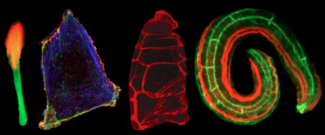
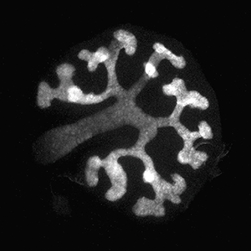
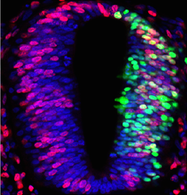
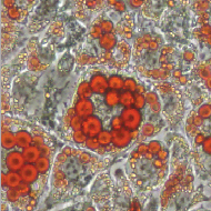
The Uhlirova lab seeks talented, creative and motivated researchers to investigatehow alterations of the essential and ubiquitous process of pre-mRNA splicing contribute to tissue-specific degeneration and cancer. The project combines state-of-the-art Drosophila genetics with studies in mammalian cells utilizing tools of cell biology, imaging, biochemistry, and genomics.
We invite applicants with a Ph.D. degree, a strong background in molecular and cell biology, expertise in imaging, and interest in utilizing high-throughput genomic technologies.
To apply, please submit your CV, a brief statement of research interests including motivation for the application, and contact information for two references toMirka.Uhlirova[at]uni-koeln.de.
Application deadline: September 13th, 2015
The successful candidates will benefit from an excellent environment within the CECAD initiative at the University of Cologne that offers diverse areas of expertise, collaboration and modern facilities.
The healthy development of an organism depends on its tissues and organs growing to the right size, stopping when they need to, and maintaining stability in their form and function. Correct development depends on the availability of nutrients to the cells in their environment, a process that is tightly controlled by signalling mechanisms that occur within and between the cells that form these structures. Disruptions in this signalling can lead to unbalanced growth within a tissue or organ, and can give rise to conditions such as cancer.
The TOR and PI3K signalling pathways regulate tissue growth according to nutrient availability, and are frequently over-activated in human cancer. In the study published yesterday in PLoS Biology, Institute for Research in Biomedicine (IRB Barcelona) PhD student Ana Ferreira and Group Leader and ICREA Research Professor Marco Milán report that the over-activation of these two pathways not only causes the excess growth of cells and tissues, but also restrict the growth of neighbouring cell populations.
They present evidence that the proteoglycan Dally, a protein that is known to modulate the spreading, stability and activity of the growth-promoting signalling molecule called Dpp (in flies) or TGF-β (in humans), is regulated by these two pathways and mediates the effects on neighbouring populations. “They do so by competing for Dpp”, says Ana Ferreira, first author of the paper and funded by a PhD fellowship from Portugal’s Fundação para a Ciência e a Tecnologia.
“PTEN, a gene that negatively regulates the PI3K pathway, is one of the most commonly lost tumour suppressors in human cancer. Understanding whether this pathway also affects TGF-β spreading in mammals may help us to gain insight into the early events of tumorigenesis and the selection of the tumour-initiating cells,” she confirms.
“Tumour initiating cells might be selected by their ability to compete for limiting growth factors and their capacity to restrict the growth of neighbouring cell populations,” says Marco Milán, head of the Development and Growth Control Laboratory at IRB Barcelona. “Seventy percent of men with prostate cancer are estimated to have lost a copy of the PTEN gene at the time of diagnosis. It will be interesting to determine whether this mechanism, identified in fruit flies, is also active in humans.”
Reference article:
Dally proteoglycan mediates the autonomous and non-autonomous effects on tissue growth caused by activation of the PI3K and TOR pathways.
This week the BioMedCentral blog put together a quiz to test how good you are at assessing the best course of action when faced with tricky peer review scenarios. These scenarios make for a fun exercise, but similar situations can and are encountered by you when invited to review manuscripts. However (like many other tasks that are asked from researchers) very few people receive formal training on how to review a manuscript. Of course, a lot of what makes a good reviewer is also what makes a good experimentalist, but how best to avoid conflict of interests, biases or becoming the dreaded third reviewer? Should formal training be provided by universities (e.g. during graduate school or postdoc) or by journals when a new reviewer is invited? Should it be the responsibility of mentors? Or should the community put together a set of guidelines that everyone agrees to follow? In other words:
What is the best way to encourage good peer reviewing?
Share your thoughts by leaving a comment below! You can comment anonymously if you prefer. We are also collating answers on social media via this Storify. And if you have any ideas for future questions please drop us an email!
Development is planning a Special Issue on Plant Development – in memory of Ian Sussex, one of the founding fathers of the plant developmental biology field. Ian passed away earlier this year (you can read more about his life in this Obituary, recently published in Developmental Biology), but his legacy lives on in his work and in the many members of the plant community he trained and worked with.
With this Special Issue, Development aims to commemorate Ian’s life and to showcase the best plant developmental biology research across the field today. Development has a long-standing history of publishing influential papers about plant developmental biology, and today our plant papers are consistently among the most highly accessed and cited articles in the journal. We are therefore excited to have the opportunity to continue in this tradition with this upcoming Special Issue.
For those interested, full details on the Special Issue, including timelines for submission, can be found here. Please contact the Development office with any queries.
The Zaidel-Bar lab (http://CellAdhesionLab.com) has an open position for a postdoctoral fellow to study the regulation of the actomyosin cortex in mammalian cells and C. elegans embryos.
The lab is part of the Mechanobiology Institute (http://mbi.nus.edu.sg), which is the world’s leading center for interdisciplinary research in tissue, cellular and molecular mechanics.
Qualifications: PhD in cell or developmental biology or in biophysics, experience working with cells and/or C. elegans, experience using fluorescence microscopy for live imaging. Experience in the cytoskeleton field is an advantage, but not required.
Application: Candidates should send Dr. Zaidel-Bar (biezbr@nus.edu.sg) their CV and list of publications along with a cover letter detailing their research interests and suitability to this position.
The Miguel-Aliaga lab investigates the plasticity of the intestine and its neurons, particularly in relation to the regulation of food intake, nutrient storage and metabolic homeostasis. We are a productive lab, well funded by the ERC, MRC and BBSRC. We belong to the MRC Clinical Sciences Centre and our lab is located in a new research building in Imperial’s Hammersmith campus in West London.
Projects involving Drosophila melanogaster, mouse and/or human cells are available, and are typically underpinned by multidisciplinary approaches combining genetics, physiological and behavioural assays, metabolic profiling and RNA-seq analyses. Our work is supported by core facilities in genomics, bioinformatics, imaging and proteomics, and by collaborations with other groups at the CSC .
Here is some developmental biology related content from other journals published by The Company of Biologists.
Deducing the stage of origin of Wilms tumours from a developmental series of Wt1 mutants
Wilms’ tumours, paediatric kidney cancers, are the archetypal example of tumours caused through the disruption of normal development. In this study, Hohenstein and colleagues compare different nephron-specific Wt1-knockout mouse models, identifying the stage of origin of human WT1-mutant Wilms’ tumours. Read the paper here [OPEN ACCESS].
Enolase 1 and PDIA3 regulate Wnt/β-catenin-driven trans-differentiation of murine alveolar epithelial cells
The alveolar epithelium represents a major site of tissue destruction during lung injury. Königshoff and colleagues conducted proteomics, expression analysis and functional studies in primary murine ATII cells and identified proteins involved in Wnt/β-catenin-driven alveolar epithelial plasticity in lung injury and repair. Read the paper here [OPEN ACCESS].
Ciliary adenylyl cyclases control the Hedgehog pathway
Protein kinase A (PKA) accumulates at the base of the cilium where it negatively regulates the Hedgehog pathway. Although PKA activity is essentially controlled by the cAMP produced by adenylyl cyclases, the influence of these enzymes on the Hh pathway remains unclear. Pons and colleagues show that ciliary adenylyl cyclases AC5 and AC6 respond to stimulatory and inhibitory GPCRs to control PKA and, hence, transduce the Hedgehog signal. Read the paper here.
Stk40 represses adipogenesis through translational control of CCAAT/enhancer-binding proteins
Stk40 is a putative serine/threonine kinase originally identified as an activator of the Erk1/2 signaling required for primitive endoderm differentiation from mouse ESCs, and later found to be important for mouse fetal lung maturation. Jin and colleagues now show that Stk40 also plays a role in repressing adipogenesis through inhibition of C/EBP protein translation. Read the paper here.
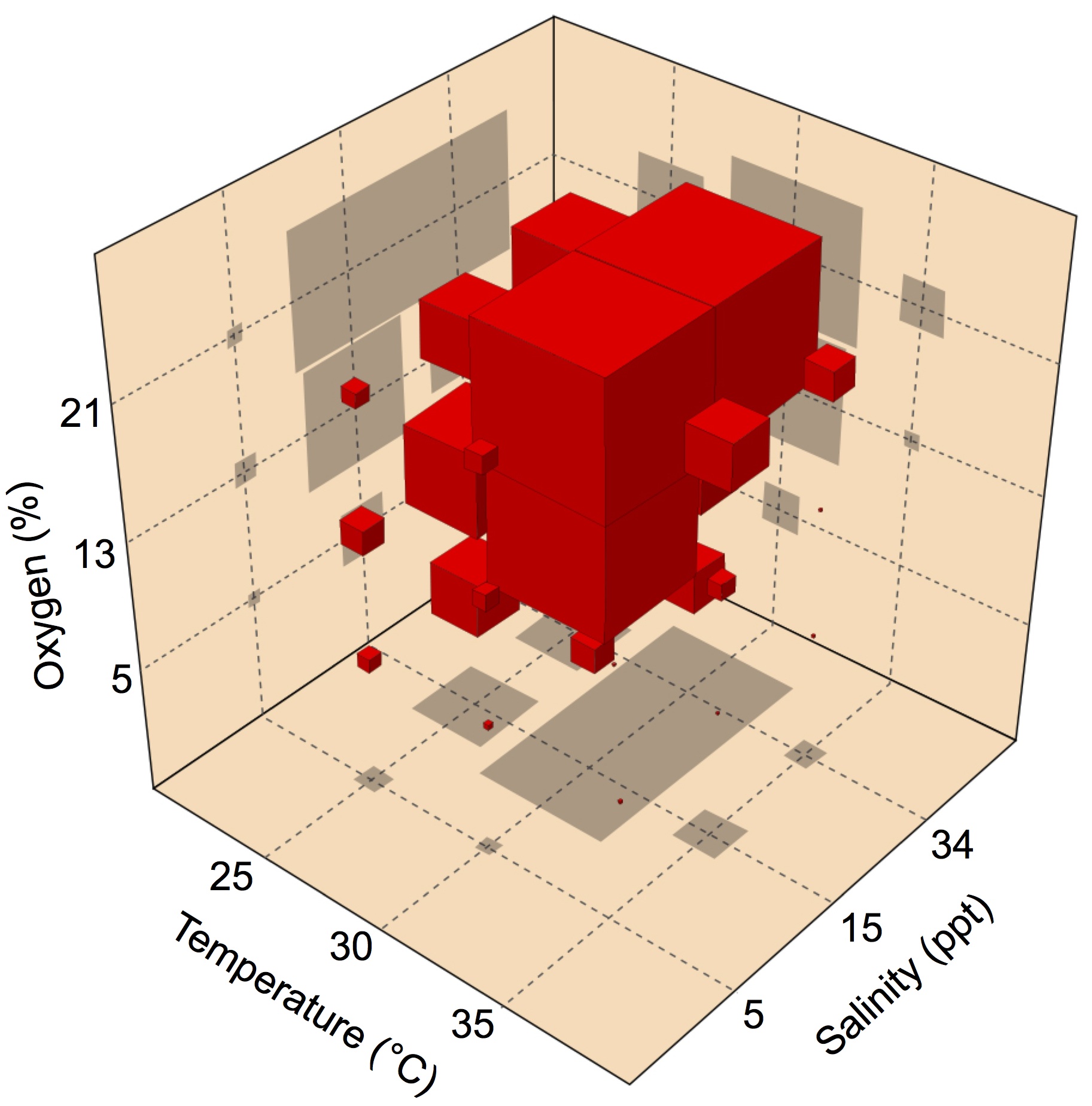
Multiple-stressor interactions influence embryo development rate in the American horseshoe crab
Fertilized eggs of the American horseshoe crab are buried in shallow nests above the high tide line, where they are exposed to variations in abiotic conditions during early development. Using a multiple-stressors approach, Julian and colleagues examined whether the rate of embryonic development in these animals is affected by exposure to temperature, salinity and oxygen. Their results show that multiple abiotic stressors can interact to affect the development rate in ways that cannot be predicted from the effects of the stressors in isolation. Read the paper here.
Cold adaptation overrides developmental regulation of sarcolipin expression in mice skeletal muscle
Sarcolipin plays an important role in muscle-based thermogenesis. Periasamy and colleagues show that it is abundantly expressed in neonatal mouse muscles and cold challenge prevents its developmental downregulation, indicating higher recruitment of muscle-based thermogenesis in neonates. Read the paper here.
Gender-bending alligators use ESR1
In crocodilian as well as several turtle species, the gender of offspring is determined by the temperature at which the eggs are exposed during a critical developmental window called the thermo-sensitive period. Hormones also contribute to this process, since exposure to estrogens feminizes alligators developing at a male-producing temperature, but the mechanisms behind this phenomenon remain elusive. Sarah Alderman highlights a recent paper by the lab of Louis Guillette Jr in Endocrinology, examining which of the two known estrogen receptos (ESCR1 and ESCR2) is responsible for this hormone-mediated switch from male to female alligators. Read this Outside JEB feature here.
The group led by ICREA Research Professor Cayetano Gonzalez at IRB Barcelona, in collaboration with the group of Professor Giuliano Callaini from the University of Siena in Italy, has published a new study in Current Biology that contributes to understanding how cilia are assembled.
Many cells in our bodies present a small structure that looks like, and as a matter of fact works as an antenna, conveying to the cell information on the extracellular environment. They are called cilia (plural) or cilium (singular). Ciliated cells play essential functions in the human body. Thus, for instance, the monitoring of fluid flow in the kidney, the detection of hormones in the brain, or the senses of hearing and smell depend on specialised neurons equipped with chemo-sensory or mechano-sensory cilia. Moreover, besides sensing, beating cilia keep fluids in motion in many parts of our bodies and are critical for human health.
A cilium can be regarded as a long and thin protrusion of the cell membrane that contains microtubules. Ciliary microtubules are arranged in a typical radial symmetry that is conserved through evolution and is templated by a small organelle that sits at the base of the cilium, known as basal body. Most animal cells contain two basal body-like structures (centrioles), but only one of them can actually work as basal body. In human cells, this is always the centriole that is said to be the “mother” because it was assembled earlier than the other, called the “daughter” centriole.
One laboratory animal model used to investigate how cilia are assembled is the vinegar fly Drosophila melanogaster. The article by the Gonzalez’s group shows that in Drosophila, as in humans, basal body fate is also reserved to the mother centriole. Moreover, through genetic manipulations that are easily performed in flies, they have been able to get a glimpse into the molecular mechanism that governs this fundamental process.
They have found that removal of the daughter-centriole specific protein Centrobin (CNB) allows daughter centrioles to serve as basal bodies. Thus CNB-depleted neurons present two cilia, the standard, which is templated by the mother centriole and a second one templated by the daughter centriole from which CNB has been removed. Conversely, mother centrioles engineered to carry CNB cannot function as basal bodies and, therefore, neurons modified this way cannot assemble cilia.
In humans, the lack of cilia, or cilia that do not work well, are the cause of a long list of disorders, known as ciliopathies, which include polydactyly, obesity, respiratory dysfunction, hearing impairment, and many others. Basic research in model organisms like the vinegar fly is helping to understand the molecular details of cilium assembly, thus paving the way to applied research in this field.
Reference article:
Loss of Centrobin enables daughter centrioles to form sensory cilia in Drosophila
M. Gottardo, G. Pollarolo, S. Llamazares, J. Reina, M. Riparbelli, G. Callaini, and C. Gonzalez
Current Biology (20 August) DOI: 10.1016/j.cub.2015.07.038
This article was first published on the 21st of August 2015 in the news section of the IRB Barcelona website
The Stowers Institute for Medical Research has two openings for postdoctoral researchers to work on projects using the cave fish system (Astyanax mexicanus) to address questions about the genetic basis of adaptation to new and extreme environments, with a particular focus on metabolic evolution.
Candidates will use RNA-sequencing, whole genome sequencing, transgenics, and metabolomics to uncover the genetic basis and underlying mechanism of the impressive metabolic adaptations cavefish have acquired to survive in nutrient poor environments.
Qualified candidates should have a background in evolutionary genetics and bioinformatics or a background in ecological genetics. Research may be field-based, lab-based, or both. Researchers who integrate genome level data with studies of ecology and evolution are encouraged to apply
One of the positions will involve substantial field work (in the caves in Mexico) to link the ecological settings in the caves with the observed and studied phenotypes.
Another position will direct the bioinformatics of the genomics of non-model organisms, and will analyze and interpret data accordingly. Experience in handling large-scale sequence data is essential.
The ideal candidate has a doctorate degree in bioinformatics, genomics, or a related field; strong command of UNIX and other programming languages; hands-on experience with genomic data; and an interest in pursuing research on emerging model organisms. In addition, the successful candidate will have a strong background in analysis of genetic and genomic data (e.g., whole-genome resequencing, RAD genotyping, QTL mapping) and/or experimental developmental biology (e.g., manipulation of gene expression, transgenesis, genome editing), and an ability and willingness to work both independently and collaboratively in a team-oriented environment. No previous experience working with fish is required.
To apply, please submit a cover letter that includes a short summary of interests, a CV, and the contact information of 2-3 professional references to nro@stowers.org
Deadline for applications is September 15, 2015. The positions are available October 1, 2015 (start date is flexible). About the Stowers Institute for Medical Research
The Stowers Institute for Medical Research in Kansas City, Missouri* is a non-profit, basic biomedical research organization dedicated to improving human health by studying the fundamental processes of life. Jim Stowers, founder of American Century Investments, and his wife, Virginia, opened the Institute in 2000. Since then, the Institute has spent over 900 million dollars in pursuit of its mission.
Currently, the Institute is home to almost 550 researchers and support personnel; over 20 independent research programs; and more than a dozen technology-development and core facilities. Kansas City is an emerging metropolitan city in the Midwest with a high quality of living and affordability.
As you may have noticed, as part of our recent redesign we created a new Resources section on the site, which you can access via the menu bar. Within this section we have created three pages with links to websites that you may find useful:
– Societies: links to national and international developmental biology societies
– Other useful links: including bioinformatics, genomics and imaging tools, as well links to educational and outreach resources
These lists are not comprehensive, and this is where we need your help! Have we missed an important database? Is there a really useful tool that we should include? Is your national society not listed? Help us make these lists as relevant as possible to the developmental biology community by leaving your suggestions as a comment here, or via our contact form.