The community site for and by
developmental and stem cell biologists

Book review: Everything you always wanted to know about auxin but were afraid to ask

Posted by , on 6 December 2012

This book review originally appeared in Development. Sabrina Sabatini reviews “Auxin Signaling: From Synthesis to Systems Biology ” (Edited by Mark Estelle, Dolf Weijers, Karin Ljung and Ottoline Leyser).

Book info:

Auxin Signaling: From Synthesis to Systems Biology. Edited by Mark Estelle, Dolf Weijers, Karin Ljung, Ottoline Leyser. Cold Spring Harbor Laboratory Press (2011) 253 pages ISBN 978-0-879698-98-0 $67.50 (hardback)


Whether you like it or not, if you work in plant science it is of utmost importance that you know what auxin is and does, since your research will, at some point, most certainly cross the auxin path. The plant hormone auxin regulates a plethora of developmental and physiological processes in plants. At the organismal level, the differential accumulation of auxin forms gradients, which, through interactions with other hormones, regulate processes as diverse as responses to external stimuli and embryonic and postembryonic development. All of these macroscopic processes are a reflection of the changes in gene expression triggered by auxin. The book Auxin Signaling tries to provide a broad coverage of each aspect of auxin signaling, from synthesis to transport and signal transduction, along with an in-depth description of auxin’s role in various events in plant development.

The book opens with a chapter by Abel and Theologis on the historical context of auxin research, which provides an evocative description of the scientific journey that brought us to the present level of knowledge of auxin signaling. This section thrills the reader with the ups and downs of auxin research, and spurs them to read the rest of the book to discover where the frontline of auxin research is now. The book then divides into four sections: auxin synthesis and transport, auxin perception and cellular responses, auxin in development, and signal integration.

The second and the third sections work together to illustrate the conceptually sequential mechanisms by which auxin is synthesized, metabolized, transported and perceived by the cell, followed by the basic cellular response. The first chapter of these describes the state of our knowledge of the auxin homeostasis pathway, despite the difficulties in the quantification of auxin and its intermediates. Apart from giving an in-depth description of the pathway, the authors have included many references to the experiments behind the facts, enticing the reader to critically assess the source of information. The next two chapters provide a unique perspective on classical topics such as auxin transport and perception. In ‘Auxin transporters – why so many?’, the authors illustrate the complexity and redundancy of the auxin transport machinery in the light of evolution, offering a refreshing (and for once non-PIN-centric) point of view. The authors of the next chapter provide a very interesting structural insight into auxin perception, cleverly describing how auxin acts differently from other hormones, the application of this mechanism as a model in drug development, and, notably, a controversial idea of what truly constitutes an auxin receptor. Perrot-Rechenmann nicely concludes this section by bringing the reader from the purely molecular to the cellular level of the auxin response, as she skillfully describes all the intricate networks that regulate the two major destinies of a non-dying cell: division or differentiation.

After explaining all the main features of auxin cellular signaling, the fourth section of the book, which might interest the readers of Development most, focuses on the role of auxin in plant development. The seven chapters that constitute this section cover the classical developmental events in which auxin has been known to play a major role: embryogenesis, shoot and root meristem activity, lateral root and vascular formation, and the differentiation of reproductive organs. The added bonus of this section is an opening chapter on mathematical modeling and a closing chapter on auxin in monocotyledon (monocot) development, which nicely broaden the horizon of the reader from the classical topics of auxin-regulated plant development.

The chapter on mathematical modeling describes recent efforts made to assist research on the role of auxin in plant development. Modeling is an invaluable tool for the study of convoluted signaling pathways, such as auxin’s, in the context of complex events, such as those that occur in development. In addition to its descriptive function, mathematical modeling is, most importantly, an invaluable instrument for the generation of new hypotheses, and any researcher working in development nowadays cannot ignore it. This chapter, by explaining each model in terms that are not overly technical, gives a good basic overview of modeling in auxin biology while also representing a good starting point for those who, after reading it, want to know more.

The role of auxin in plant development is complex, but the chapters that constitute the core of this section do an excellent job at summarizing the vast literature on the topic. Moreover, each piece covers the most advanced data and theories of their topic, making this section a very good read, even, if not especially, for those that already work in the auxin field. Despite the clarity and the brief introductory remarks in all chapters, it will be better for the novice reader to have grasped the notions illustrated in the previous sections in order to get the most from these chapters.

The concluding chapter of this section not only describes the role of auxin in monocot development, but also gives a detailed overview on monocot development itself. Some plant researchers tend to focus their attention, for practical reasons, on dicots. This chapter, although explicative and comprehensive, reminds us that the comparative study of auxin function can, in the light of evolution, tell us more about auxin signaling than independent study in a single phylum.

The last section of the book deals with signal integration. The introductory chapter offers an unusual view of auxin as the currency of the plant cell. Beyond this singular metaphor, the chapter gives a broad overview of auxin pathway interaction with the light response, the circadian clock, other hormones and pathogens. The chapter that follows neatly describes all the molecular principles underlying the complexity that can be generated within the auxin pathway. In my opinion, this piece should be read before the chapters on development, as it lays fundamental concepts that would help the reader to better interpret the events covered in the previous section. This is actually easy to do, since, as can be said for the whole book, each chapter is quite autonomous and can be read independently. The remaining chapters in this section cover the interplay between the auxin pathway and the response to light, and the importance of auxin in the plant-microbe interaction. Despite the high quality of this section, I believe that it lacks chapters on the role of auxin in the response to nutrient abundance, drought and the effects of other hormones on the auxin pathway. Although hints on these topics are given throughout, dedicated chapters would have made this book truly comprehensive.

Overall, this volume suffers slightly from being a subject collection, rather than a book. In particular, even though there is a preface from the editors and an excellent opening chapter, there is no concluding chapter, which leaves the reader slightly baffled at the end of the book. This is a common problem in scientific texts, but in a volume on a topic as complex as auxin signaling, a concluding chapter would have made the work feel more like a book and less like a well-edited collection of reviews.

However, each chapter in Auxin Signaling is truly remarkable: well written and easy to follow, providing sufficiently detailed yet understandable descriptions of complex concepts, and allowing a glimpse at the experimental procedures behind them. The authors have succeeded in producing a volume that could be extremely useful for a graduate student pursuing a research career in plant biology and, most certainly, for more experienced scientists who wish to gain comprehensive knowledge of the current state of auxin research. Indeed, the book is not only descriptive, but also provides the personal perspectives of the authors and thought-provoking discussions, making it of great interest for those who work in the auxin field. Although Auxin Signaling might be too specific for an undergraduate reader, its clarity makes it a good tool for lecturers and for those curious students who could still extract important notions and concepts from it.

Thumbs up (1 votes)
Loading...

Categories: Book Reviews

Navigate the archive

Use our Advanced Search tool to search and filter posts by date, category, tags and authors.

December 6

Posted by , on 6 December 2012

Today’s recommended paper is:

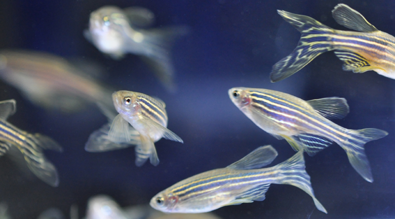
Coupling Mechanical Deformations and Planar Cell Polarity to Create Regular Patterns in the Zebrafish Retina
Guillaume Salbreux et al. (2012)
PLOS Computational Biology 8 (8), e1002618

Submitted by Eva Amsen:
“This interdisciplinary paper combines physics, mathematics and biology to show how computational modeling can describe the formation of complex biological structures.”

From December 1 to 24 we are featuring Node readers’ favourite papers of the past year. Click the calendar in the side bar each day to see a new paper. To see all papers submitted so far, see the calendar archive.

Thumbs up (No Ratings Yet)
Loading...

Tags:
Categories: Research

December 5

Posted by , on 5 December 2012

Today’s recommended paper is:

In toto live imaging of mouse morphogenesis and new insights into neural tube closure
R’ada Massarwa and Lee Niswander (2013)
Development 140 (1), 226-236

Submitted by Bob Goldstein:
“A clearer view of mammalian neural tube closure… an important step forward, and beautiful!”

From December 1 to 24 we are featuring Node readers’ favourite papers of the past year. Click the calendar in the side bar each day to see a new paper. To see all papers submitted so far, see the calendar archive.

Thumbs up (2 votes)
Loading...

Tags:
Categories: Research

Reprogramming development

Posted by , on 4 December 2012

This editorial by Development Editor-in-Chief Olivier Pourquié appears in the current issue of Development.

It seems most appropriate to start this editorial by congratulating the 2012 winners of the Nobel prize, John Gurdon and Shinya Yamanaka, for their work on stem cells and reprogramming. We at Development are particularly proud of this prize as John Gurdon published his original paper on the reprogramming of the frog oocyte nucleus to a totipotent state – the work that led to this award – in 1962 in the Journal of Embryology and Experimental Morphology (Gurdon, 1962), which was to become Development in 1987. Furthermore, John Gurdon was, until 2011, the Chair of The Company of Biologists, the charity that runs our journal. John Gurdon has been a role model for many of us and his original thinking has stimulated the field for many years and continues to do so.

The discovery that the nucleus of an adult cell can be reprogrammed to a totipotent embryonic state by nuclear transplantation in the frog revealed an unsuspected degree of plasticity of the genome and opened the way to the studies by Shinya Yamanaka on the reprogramming of adult somatic cells by a set of transcription factors (Takahashi and Yamanaka, 2006). Indeed, the experiments of Gurdon and Yamanaka clearly established that the information contained in the DNA of all somatic cells is sufficient to recreate an adult organism. That the nucleus of differentiated cells can be reprogrammed to the pluripotent state of early embryonic cells shows that it is possible to reverse the course of development and differentiation. This work opened tremendous new research avenues into the genetic control of pluripotency and differentiation. The ability to reverse-engineer embryonic cells and their derivatives from an adult counterpart, combined with the recent demonstration that sophisticated organs, such as the retina or hypophysis, can be grown from embryonic stem cells in vitro (Eiraku et al., 2011; Suga et al., 2011), means that recreating human organs in vitro is a real and achievable goal. This should open a new era in which the uncharted territory of human developmental biology will be explored. In addition, it will allow us to produce differentiated cells of all human lineages at all stages of differentiation, raising the possibility of establishing in vitro models of human diseases to study their pathophysiology and to screen for new treatments or cures. Finally, these advances should favour the development of cell therapy and regenerative medicine, potentially allowing the replacement of missing cells of body parts with cells from organs engineered in vitro. These are some of the major challenges that are now within reach thanks to Gurdon and Yamanaka’s discoveries.

In my view, this Nobel prize also beautifully illustrates the mutation that developmental biology is currently experiencing. Both Gurdon and Yamanaka clearly tackled a central question of developmental biology: how the genetic information is deployed during embryonic development to generate the variety of cell types and structures that will compose the adult body, and how this process might be reversed. Still, many scientists, particularly among our younger colleagues, will consider Gurdon and Yamanaka as stem cell biologists rather than developmental biologists. Although this might seem a semantic argument, it has a strong impact on the perception of a journal such as Development. The rapidly growing field of stem cell biology is largely an offshoot of developmental biology. However, it is now becoming independent from the more traditional developmental biology, creating its own structures with new societies and new journals. This reflects a healthy growth in the numbers of stem cell scientists, but it is occurring partly at the expense of the developmental biology community. Thus, while Development is clearly viewed as a community journal for those involved in core areas of developmental biology, feedback suggests that stem cell scientists do not necessarily regard Development in the same way.

I see engagement with the stem cell community as crucial to the future success of Development. As Editor in Chief, I have initiated several actions to raise the profile of the journal within this field. These include the recruitment of expert editors in the field, and creation of the ‘Development and Stem Cells’ section of the journal. Notably, many of our most highly downloaded and cited papers of the past 2 years appeared in this section – a preliminary indication that this move has been a success. In 2013, we hope to intensify our actions to reach out to the stem cell community and establish Development as an important forum for the publication of outstanding papers in this field.

Of course, while I believe that stem cell science is an important and expanding field within developmental biology, we will continue to be the home for more traditional disciplines, as well as for other growing areas, such as quantitative and systems biology, and evo-devo. In addition, when I took over as Editor in Chief, I felt that there was a need for Development to publish papers dealing with techniques of importance to the community. Over the past 2 years, we have published a number of such technical papers, and we are now expanding and renaming this section ‘Techniques and Resources’. We hope that this will provide an ideal home for high-quality papers of a technical nature, or those that describe a new resource, and we welcome your submissions.

Importantly, at the heart of our evolution as a journal, and running through this editorial, is the concept of community. Development is a not-for-profit journal run by scientists for scientists, and it is vital for us to make sure that we are efficiently serving the community of developmental biologists. We are very open to your feedback on what we are doing well (or badly) at the journal, and we will greatly benefit from your support while we move towards these new areas. Our community website the Node (thenode.biologists.com), launched in 2010, is one place where you can share your thoughts on the state of the developmental biology field, on scientific publishing and how Development is doing, or on anything else of relevance to developmental biologists. For those of you not yet familiar with the Node, I encourage you to visit the site and to contribute. For those happier in the real world than the virtual one, you will also find Development editors and staff at meetings throughout the globe and the year, and we’re always happy to talk about the journal and to receive your input.

This year has seen little change in the composition of the team of editors. Ken Zaret decided to step down as an editor and we are extremely grateful for his many years of service at Development and for his hard work to maintain the high quality standards of the journal. Although we are very sorry to see him leave, we were happy to welcome a new editor, Haruhiko Koseki from the Riken Center for Immunology in Yokohama. Haruhiko is a specialist in mouse development and epigenetics. He has joined the group of current editors, which also includes Magdalena Götz, Alex Joyner, Gordon Keller, Thomas Lecuit, Ottoline Leyser, Rong Li, Shin-Ichi Nishikawa, Nipam Patel, Liz Robertson, Geraldine Seydoux, Austin Smith, Patrick Tam and Steve Wilson. I congratulate them for their outstanding work in the past year. I also thank the in-house Development team – particularly Katherine Brown, our Executive Editor, and Claire Moulton, our Publisher – for their excellent support. We look forward to a successful 2013.

Thumbs up (4 votes)
Loading...

Tags: ,
Categories: News

In Development this week (Vol. 140, Issue 1)

Posted by , on 4 December 2012

Here are the highlights from the current issue of Development:


Gut feeling about Wnt doses

The intestinal epithelium continuously renews throughout life. Canonical Wnt signalling – a major player in this renewal – both controls intestinal epithelial proliferation and maintains intestinal stem cells. The existence of a gradient of β-catenin expression along the colonic crypt axis, with the highest β-catenin levels at the bottom where the intestinal stem cells reside, suggests that Wnt signalling might have dose-dependent roles in the colonic epithelium. On p. 66, Yasuhiro Yamada, Konrad Hochedlinger and colleagues use a β-catenin-inducible mouse model to investigate this possibility. High levels of β-catenin expression induce crypt formation but reduce cell proliferation among progenitor cells, they report, whereas lower levels have the opposite effect. Notably, Notch signalling is activated in the slow-cycling crypt cells produced by β-catenin overexpression, and treatment of β-catenin-expressing mice with a Notch inhibitor turns the slow-cycling cells into actively proliferating cells. Together, these results suggest that different levels of Wnt signalling, in cooperation with Notch signalling, control the differentiation and proliferation of the colonic epithelium.


Micro-(RNA)managing muscle repair

Following muscle injury, quiescent muscle stem cells (satellite cells) are activated and then proliferate and differentiate to regenerate myofibres. Wnt signalling regulates the differentiation of activated satellite cells, but what other factors control skeletal muscle regeneration? On p. 31, Francisco Naya and co-workers report that the transcription factor myocyte enhancer factor 2A (MEF2A) plays an essential role in skeletal muscle regeneration in adult mice. Myofibre formation is impaired in injured Mef2a knockout mice, the researchers report, and this impaired injury response is associated with downregulation of the Gtl2-Dio3 locus, the largest known mammalian microRNA (miRNA) cluster. Notably, a subset of the miRNAs in this locus represses secreted Frizzled-related proteins (sFRPs), which are inhibitors of Wnt signalling. Consistent with these data, sFRP expression is upregulated and Wnt activity is attenuated in injured Mef2a knockout muscle. These and other results suggest that miRNA-mediated modulation of Wnt signalling by MEF2A is required for muscle regeneration and suggest that targeting this pathway might enhance the regeneration of diseased muscle.


Epigenetic maintenance via DNA replication machinery

During development, cell type-specific epigenetic states must be duplicated accurately during mitosis to maintain cellular memory. The maintenance of epigenetic memory seems to involve the DNA replication machinery but is poorly understood. Here (p. 156), Yeonhee Choi and co-workers show that INCURVATA2 (ICU2), the catalytic subunit of DNA polymerase α in Arabidopsis, ensures the stable maintenance of histone modifications. Vernalisation, the acquisition of floral competence though exposure to prolonged cold, requires repression of Flowering Locus C (FLC) through accumulation of the histone mark H3K27me3. The researchers show that the missense mutant allele icu2-1 does not affect the recruitment of CLF, a component of polycomb repressive complex 2 (PRC2), and the resultant deposition of the H3K27me3 mark on the FLC locus. However, icu2-1 mutants do not stably maintain this epigenetic mark, which results in mosaic FLC de-repression after vernalisation. ICU2 also maintains repressive histone modifications at other PRC2 targets and at retroelements, and it facilitates histone assembly in dividing cells, which suggests a possible mechanism for ICU2-mediated epigenetic maintenance.


Otx2 switches early pluripotency states

Mouse embryonic stem cells (ESCs) and epiblast stem cells (EpiSCs) correspond to the naïve ground state of the preimplantation epiblast and the primed state of the post-implantation epiblast, respectively. The ESC state is characterised by spontaneous, reversible differences among the ESCs in their susceptibility to self-renewal and differentiation signals. Here (p. 43), Antonio Simeone and colleagues investigate the mechanisms that control this metastable state and the transition from ESCs to EpiSCs. The researchers report that Otx2, a transcription factor that is required for brain development, is expressed in a large subset of ESCs and maintains the ESC metastable state by antagonising ground state pluripotency and promoting commitment to differentiation. Otx2 is also needed for ESC transition into EpiSCs, they report, and stabilises the EpiSC state in cooperation with BMP4 and Fgf2. Finally, they show that Otx2 is required for the differentiation of ESC-derived neural progenitors. Thus, Otx2 is an intrinsic determinant of the ESC state and of the ESC to EpiSC pluripotency transition.


Sphingosine 1-phosphate stops muscle wasting

Duchenne muscular dystrophy – a lethal disease caused by dystrophin mutations – is characterised by age-dependent muscle wasting. Mario Pantoja and co-workers now report that increased intracellular sphingosine 1-phosphate (S1P) suppresses dystrophic muscle phenotypes in a Drosophila Dystrophin mutant (see p. 136). The researchers use localisation of Projectin protein (a titin homolog) in sarcomeres, as well as muscle morphology and movement assays, to show that reduction of wunen (a homolog of lipid phosphate phosphatase 3, which dephosphorylates several phospholipids in higher animals) suppresses dystrophic muscle defects; wunen is known to suppress a wing vein defect also seen in Drosophila Dystrophin mutants. Hypothesising that wunen-based suppression may be through elevation of S1P, which promotes cell proliferation and differentiation in muscle, the researchers use several genetic approaches and pharmacological agents to raise S1P levels in the dystrophic flies. In each case, increased S1P levels suppress dystrophic muscle phenotypes. The researchers suggest, therefore, that Drosophila could be used to identify small molecules that might suppress muscle wasting in human patients.

A Rosa future for Fucci cell-cycle indicators

Visualising cell-cycle progression in living embryos is essential for improving our understanding of developmental processes. The fluorescent ubiquitylation-based cell-cycle indicator (Fucci), which was generated by fusing the ubiquitylation domains of Cdt1 and Geminin to different fluorescent proteins, has been used to label G1 and S/G2/M phase nuclei orange and green, respectively. Cell cycle dynamics during development have been studied in transgenic mice expressing Fucci under the control of the CAG promoter but Fucci expression levels are somewhat variable. Now, Shinichi Aizawa and colleagues (p. 237) describe two new mouse cell-cycle reporter lines that use Fucci2, a Fucci derivative that emits red and green fluorescence. The R26p-Fucci2 transgenic line uses the Rosa26 promoter and harbours the G1 and S/G2/M phase probes in a single transgene to ensure their co-inheritance. In the R26R-Fucci2 transgenic line, the two probes are incorporated into the Rosa26 locus conditionally to allow cell-cycle analysis in specific cell types. Time-lapse imaging experiments suggest that both reporter lines hold great promise for studying cell-cycle behaviour in vivo.


Plus…


Endoreplication and polyploidy: insights into development and disease

Polyploid cells have genomes that contain multiples of the typical diploid chromosome number and are found in many different organisms. As part of the “Development: The Big Picture” series, Fox and Duronio review the conserved mechanisms that control the generation of polyploidy and highlight studies that have  begun to provide clues into the physiological function of polyploidy. See the Primer on p. 3

Click here to see other articles in the ‘Development: the Big Picture” series.


Cell polarity: models and mechanisms from yeast, worms and flies

Determinants of cell polarity dictate the behaviour of many cell types during development. Recent work in yeast, worms and flies, reviewed here by Barry Thompson, has combined computer modelling with experimental analysis to reveal the mechanisms that drive the polarisation of such determinants. See the Review on p. 13


Thumbs up (No Ratings Yet)
Loading...

Categories: Research

December 4

Posted by , on 4 December 2012

Today’s recommended paper is:

NuRD Suppresses Pluripotency Gene Expression to Promote Transcriptional Heterogeneity and Lineage Commitment
Nicola Reynolds et al. (2012)
Cell Stem Cell 10 (5), 583-594

Submitted by Mary Todd Bergman:
“Paul Bertone’s lab at EMBL-EBI discovered a key component of the ‘go’ signal that tells stem cells to commit already and become one type of cell or another. In collaboration with Brian Hendrich at the University of Cambridge, Paul’s group studied differences in gene expression patterns between self-renewing and differentiating stem cells, specifically focusing on the Nucleosome Remodelling and Deacetylation (NuRD) corepressor complex. They found that NuRD directly regulates how pluripotency genes are expressed in stem cells, and that NuRD is required for their exit from pluripotency. They also investigated the binding patterns of the NuRD complex to understand how it regulates its target genes. Combined with transcriptional profiling, a picture emerged of NuRD as a gatekeeper of cell differentiation.”

From December 1 to 24 we are featuring Node readers’ favourite papers of the past year. Click the calendar in the side bar each day to see a new paper. To see all papers submitted so far, see the calendar archive.

Thumbs up (No Ratings Yet)
Loading...

Tags:
Categories: Research

EVODEVO WIKI EDIT EVENT.

Posted by , on 4 December 2012

EVODEVO WIKIPAEDIA EDIT-ATHON. Friday December 7th 2012 a the Department of Zoology, South Parks Road, University of Oxford

We are spending a day editing and updating EVODEVO related pages on Wikipaedia. If you would like to join us contact peter.holland@zoo.ox.ac.uk or aziz.aboobaker@zoo.ox.ac.uk

We will start about 10.30 am and two official trainers from Wikimedia UK will joins us at around 11 to help us get started.

Thumbs up (3 votes)
Loading...

Categories: Events, News

December 3

Posted by , on 3 December 2012

Today’s recommended paper is:

Bimodal control of Hoxd gene transcription in the spinal cord defines two regulatory subclusters
Patrick Tschopp, Alix J. Christen and Denis Duboule (2012)
Development 139 (5), 929-939

Submitted by Rachael Inglis:
“This is a great example of new results enriching our understanding of a classical developmental phenomenon: the co-linear expression of the Hox gene cluster from anterior to posterior along the spinal cord, which actually turns out to be two sub-clusters that are regulated independently and correspond to the regions of the spinal cord that innervate the forelimb and the hindlimb.”

From December 1 to 24 we are featuring Node readers’ favourite papers of the past year. Click the calendar in the side bar each day to see a new paper. To see all papers submitted so far, see the calendar archive.

Thumbs up (1 votes)
Loading...

Tags:
Categories: Research

December 2

Posted by , on 2 December 2012

Today’s recommended paper is:

Polyploidization of glia in neural development links tissue growth to blood–brain barrier integrity
Yingdee Unhavaithaya and Terry L. Orr-Weaver (2012)
Genes & Development 26, 31-36

Submitted by Tohru Yano:
“This paper tell us the link between cell properties and tissue
growth and biological functions.”

From December 1 to 24 we are featuring Node readers’ favourite papers of the past year. Click the calendar in the side bar each day to see a new paper. To see all papers submitted so far, see the calendar archive.

Thumbs up (No Ratings Yet)
Loading...

Tags:
Categories: Research

December 1

Posted by , on 1 December 2012

Today’s recommended paper is:

The accessible chromatin landscape of the human genome
Robert E. Thurman, Eric Rynes, Richard Humbert, et al. (2012)
Nature 489, 75–82

Submitted by Nishal Patel:
“Whilst humans aren’t exactly a model organism that most developmental biologists work on, the ENCODE project is ground breaking and transcription factors and chromatin accessibility are important in development.”

From December 1 to 24 we are featuring Node readers’ favourite papers of the past year. Click the calendar in the side bar each day to see a new paper. To see all papers submitted so far, see the calendar archive.

Thumbs up (3 votes)
Loading...

Tags:
Categories: Research