

In Development this week (Vol. 140, Issue 9)
Posted by Seema Grewal, on 9 April 2013
Here are the highlights from the current issue of Development:
Dynamics of human thymus development
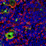 The thymus is the primary organ responsible for generating T cells. Although thymus development has been studied in mice, little is known about how the human thymus develops. Here (p. 2015), Clare Blackburn and colleagues provide a comprehensive analysis of human thymus organogenesis. Using gene expression analyses, the researchers show that the spatial and temporal expression patterns of factors involved in thymus development in mice are conserved in humans. They also demonstrate that the human thymus arises from the third pharyngeal pouch, as in mice and in contrast to previous suggestions. Furthermore, they report, thymic epithelial cell differentiation as well as the immigration of mesenchymal cells and vascular progenitors into the human thymus occur after the onset of FOXN1 expression, consistent with the timing of these events in mice. Finally, the authors define precisely when the human thymus becomes colonised with haematopoietic cells, which are CD45+/CD7+/CD34int cells. Collectively, this study provides key insights into the conservation of thymus development between mice and humans, which has major clinical implications for enhancing or replacing thymus function.
Coordinating head-to-tail with front-to-back
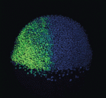 During embryogenesis, the anterior-posterior (AP) and dorsal-ventral (DV) axes are specified by the activity of key signalling pathways. FGF, Wnt and retinoic acid together pattern the AP axis: high activity defines more posterior tissues, which are specified later in development than anterior tissues. The BMP pathway specifies ventral fate; low BMP activity defines dorsal. Whether and how these pathways intersect to coordinate patterning of the two axes is poorly understood. On p. 1970, Megumi Hashiguchi and Mary Mullins provide evidence for synchronisation of DV and AP patterning in the zebrafish embryo. Upon temporally restricted inhibition of BMP in embryos anteriorised by inhibition of FGF or Wnt, the dorsalised tissue takes on the AP fate appropriate to the anteriorised embryo, rather than that corresponding to the time of BMP inhibition. The authors identify one mechanism mediating pathway cross-talk: MAPK, activated downstream of FGF, phosphorylates and inhibits the BMP effector Smad5. Thus, this work establishes the close temporal coordination of AP and DV patterning, and provides insights into how this is achieved.
miR-203 drives progenitor cell differentiation
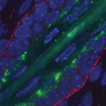 MicroRNAs are important for the regulation of gene expression in a vast array of processes. In the skin, miR-203 has been shown to be crucial for the proper differentiation of the interfollicular progenitor cells, although the specific mechanism of this has remained elusive. In this issue (p. 1882), Rui Yi and colleagues investigate the precise timing of miR-203 activation during epidermal differentiation. They show that miR-203 is transcriptionally activated in the differentiating progeny of interfollicular progenitor cells upon asymmetric cell division. Using keratinocytes derived from miR-203-inducible mice, the authors found that miR-203 functions to promote an immediate exit from the cell cycle, leading to a complete loss of self-renewal after just 72 hours. They further identify a multitude of novel miR-203 targets in vivo, and demonstrate that co-repression by miR-203 of many of these, including Skp2, Msi2 and p63, is necessary for the function of miR-203 in inhibiting self-renewal. These data provide novel insights into the widespread role of miR-203 in differentiating interfollicular progenitor cells in the skin.
Leaf patterning: AS1 far AS2 we know
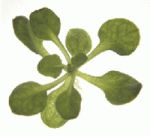 The correct establishment of adaxial-abaxial patterning is crucial for leaf expansion and growth. The AUXIN RESPONSE FACTOR (ARF) family of proteins are key determinants of organ symmetry and abaxial patterning in Arabidopsis thaliana and are subject to complex regulatory control at both the transcriptional and translational level. Here (p. 1958), Chiyoko Machida and colleagues uncover an additional, dual mechanism for the regulation of ETT (also known as AFR3) by an ASYMMETRIC LEAVES1 (AS1)-AS2 complex. First, the authors show that the AS1-AS2 complex directly represses ETT expression via binding of AS1 to the promoter region. Second, they provide evidence for an indirect mechanism of regulation via miR390- and RDR6-dependent post-transcriptional gene silencing of both ETT and ARF4. The authors also suggest a possible epigenetic mechanism, as AS1-AS2 maintains the methylation status of ETT within the coding region. These discoveries shed light on the molecular framework of early leaf patterning events and help to uncover the events that lead to the specification of distinct adaxial and abaxial fates during leaf development.
PAR-alell pathways for polarity
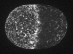 In the C. elegans embryo, anterior-posterior polarity is defined at the one-cell stage, via asymmetric and reciprocal localisation of cortex-associated PAR protein complexes: PAR-3, PAR-6 and aPKC localise to the anterior, whereas PAR-1, PAR-2 and LGL-1 are enriched at the posterior. Polarity maintenance involves mutual antagonism between the anterior and posterior complexes and may also involve CDC-42-dependent regulation of myosin activity. Kenneth Kemphues and co-workers (p. 2005) now provide evidence for multiple and partially redundant pathways acting at the posterior to maintain polarity once it has been established. Both PAR-2 and CDC-42, acting in separate pathways, have dual functions in independently regulating both anterior PAR complex localisation and myosin activity, whereas LGL-1 appears to have a buffering role in controlling PAR-6 protein levels. Although the molecular details of these pathways remain incomplete, the complex and overlapping mechanisms operating to maintain polarity in the early embryo underscore the importance of robust and efficient polarisation for subsequent development.
Stem cells go out of bounds
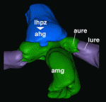 Many animal tissues maintain populations of slowly proliferating stem cells that contribute to tissue homeostasis and repair. In Drosophila, for example, stem cells reside throughout the midgut and within the hindgut and renal tubules. But how and when do these cells arise? Volker Hartenstein and colleagues now show that Drosophila gut progenitors migrate across tissue boundaries and adopt the fate of the organ in which they come to reside (p. 1903). Using lineage tracing, the researchers demonstrate that a subset of adult midgut progenitors, which are initially located in the larval midgut, migrate posteriorly during development and contribute to the adult ureter and, subsequently, the renal stem cell population. In addition, they report, a population of hindgut progenitors migrates anteriorly into the midgut territory to differentiate and give rise to midgut enterocytes. These findings suggest that a stable boundary between the midgut (an endodermal tissue) and the hindgut/renal tubules (ectodermal tissues) does not exist and instead multipotent progenitors are able to cross the boundaries between these domains.
Plus:
Molecular pathways regulating mitotic spindle orientation in animal cells
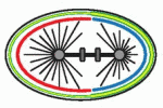 Orientation of the cell division axis is essential for both symmetric cell divisions and for the asymmetric distribution of fate determinants during, for example, stem cell divisions. Lu and Johnston review both the well-established spindle orientation pathways and recently identified regulators to provide a updated view of how positioning of the mitotic spindle occurs. See the Review article on p. 1483
Getting out and about: the emergence and morphogenesis of the vertebrate lymphatic vasculature
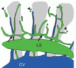 New insights into lymphatic vascular development have recently been achieved thanks to the use of alternative model systems, new molecular tools, novel imaging technologies and a growing interest in the role of lymphatic vessels in human disorders. Here. Hogan and colleagues review the most recent advances in lymphatic vascular development, with a major focus on mouse and zebrafish model systems. See the Review article on p. 1857
 Voting for the stem cell image contest
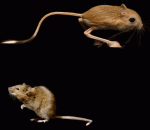 – Over the last couple of years, Kim Cooper (a post-doc in the Tabin lab) has been providing us with updates (see her intro post
 An unproven stem cell therapy has taken centre stage in Italy after patients successfully lobbied the Italian government to allow its use in public hospitals. The highly controversial and untested procedure was created by the privately owned Stamina Foundation, but blocked by the Italian Medicine’s Agency, AIFA. Last week’s decision by the Ministry of Health to override AIFA’s block has horrified Italy’s leading stem cell scientists. In a letter to the Ministry, they describe the decision as providing “a dangerous short circuit between patients’ hopes and lucrative commercial practices” of organisations operating outside the “scientific and moral foundations” of medicine.
  Our
Our 
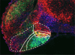 The hypothalamus is a key integrative centre in the vertebrate brain that regulates many essential functions, including homeostasis and stress responses. Several transcription factors that are essential for hypothalamic development have been identified but the production of diverse neuron types in this complex brain region is poorly understood. Here (
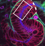 The formation and elongation of polarised epithelial tubules is essential for the structure and function of several metazoan organ systems but the molecular mechanisms that regulate tubulogenesis are largely unknown. Here (
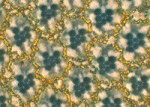 Planar cell polarity depends on the asymmetric localisation of core planar polarity proteins at apicolateral junctions. This asymmetric distribution probably develops through amplification of an initial asymmetry and seems to require the regulation of core protein levels. Now, Helen Strutt, Elizabeth Searle and co-workers (
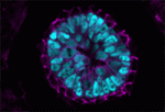 During mammalian embryogenesis, substantial cell proliferation occurs before the establishment of the body plan during gastrulation. Thus, before gastrulation, individual embryonic cells must be pluripotent. In vitro experiments with embryonic stem cells (ESCs) have indicated that the transcription factors Oct4, Sox2 and Nanog are components of a gene regulatory network (GRN) that stimulates self-renewal of pluripotent cells and have identified Tcf7l1 (Tcf3) as an inhibitor of GRN activity. But what is Tcf7l1’s function during embryonic development? To find out, Bradley Merrill and colleagues have been examining embryogenesis in Tcf7l1-/- mouse embryos (see
 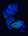 Canonical Wnt signalling and E-cadherin-mediated cell adhesion are both involved in mouse embryonic stem (mES) cell maintenance. β-catenin (Ctnnb1) is central to both these processes – it mediates the transactivation of Wnt target genes and also connects E-cadherin to the actin cytoskeleton via α-catenin. But which β-catenin function is absolutely required for mES cell self-renewal and pluripotency? On
 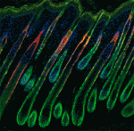 Adult tissue-specific stem cells both self-renew and generate functional progeny. Mammalian hair follicles, which are characterised by cyclical phases of growth (anagen), regression (catagen) and rest (telogen), are an ideal system in which to investigate the homeostasis of an adult stem cell population. On
 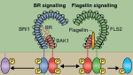 Zhi-Yong Wang and colleagues provide an overview of the highly integrated BR signalling network and explain how this steroid hormone functions as a master regulator of plant growth, development and metabolism.
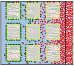 Alex Schier et al analyze various morphogen transport models using the morphogens Nodal, fibroblast growth factor and Decapentaplegic as case studies. They propose that most of the available data support the idea that morphogen gradients form by diffusion that is hindered by tortuosity and binding to extracellular molecules.
 This workshop is geared toward university faculty, postdocs, and graduate students interested in teaching developmental biology. It provides basic hands-on experience with organisms commonly studied in teaching laboratories. These include sea urchins and sand dollars, planaria, Drosophila, chick embryos, Spirostomum, Hydra, Lumbriculus, and flowering plants. Techniques will range from classical microsurgical techniques to fluorescence microscopy and applications of reporter gene technology.