This editorial by Development Editor-in-Chief Olivier Pourquié appears in the current issue of Development.
It seems most appropriate to start this editorial by congratulating the 2012 winners of the Nobel prize, John Gurdon and Shinya Yamanaka, for their work on stem cells and reprogramming. We at Development are particularly proud of this prize as John Gurdon published his original paper on the reprogramming of the frog oocyte nucleus to a totipotent state – the work that led to this award – in 1962 in the Journal of Embryology and Experimental Morphology (Gurdon, 1962), which was to become Development in 1987. Furthermore, John Gurdon was, until 2011, the Chair of The Company of Biologists, the charity that runs our journal. John Gurdon has been a role model for many of us and his original thinking has stimulated the field for many years and continues to do so.
The discovery that the nucleus of an adult cell can be reprogrammed to a totipotent embryonic state by nuclear transplantation in the frog revealed an unsuspected degree of plasticity of the genome and opened the way to the studies by Shinya Yamanaka on the reprogramming of adult somatic cells by a set of transcription factors (Takahashi and Yamanaka, 2006). Indeed, the experiments of Gurdon and Yamanaka clearly established that the information contained in the DNA of all somatic cells is sufficient to recreate an adult organism. That the nucleus of differentiated cells can be reprogrammed to the pluripotent state of early embryonic cells shows that it is possible to reverse the course of development and differentiation. This work opened tremendous new research avenues into the genetic control of pluripotency and differentiation. The ability to reverse-engineer embryonic cells and their derivatives from an adult counterpart, combined with the recent demonstration that sophisticated organs, such as the retina or hypophysis, can be grown from embryonic stem cells in vitro (Eiraku et al., 2011; Suga et al., 2011), means that recreating human organs in vitro is a real and achievable goal. This should open a new era in which the uncharted territory of human developmental biology will be explored. In addition, it will allow us to produce differentiated cells of all human lineages at all stages of differentiation, raising the possibility of establishing in vitro models of human diseases to study their pathophysiology and to screen for new treatments or cures. Finally, these advances should favour the development of cell therapy and regenerative medicine, potentially allowing the replacement of missing cells of body parts with cells from organs engineered in vitro. These are some of the major challenges that are now within reach thanks to Gurdon and Yamanaka’s discoveries.
In my view, this Nobel prize also beautifully illustrates the mutation that developmental biology is currently experiencing. Both Gurdon and Yamanaka clearly tackled a central question of developmental biology: how the genetic information is deployed during embryonic development to generate the variety of cell types and structures that will compose the adult body, and how this process might be reversed. Still, many scientists, particularly among our younger colleagues, will consider Gurdon and Yamanaka as stem cell biologists rather than developmental biologists. Although this might seem a semantic argument, it has a strong impact on the perception of a journal such as Development. The rapidly growing field of stem cell biology is largely an offshoot of developmental biology. However, it is now becoming independent from the more traditional developmental biology, creating its own structures with new societies and new journals. This reflects a healthy growth in the numbers of stem cell scientists, but it is occurring partly at the expense of the developmental biology community. Thus, while Development is clearly viewed as a community journal for those involved in core areas of developmental biology, feedback suggests that stem cell scientists do not necessarily regard Development in the same way.
I see engagement with the stem cell community as crucial to the future success of Development. As Editor in Chief, I have initiated several actions to raise the profile of the journal within this field. These include the recruitment of expert editors in the field, and creation of the ‘Development and Stem Cells’ section of the journal. Notably, many of our most highly downloaded and cited papers of the past 2 years appeared in this section – a preliminary indication that this move has been a success. In 2013, we hope to intensify our actions to reach out to the stem cell community and establish Development as an important forum for the publication of outstanding papers in this field.
Of course, while I believe that stem cell science is an important and expanding field within developmental biology, we will continue to be the home for more traditional disciplines, as well as for other growing areas, such as quantitative and systems biology, and evo-devo. In addition, when I took over as Editor in Chief, I felt that there was a need for Development to publish papers dealing with techniques of importance to the community. Over the past 2 years, we have published a number of such technical papers, and we are now expanding and renaming this section ‘Techniques and Resources’. We hope that this will provide an ideal home for high-quality papers of a technical nature, or those that describe a new resource, and we welcome your submissions.
Importantly, at the heart of our evolution as a journal, and running through this editorial, is the concept of community. Development is a not-for-profit journal run by scientists for scientists, and it is vital for us to make sure that we are efficiently serving the community of developmental biologists. We are very open to your feedback on what we are doing well (or badly) at the journal, and we will greatly benefit from your support while we move towards these new areas. Our community website the Node (thenode.biologists.com), launched in 2010, is one place where you can share your thoughts on the state of the developmental biology field, on scientific publishing and how Development is doing, or on anything else of relevance to developmental biologists. For those of you not yet familiar with the Node, I encourage you to visit the site and to contribute. For those happier in the real world than the virtual one, you will also find Development editors and staff at meetings throughout the globe and the year, and we’re always happy to talk about the journal and to receive your input.
This year has seen little change in the composition of the team of editors. Ken Zaret decided to step down as an editor and we are extremely grateful for his many years of service at Development and for his hard work to maintain the high quality standards of the journal. Although we are very sorry to see him leave, we were happy to welcome a new editor, Haruhiko Koseki from the Riken Center for Immunology in Yokohama. Haruhiko is a specialist in mouse development and epigenetics. He has joined the group of current editors, which also includes Magdalena Götz, Alex Joyner, Gordon Keller, Thomas Lecuit, Ottoline Leyser, Rong Li, Shin-Ichi Nishikawa, Nipam Patel, Liz Robertson, Geraldine Seydoux, Austin Smith, Patrick Tam and Steve Wilson. I congratulate them for their outstanding work in the past year. I also thank the in-house Development team – particularly Katherine Brown, our Executive Editor, and Claire Moulton, our Publisher – for their excellent support. We look forward to a successful 2013.
 (4 votes)
 Loading...


 (No Ratings Yet)
 (2 votes)
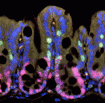 The intestinal epithelium continuously renews throughout life. Canonical Wnt signalling – a major player in this renewal – both controls intestinal epithelial proliferation and maintains intestinal stem cells. The existence of a gradient of β-catenin expression along the colonic crypt axis, with the highest β-catenin levels at the bottom where the intestinal stem cells reside, suggests that Wnt signalling might have dose-dependent roles in the colonic epithelium. On
 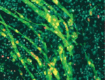 Following muscle injury, quiescent muscle stem cells (satellite cells) are activated and then proliferate and differentiate to regenerate myofibres. Wnt signalling regulates the differentiation of activated satellite cells, but what other factors control skeletal muscle regeneration? On
 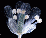 During development, cell type-specific epigenetic states must be duplicated accurately during mitosis to maintain cellular memory. The maintenance of epigenetic memory seems to involve the DNA replication machinery but is poorly understood. Here (
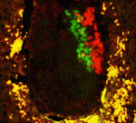 Mouse embryonic stem cells (ESCs) and epiblast stem cells (EpiSCs) correspond to the naïve ground state of the preimplantation epiblast and the primed state of the post-implantation epiblast, respectively. The ESC state is characterised by spontaneous, reversible differences among the ESCs in their susceptibility to self-renewal and differentiation signals. Here (
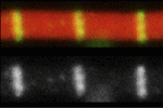 Duchenne muscular dystrophy – a lethal disease caused by dystrophin mutations – is characterised by age-dependent muscle wasting. Mario Pantoja and co-workers now report that increased intracellular sphingosine 1-phosphate (S1P) suppresses dystrophic muscle phenotypes in a Drosophila Dystrophin mutant (see
 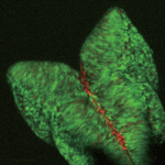 Visualising cell-cycle progression in living embryos is essential for improving our understanding of developmental processes. The fluorescent ubiquitylation-based cell-cycle indicator (Fucci), which was generated by fusing the ubiquitylation domains of Cdt1 and Geminin to different fluorescent proteins, has been used to label G1 and S/G2/M phase nuclei orange and green, respectively. Cell cycle dynamics during development have been studied in transgenic mice expressing Fucci under the control of the CAG promoter but Fucci expression levels are somewhat variable. Now, Shinichi Aizawa and colleagues (
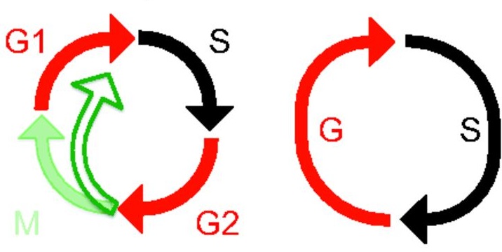 Polyploid cells have genomes that contain multiples of the typical diploid chromosome number and are found in many different organisms. As part of the “Development: The Big Picture” series, Fox and Duronio review the conserved mechanisms that control the generation of polyploidy and highlight studies that have begun to provide clues into the physiological function of polyploidy. See the Primer on p.
 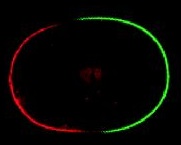 Determinants of cell polarity dictate the behaviour of many cell types during development. Recent work in yeast, worms and flies, reviewed here by Barry Thompson, has combined computer modelling with experimental analysis to reveal the mechanisms that drive the polarisation of such determinants. See the Review on p.
  (3 votes)
