Cellular differentiation describes the process by which embryonic cells become different from one another, acquiring distinct identities and specialised functions. These cells are responding to signals that modify the dynamics of the interacting key genes controlling the cell’s state. The arrival of more and better single cell data opens up new opportunities for better understanding of this cellular decision-making. However, to realise these opportunities, new mathematical and statistical tools are needed to characterise the cellular dynamics and to organise, analyse and visualise such data. This workshop aims at reviewing the current work in this area.
Announcement of post-doc scholarship at the Division of Pediatrics, Lund University, Sweden
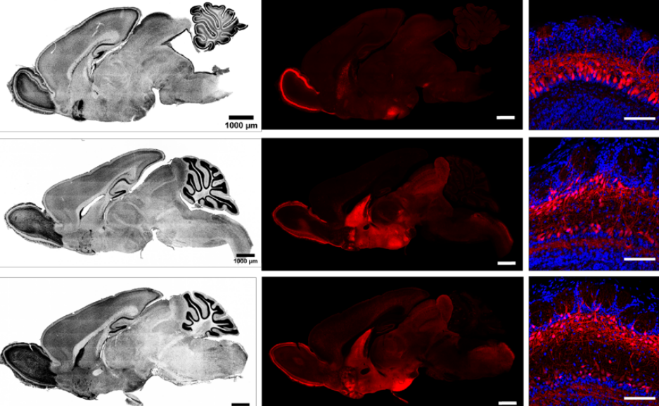
The importance of HIF-2afor neural crest cell migration and its connection to tumor initiation
It is well known that hypoxia, i.e. low oxygen levels, predicts poor outcome and metastatic burden in a variety of solid tumors. Transcriptionally active Hypoxia Inducible Factors (HIFs) and their downstream target genes orchestrate the cellular adaptation to increased oxygen demands in both normal and tumor cells (Hammarlund, Flashman, Mohlin & Licausi, Science 2020). We have recently shown that the cancer-associated protein HIF-2a is important for trunk neural crest stem cell development (Niklasson et al., Dev Dyn 2020). In this project, we are interested in investigating how HIF-2a affects early and late neural crest cell migration, population of organs and how this connects to childhood tumor initiation.
The purpose of this scholarship is for the candidate to deepen their skills in the field of molecular physiology. The candidate will acquire theoretical knowledge within the areas of developmental and tumor biology as well as learn a set of methodologies including microdissections, cell culture, cloning, virus work and analyzing omics data. The candidate will learn to handle several different research organisms, including chick embryos and in vivo mouse models.
Preliminary start date: 1st of March 2021 (to be discussed)
– To be eligible for a post-doc scholarshop at Lund University the recipient must hold a PhD degree within a relevant field. The PhD degree must not be from Lund University. The PhD degree must not be older than three years. The applicant must not have been employed at Lund University in the past two years.
– Obtained PhD within cancer research, developmental biology or equivalent.
– Published papers in peer-reviewed international journals
– Fluent in written and spoken English
The following qualifications are advantageous but not necessary:
Experience in:
Analyzing omics data (e.g., RNA sequencing and mass spectrometry)
In vivo models (e.g., chick embryo, mice, rats, zebrafish)
Wet lab techniques
Imaging
The candidate will be part of a team, but at the same time needs to be independent enough to lead their own projects. The candidate should be fluent in English and have experience from oral and written scientific communication.
We put great emphasis on personal suitability.
Written application, including reference number, is to be sent via e-mail to the supervisor and must include the following:
– CV
– Personal letter stating the reasons why the study suits the applicant (maximum one page)
This is the first in a new series on the Node profiling scientists who do art (or artists who do science). We’ll learn about why they do it, their artistic influences, their techniques and tips. We’ll be looking for new people to feature in this series throughout the year – whatever kind of art you do, from sculpture to embroidery to music to drawing, if you want to share it with the community just email thenode@biologists.com (nominations are also welcome!).
Our first profile is of Suyash Naik, a graduate student from the IST Austria, whose art we first saw on Twitter.
Where do you work and what are you currently working on?
Suyash
I study at the Institute of Science and Technology Austria, with the supervision of Dr Carl-Phillip Heisenberg and Dr Edouard Hannezo. I’m currently in the third year of my graduate studies working to understand the mechanics of epithelium movement during zebrafish embryonic development. I am especially interested in understanding the intermediate filament cytoskeleton’s role as the epithelium spreads during gastrulation. For this I am learning to apply various interesting biophysical techniques that our labs have developed, such as laser cutting and imaging.
Has science always been an important part of your life?
I’d say curiosity and learning new things have been important aspects of my life. I grew up with an interest in natural history and dinosaur fossils, and also liked watching and reading popular science fiction. Naturally I was always interested in learning more about science and understanding the world we live in. This interest was fuelled further by great teachers and incredible opportunities that allowed me to do the things I do day to day now. For instance, in school I took part in a group that used to plan and do simple experiments revolving around DNA. This led me to pursue an education at IISER where I had the opportunity to explore and work in different labs.
And what about art – were you always interested in drawing? What or who are your artistic influences?
I have been interested in sketching and photography but I have never had the opportunity to pursue any formal education in them. But recently I made more of an effort to consistently create and also be more forthcoming with the things I’m doing and have done in the past, as a way to have an outlet for creativity and to track my mental health during these stressful times. For me what is most interesting and important is the change of perspective I gain when viewing something as an artistic inspiration rather than a scientific curiosity. I have always enjoyed visual art like paintings and animations. The lockdowns this year also greatly increased my appreciation for street art. Watching great pieces from some of my favourite artists like Nekro and Illunis has been a real joy during lockdown walks along the Danube canal this year. But most importantly of all I draw inspiration from my creative friends who make some truly amazing artwork!
“For me what is most interesting and important is the change of perspective I gain when viewing something as an artistic inspiration rather than a scientific curiosity.”
How do you approach your art?
I mostly use digital mediums currently as I have some familiarity with them, but I am still hoping to learn different types of techniques to create sciart. I am looking forward to learning more about art techniques in the coming years and creating/experimenting different styles. I usually enjoy colorful and futuristic looking artwork and that is something I especially want to learn how to create.
Tell me about your zebrafish artwork?
For the zebrafish artwork, I had thought of the design long ago to sort of combine the ideas that I wanted to base my PhD around and things that were ongoing in my lab at the moment.
Zebrafish embryo
The embryonic part of the embryo is an artistic representation of a model epithelium. Recently in my lab a few great publications (such as this article by Schwayer et.al, this article by Boocock et. al., this and this review by Shamipour et.al.) explore how flows of different cytoskeletal proteins play an important role during development. This made me think of some acrylic pour that a friend of mine had made, and I wanted to incorporate that as a component of the artwork. That leads to the flowy backdrop of color that exists in the picture. The yolk over which the epithelium moves is a collection of nutrition for the developing embryo in the form of collected yolk granules. These were represented in spirals depicting these granules.
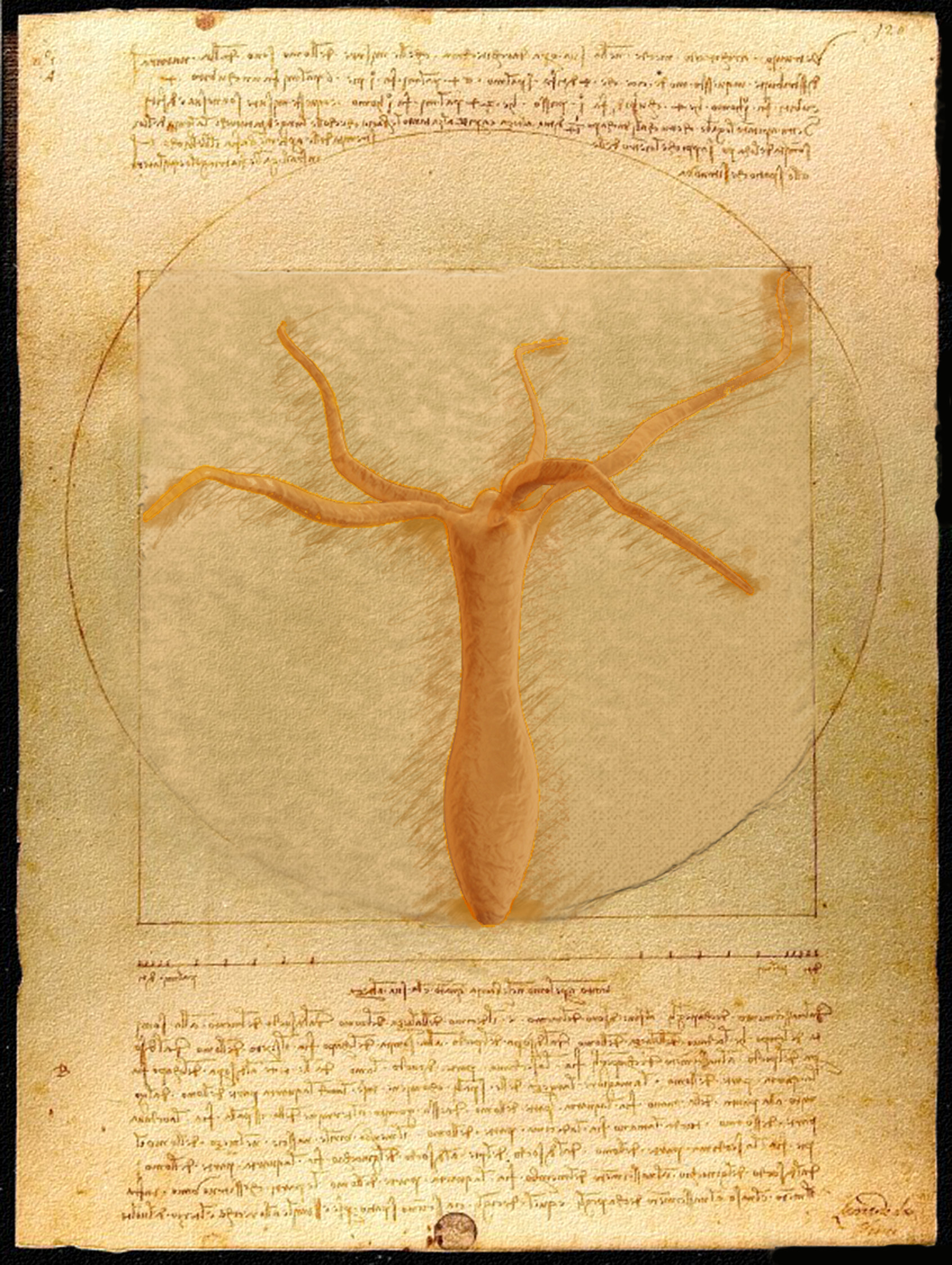
And what about the Hydra pictures – I think both have reference to historical pieces or myths?
The Hydra pictures were some that I had created working on a project during my undergraduate studies.
Hydra vitruvitous
I was fascinated by the role of mechanics in the somersault motion of the organism. Of course it was a running joke in my group that the organism had gotten its name from its incredible ability to regenerate and hence was mythical. This along with the fact that I was enjoying working alongside Leonardo da Vinci as an assassin in virtual gamespace (Assassin’s creed series) made me want to depict Hydra as a part of something you would find in his work. I felt drawn to the Vitruvian man as it was very a mechanical description of the human body.
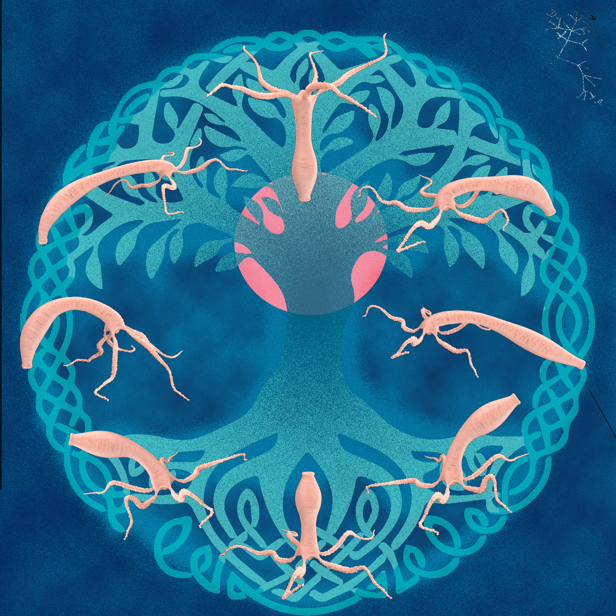
Hydrassil
When the work was published we also wanted to create something to celebrate the occasion and for trying to get selected as a cover article. For that I made another similarly mythological/gaming themed piece as a succession for the vitruvian Hydra. Hence I decided to go again along similar themes and went with a celtic Norse mythological reference. I hence depicted the major steps in the somersault movement at the branches and roots of the world tree, Yggdrasil. The sun rises in the background shining light onto the pioneering steps in a ‘primitive’ organism performing a motion on a solid surface.
What are you thinking of working on next?
I am looking forward to getting back to some exciting experiments I have planned for next year, maybe those would drive some new interesting ideas. I do not have a concrete plan of where I am going next with my art studies but what I do know is that I want to create and learn as much as I can!
The Novo Nordisk Foundation Center for Stem Cell Biology (DanStem) addresses basic research questions in stem cell and developmental biology and has activities focused on the translation of promising basic research results into new strategies and targets for the development of new therapies for cancer and chronic diseases such as diabetes and liver failure. Learn more about DanStem at https://danstem.ku.dk/.
The Novo Nordisk Foundation Center for Protein Research (CPR) promotes basic and applied research on proteins of medical relevance. The research scope spans in silico biology, proteomics, biochemistry and cell biology, and the unifying theme is the identification and functional exploration of proteins and protein pathways involved in disease mechanisms. Learn more about CPR at www.cpr.ku.dk.
Both DanStem and CPR are vibrant, internationally diverse, ambitious research centers, housing modern laboratories with state-of-the-art facilities located in close proximity to one another at the Faculty of Health and Medical Sciences, University of Copenhagen. The setting is ideally suited for seamless exchange of expertise, material and lively scientific communication.
Advanced light microscopy is a key technology at DanStem and CPR. The Centers host approximately 15 research groups with over 50 users (Master and PhD students, Postdocs, Assistant and Associate Professors). Applications in cellular imaging include a variety of model systems (cell lines, 3D cell cultures, tissues, and organisms) and microscopy techniques (wide-field, confocal, high-throughput, light-sheet, FCS, FRAP, live-cell imaging). User support for microscopy and image analysis is provided by dedicated technology platforms at the Centers.
We are now seeking a highly motivated and qualified image analysis specialist to work within the Imaging Research Platform at DanStem and the Protein Imaging Facility within CPR, alongside the light microscopy specialists at both platforms.
Responsibilities
The image analysis specialist works in a team with dedicated microscopy specialists to provide service and support for multiple research projects of broad biomedical scope throughout the two research centers. The image analysis specialist at CPR and DanStem is expected to
Provide support and training for users of both Centers on a variety of image analysis software packages (e.g. Fiji/Image J, Bitplane/Imaris, Perkin Elmer/Harmony, Olympus/ScanR Analysis, Zeiss ZEN, Huygens)
Provide guidance in identifying optimal software and image analysis solutions for a given task
Help users with analysis and graphic presentation of multidimensional data (e.g. Tibco Spotfire)
Organize and participate in image analysis workshops (including practical demonstrations) for graduate and undergraduate students
Manage computer workstations for offline image analysis and take care of software purchases, installation, maintenance and updates (local IT-support is provided)
Establish a fully virtualized analysis environment for the above-mentioned analysis packages using our newly established HPC server (incl. dedicated GPU processing)
Initiative for and collaborations utilizing project-oriented coding of new image analysis tools using our HPC platform for high-content or light-sheet applications is welcome and will be supported.
Network actively with the global image analysis community and stay up-to-date with current trends in the field
Qualifications and competences
Candidates are expected to have a Master’s or PhD degree in a biological sciences-related discipline with at least three years of experience in bio-image analysis. Previous experience with user support in a facility environment is of advantage. Alternatively, candidates with relevant expertise, such as in computer science and data analysis or physics, coupled with a strong interest in cellular imaging and experience in working with biologists can be considered. In addition, we wish for the candidate to have:
A broad overview of image analysis tools and expertise with a variety of image analysis software packages
Experience with 2D, 3D and 4D image analysis and segmentation of structures
Experience with downstream data handling and analysis, including statistical evaluation
Expertise in development of custom image analysis solutions with Matlab, Fiji/ImageJ, Python, Java, R or similar programming languages
A creative mind as well as an active network to tackle challenging tasks in image analysis
Ideally, a good level of practical experience with advanced light microscopy, including wide-field, confocal and high-content microscopy
Excellent communication skills and fluency in English
A service-minded and team player attitude
Ability to work in a multidisciplinary and international environment
Employment conditions
The position, with formal employment title of special consultant at the University of Copenhagen, will be in accordance with the provisions of the collective agreement between the Danish Government and AC (the Danish Confederation of Professional Associations). To the salary is added a monthly contribution to a pension fund according to the collective agreement, and a supplement could be negotiated, depending on the candidate’s experiences and qualification. In all cases, the ability to perform the job will be the primary consideration, and thus we encourage all, regardless of their personal background and status, to apply.
Informal inquiries can be directed to Malte Paulsen (Head of Research Platforms at DanStem, malte.paulsen@sund.ku.dk) and Claudia Lukas (Head of Protein Imaging Facility, CPR, claudia.lukas@cpr.ku.dk).
To apply
To apply, please submit your application, in English, via https://employment.ku.dk/administrative/ by clicking “Apply online”. Applications must include a statement of motivation, curriculum vitae, and copies of relevant diplomas. Only applications in English and submitted online and be accepted.
The Centers highly value diversity and encourage applications from people of all backgrounds.
The closing date for applications is 28 February 2021.
Part of the International Alliance of Research Universities (IARU), and among Europe’s top-ranking universities, the University of Copenhagen promotes research and teaching of the highest international standard. Rich in tradition and modern in outlook, the University gives students and staff the opportunity to cultivate their talent in an ambitious and informal environment. An effective organisation – with good working conditions and a collaborative work culture – creates the ideal framework for a successful academic career.
We are looking for a highly motivated PhD student interested in zebrafish brain development, RNA metabolism, gene editing and transcriptomic analyses to join the Nikolaou Lab at the University of Bath. The deadline for applications is 31st January 2021. The anticipated start date for this project is early October 2021. Applicants should hold, or expect to receive, a First Class or good Upper Second-Class Honours degree (or the equivalent). A master’s level qualification would also be advantageous. UK and EU candidates (with settled or pre-settled status in the UK) applying for this project will be considered for a University Research Studentship which will cover UK/EU tuition fees, a training support grant of £1,000 per annum and a tax-free maintenance allowance at the UKRI Doctoral Stipend rate (£15,285 in 2020-21) for a period of up to 3.5 years.
Brief description of the project:
Regulation of pre-mRNA splicing plays a significant role in neurons by diversifying the proteome and modulating gene expression during development and in response to physiological cues. Although most pre-mRNA processing reactions are thought to occur in the nucleus, numerous RNA splicing regulators are also found in neurites, however, very little is known about their extra-nuclear functions. Our recent work showed for the first time that the non-nuclear pool of a major spliceosome component (SNRNP70) modulates the production of alternative spliced mRNA isoforms essential for motor connectivity and protects transcripts from degradation.
This project aims to investigate further the extra-nuclear activities of SNRNP70 in the context of neuronal connectivity in zebrafish. The ease of genetic manipulations together with the translucency and small size of their offspring allows us to monitor neural cell behaviour and function and observe changes in neuronal connectivity. We will use a range of genetic tools, including transgenic over-expression of cytoplasmic SNRNP70 and nuclear-only SNRNP70 zebrafish knock-in lines to establish developmental functions attributed to the cytoplasmic pool of SNRNP70. The results from this project will contribute to our understanding of how local RNA metabolism in axons contributes to the normal development of neural connections in the brain.
The International C. elegans Conference takes place every two years and features cutting-edge research in a diverse array of topics, including physiology, neurobiology, development, evolution, behavior, aging, ecology, gene regulation, genomics, and more. For 2021, #Worm21 has been reimagined for a virtual platform and will focus on early career researchers at every stage.
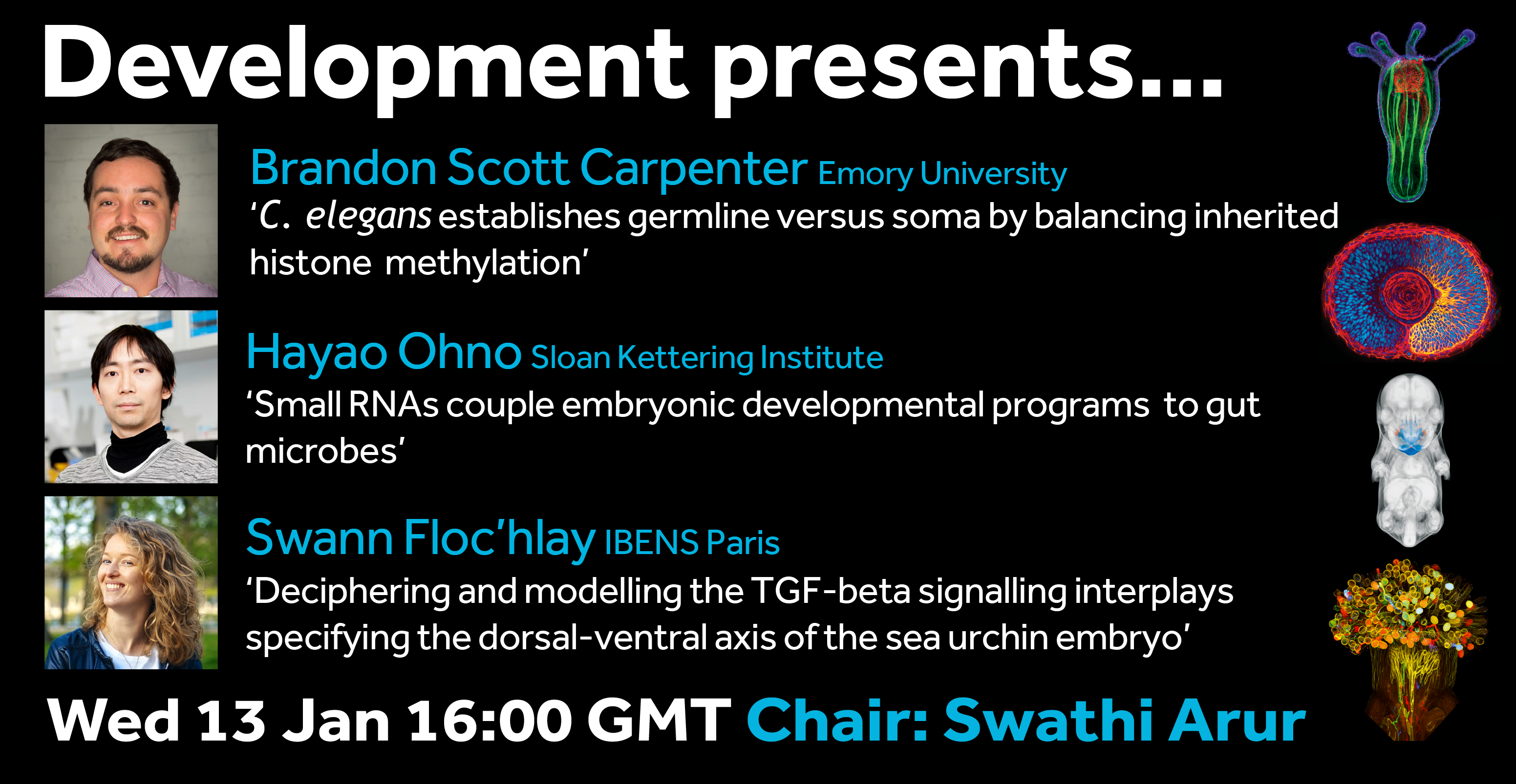
We’re happy to confirm the next webinar in our Development presents… series will be chaired by our Editor Swathi Arur (The University of Texas MD Anderson Cancer Center), who has brought together talks that span her interests in C. elegans development, the germline and cell signalling.
The webinar will be held in Remo, our browser-based conferencing platform – after the talks you’ll have the chance to meet the speakers and other participants at virtual conference tables. If you can’t make it on the day, talks will be available to watch for a couple of weeks after the event (look out for details on the Node).
For more information about what to expect in Remo, go to
Updated 11 January. Let us know if we missed anything
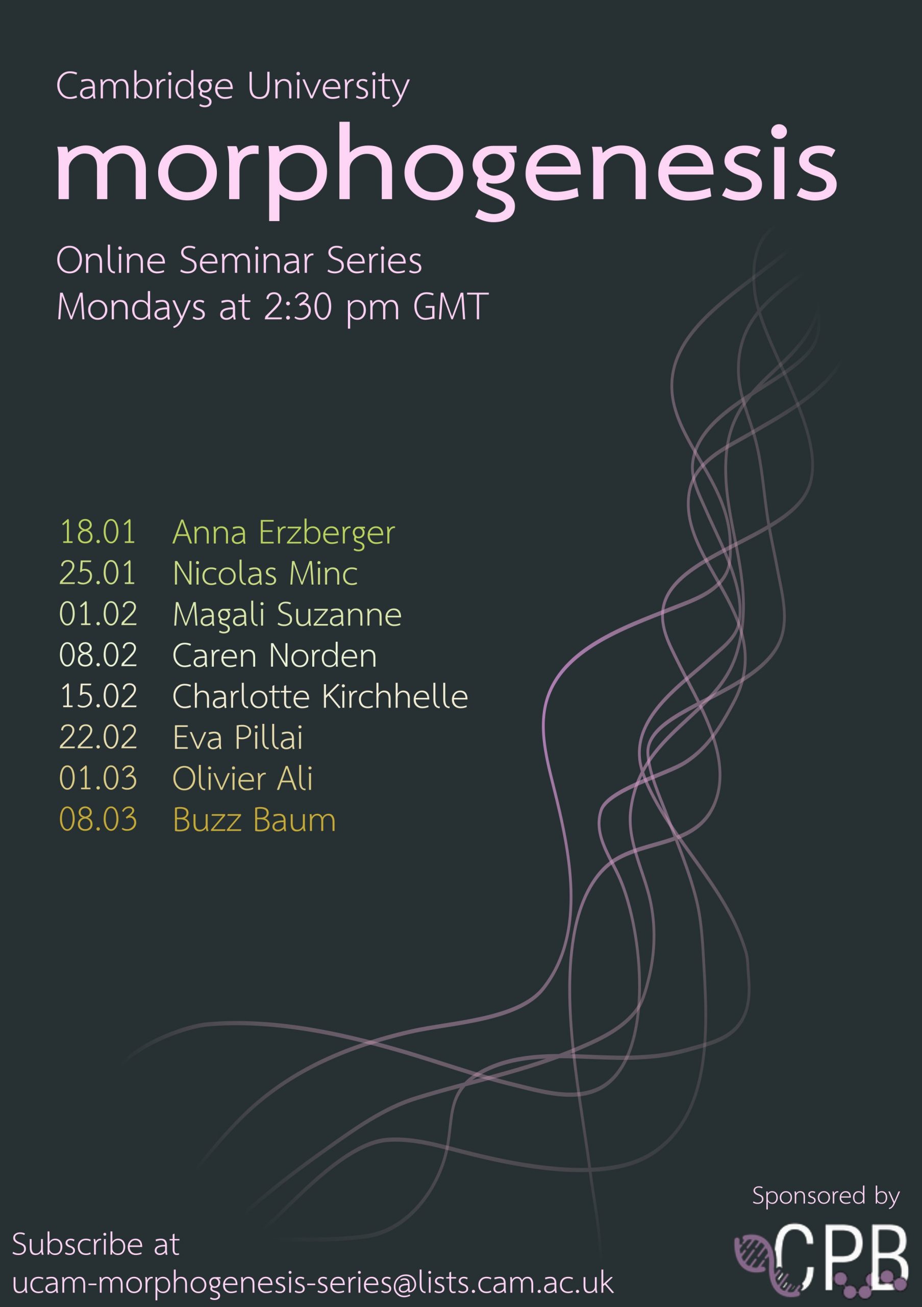
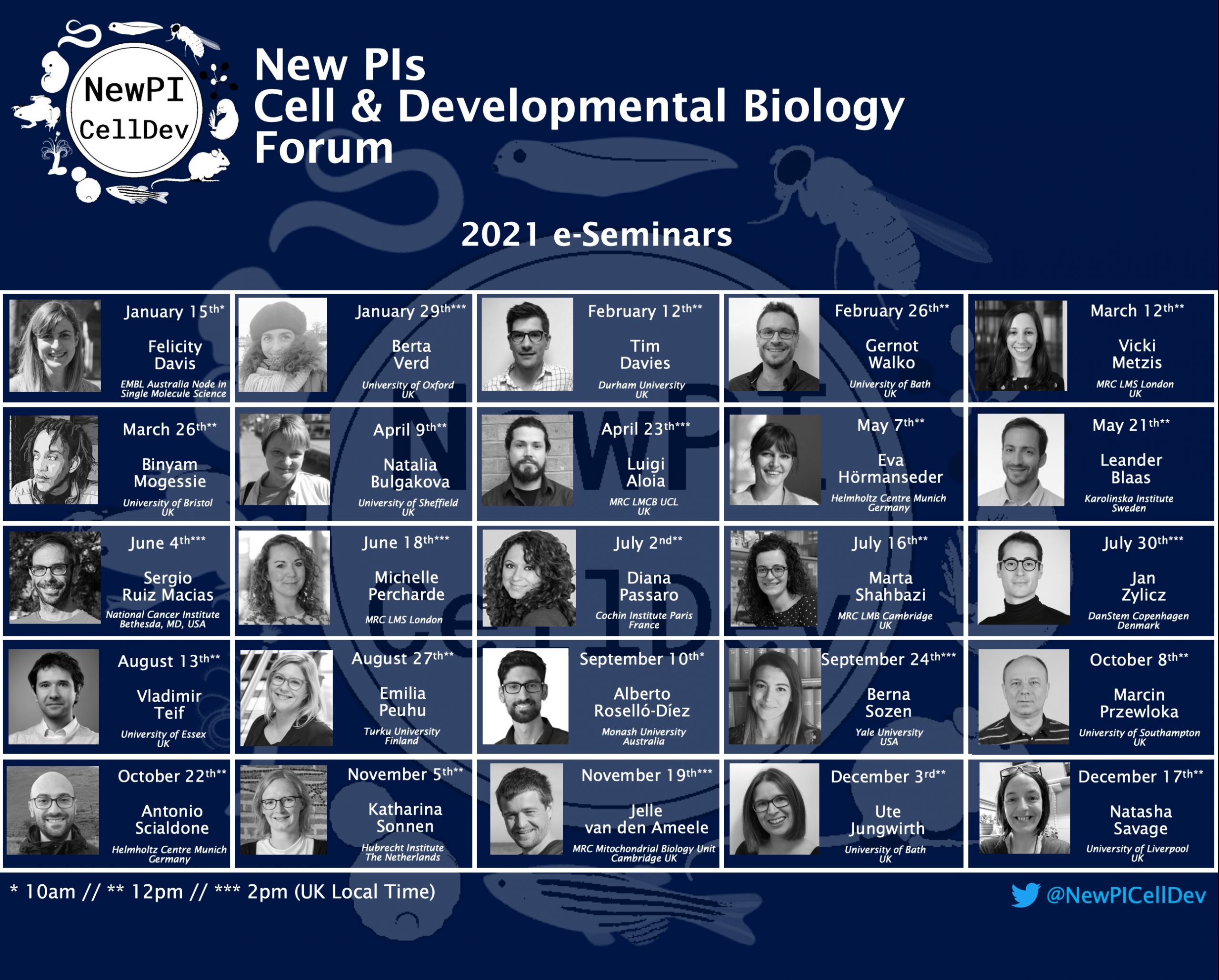
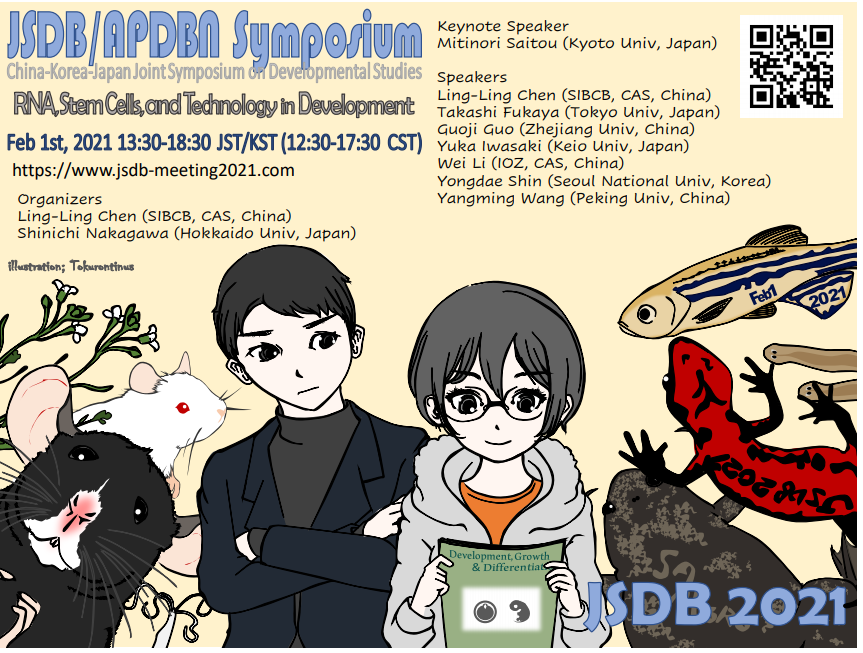
Various organisations and looser assemblies of locked down researchers have begun to put together online seminar and talk series, many of which are open to anyone (usually with registration), and many of which also have previous talks recorded.
Here’s a list of what we’ve found recently, developmental biology and adjacent – please let us know if we missed anything so we can keep it up to date. For upcoming virtual developmental biology conferences/symposia, see our Events calendar page.
First up from us is Development presents…, the webinar series hosted each month by a different Development Editor which will be a platform for early career researchers to share their work. As well as the talks, you also have the chance to meet the speakers and other participants at interactive video tables – giving the developmental biology community the chance to network virtually.
The next webinar will be Wednesday 13 January, 16.00 GMT, hosted by Swathi Arur and featuring talks from Brandon Scott Carpenter, Hayao Ohno and Swann Floc’hlay.
Next talk: January 14, Evolutionary Studies:
Just Under the Surface: Leveraging Zebrafish to Understand the Interplay Between Evolution and Development
We currently have an opening for a Reviews Editor as a maternity cover position on Development. As this is a temporary position, we are specifically looking for candidates with editorial experience.
Core responsibilities of the position include:
Commissioning, handling peer review and developmental editing of material for the front section of the journal
Representing the journal at international conferences and within the wider scientific community
Writing press releases, article highlights and material for Development’s community website ‘the Node’
Creative involvement in the journal’s development
For further details and instructions on how to apply, please see the full job advert here. If you are interested in applying, but would like further information or have any questions, please feel free to drop me an email.
Naa12 rescues embryonic lethality in Naa10-Deficient Mice in the amino-terminal acetylation pathway
Hyae Yon Kweon, Mi-Ni Lee, Max Dörfel, Seungwoon Seo, Leah Gottlieb, Thomas Papazyan, Nina McTiernan, Rasmus Ree, Andrew Garcia, Michael Flory, Jonathan Crain, Alison Sebold, Scott Lyons, Ahmed Ismail, Elaine Marchi, Seong-keun Sonn, Se-Jin Jeong, Sejin Jeon, Shinyeong Ju, Simon J. Conway, TaeSoo Kim, Hyun-Seok Kim, Cheolju Lee, Tae-Young Roh, Thomas Arnesen, Ronen Marmorstein, Gholson J. Lyon, Goo Taeg Oh
Serine Palmitoyltransferase Controls Stemness of Intestinal Progenitors
Ying Li, Bhagirath Chaurasia, Vincent Kaddai, Joseph L. Wilkerson, J. Alan Maschek, James Cox, Peng Wei, Claire Bensard, Peter J Meikle, Hans Clevers, James A Shayman, Yoshio Hirabayashi, William L. Holland, Jared Rutter, Scott A. Summers
CTCF is a Barrier for Totipotent-like Reprogramming
Teresa Olbrich, Maria Vega-Sendino, Desiree Tillo, Wei Wu, Nicholas Zolnerowich, Andy D. Tran, Catherine N. Domingo, Mariajose Franco, Marta Markiewicz-Potoczny, Gianluca Pegoraro, Peter C. FitzGerald, Michael J. Kruhlak, Eros Lazzerini-Denchi, Elphege P. Nora, Andre Nussenzweig, Sergio Ruiz
Robust integrated intracellular organization of the human iPS cell: where, how much, and how variable?
Matheus P. Viana, Jianxu Chen, Theo A. Knijnenburg, Ritvik Vasan, Calysta Yan, Joy E. Arakaki, Matte Bailey, Ben Berry, Antoine Borensztejn, Jackson M. Brown, Sara Carlson, Julie A. Cass, Basudev Chaudhuri, Kimberly R. Cordes Metzler, Mackenzie E. Coston, Zach J. Crabtree, Steve Davidson, Colette M. DeLizo, Shailja Dhaka, Stephanie Q. Dinh, Thao P. Do, Justin Domingus, Rory M. Donovan-Maiye, Tyler J. Foster, Christopher L. Frick, Griffin Fujioka, Margaret A. Fuqua, Jamie L. Gehring, Kaytlyn A. Gerbin, Tanya Grancharova, Benjamin W. Gregor, Lisa Harrylock, Amanda Haupt, Melissa C. Hendershott, Caroline Hookway, Alan R. Horwitz, Chris Hughes, Eric J. Isaac, Gregory R. Johnson, Brian Kim, Andrew N. Leonard, Winnie Leung, Jordan J. Lucas, Susan A. Ludmann, Blair M. Lyons, Haseeb Malik, Ryan McGregor, Gabe E. Medrash, Sean L. Meharry, Kevin Mitcham, Irina A. Mueller, Timothy L. Murphy-Stevens, Aditya Nath, Angelique M. Nelson, Luana Paleologu, T. Alexander Popiel, Megan M. Riel-Mehan, Brock Roberts, Lisa M. Schaefbauer, Magdalena Schwarzl, Jamie Sherman, Sylvain Slaton, M. Filip Sluzewski, Jacqueline E. Smith, Youngmee Sul, Madison J. Swain-Bowden, W. Joyce Tang, Derek J. Thirstrup, Daniel T. Toloudis, Andrew P. Tucker, Veronica Valencia, Winfried Wiegraebe, Thushara Wijeratna, Ruian Yang, Rebecca J. Zaunbrecher, Allen Institute for Cell Science, Graham T. Johnson, Ruwanthi N. Gunawardane, Nathalie Gaudreault, Julie A. Theriot, Susanne M. Rafelski
Dog color patterns explained by modular promoters of ancient canid origin
Danika L. Bannasch, Christopher B. Kaelin, Anna Letko, Robert Loechel, Petra Hug, Vidhya Jagannathan, Jan Henkel, Petra Roosje, Marjo K. Hytönen, Hannes Lohi, Meharji Arumilli, DoGA consortium, Katie M. Minor, James R. Mickelson, Cord Drögemüller, Gregory S. Barsh, Tosso Leeb
Molecular topography of an entire nervous system
Seth R Taylor, Gabriel Santpere, Alexis Weinreb, Alec Barrett, Molly B. Reilly, Chuan Xu, Erdem Varol, Panos Oikonomou, Lori Glenwinkel, Rebecca McWhirter, Abigail Poff, Manasa Basavaraju, Ibnul Rafi, Eviatar Yemini, Steven J Cook, Alexander Abrams, Berta Vidal, Cyril Cros, Saeed Tavazoie, Nenad Sestan, Marc Hammarlund, Oliver Hobert, David M. Miller III
Automated hiPSC culture and sample preparation for 3D live cell microscopy
Mackenzie E. Coston, Benjamin W. Gregor, Joy Arakaki, Antoine Borensztejn, Thao P. Do, Margaret A. Fuqua, Amanda Haupt, Melissa C. Hendershott, Winnie Leung, Irina A. Mueller, Angelique M. Nelson, Susanne M. Rafelski, Madison J. Swain-Bowden, W. Joyce Tang, Derek J. Thirstrup, Winfried Wiegraebe, Calysta Yan, Ruwanthi N Gunawardane, Nathalie Gaudreault
Scalable production of tissue-like vascularised liver organoids from human PSCs
Sean P Harrison, Richard Siller, Yoshiaki Tanaka, Yangfei Xiang, Benjamin Patterson, Henning Kempf, Espen Melum, Kathrine S Åsrud, Maria E Chollet, Elisabeth Andersen, Per Morten Sandset, Saphira Baumgarten, Flavio Bonanini, Dorota Kurek, Santosh Mathapati, Runar Almaas, Kulbhushan Sharma, Steven R Wilson, Frøydis S Skottvoll, Ida C Boger, Inger L Bogen, Tuula A Nyman, Jun J Wu, Ales Bezrouk, Dana Cizkova, Jaroslav Mokry, Robert Zweigerdt, In-Hyun Park, Gareth J Sullivan
 (3 votes)
 (No Ratings Yet)


 (1 votes)