Closing date for applications 19th of January 2021
Starting date end of 2021, beginning of 2022
The 2020 call for the Spanish “Juan de la Cierva Postdoctoral Fellowships” is now open. This is a fantastic opportunity to join the Araújo group, who are focused on identifying the molecular mechanisms underlying single-cell branching during development and disease. We study this during morphogenesis of the Drosophila melanogaster tracheal and nervous systems (Ricolo et al. Current Biology 2016 and Ricolo and Araújo, eLife, 2020), and are also interested in knowing how single-cell branching affects the whole organismal behaviour. We are combining cell analysis and confocal tissue live imaging, with genetic approaches and CRISPR/Cas9 technologies.
We are based at the Institute of Biomedicine of the University of Barcelona (IBUB) and are looking for a motivated and enthusiastic candidate who will play a central role in the lab. You must have a Ph.D. in areas relevant to cell/developmental biology and/or in computational biology (and have defended your Ph.D. thesis after the 1st of January 2016). Applicants are expected to have excellent interpersonal and communication skills, be highly independent and committed to research in a fast-moving and exciting field.
If you’re interested, please write to sofiajaraujo [at] ub.edu
Nutrients such as phosphate are limiting in most soils. This has a great impact on crop yield in agriculture. Approaches to overcome these challenges often include using fertilizers, that in turn have a detrimental effect on the environment and our climate. Plants have developed adaptation mechanisms to efficiently forage the soil for phosphate such as modifying root system architecture and root hair density. In Wendrich et al., we unexpectedly identified a vascular transcription factor complex that controls root hair formation in response to limiting phosphate conditions.
One of the main interests of our lab is identifying the molecular pathways controlled by the TARGET OF MONOPTEROS 5 and LONESOME HIGHWAY (TMO5/LHW) transcription factor heterodimer. In the root meristem, this complex is active in the inner most cells of the vasculature called the xylem. Here it controls the expression of LONELY GUY 4, which encodes an enzyme that is rate-limiting for the conversion of the phytohormone cytokinin into its bioactive form. Cytokinin is involved in many aspects of vascular development including patterning and proliferation. Using bulk transcriptomics, we previously revealed 273 target genes of the TMO5/LHW complex, but given the downstream intermediate cytokinin is mobile, we expected that many of these target genes would be expressed outside of the xylem cells. This is where this project started.
At that time, single-cell technologies were quickly becoming important in other fields and proved ideal to determine gene expression at unprecedented resolution. We thus decided we needed to get this technology up and running to answer our research question. Our institute had recently invested heavily to establish the technique, with commercial microfluidics systems and custom bioinformatic pipelines, but it remained unclear how reliable and efficient this would work on isolated plant cells. We teamed up with the group of Yvan Saeys in our neighboring VIB research center to generate a reference dataset for the Arabidopsis root meristem. We first needed to optimize the workflow leading up to the microfluidics single cell droplet emulsion. With their tough cell walls, plant cells are not easily dissociated from their tissue, requiring enzymatic digestion of the cell walls, called “protoplasting”. Cells in the outer layers are more accessible for these enzymes and tend to be overrepresented after in protoplast suspensions. We therefore first made sure that our single cell protoplast suspensions contained a good representation of each cell type, inner and outer layers, and was free of most debris and impurities, such as dead cells.
Unfortunately, this was the easy part. Now came the analysis, annotation and, importantly, full validation of the technology. This was where our collaboration effort truly started to pay off. The analysis pipelines were custom redesigned for our plant samples. This included building a visualization tool, for quick access to the data. Endless discussions, quick face-to-face check-ups and back-and-forth emailing ensured that we quickly learned from each other and from the dataset. Because of the rich resources available for Arabidopsis, we were able to annotate the main cell types of the root such as epidermis, ground tissues and vascular tissues. We however decided to take it a step further and zoom in to the level of sub-cell-types. Especially the different cell types of the vascular system were more difficult to annotate, as several cell types, like for example the procambium, only have limited known marker genes that are specifically expressed. Sub-cell-type predictions were confirmed by cell ploidy analysis and an extensive trajectory inference. By iteratively applying these trajectory methods, and having an open communication line between wet- and drylab, we were able to assess the robustness of the predicted trajectories and the resulting predicted subcluster annotations.
We next set out to evaluate the predictive power of this dataset, by construction of promoter reporter lines of genes with hitherto unknown expression patterns. All group members chipped in and a remarkable 100% of the lines with stable expression patterns validated the predicted cell type and developmental states. We were thus eager to apply this rich dataset on the 273 target genes of the TMO5/LHW complex previously identified. We identified an unexpected overrepresentation of genes expressed in root hairs (trichoblasts), suggesting that these vascular transcription factors may play a role in the development of the outermost trichoblast cells. Indeed, when overexpressing TMO5 and LHW in all cells of the root meristem, this dramatically increased the density of root hairs, resembling wild type roots grown in phosphate limiting conditions. Extensive follow-up genetic work allowed us to conclude that cytokinin, produced in inner vascular cells, acts as a mobile intermediate to control root hair response to low phosphate conditions in the outer most epidermal cell.
In our opinion, this is a good example of how collaborations within and between teams with different expertise can lead to new biological insights.
Postdoctoral Fellow (m/f/d) | RNA/Protein Biochemistry and Molecular Biology
MAX PLANCK INSTITUTE FOR HEART AND LUNG RESEARCH – W. G. KERCKHOFF INSTITUTE, BAD NAUHEIM
TYPE OF JOB
SCIENTIST
Job offer from December 08, 2020
The Max Planck Institute for Heart and Lung Research in Bad Nauheim (near Frankfurt, Germany), Department of Developmental Genetics (Prof. Dr. Didier Stainier) invites applications for a Postdoctoral Fellow (m/f/d) in RNA/Protein Biochemistry and Molecular Biology to study Genetic CompensationReference Number 2020_30.
A highly motivated postdoctoral candidate is invited to lead new projects to address fundamental questions in Genetic Compensation and Transcriptional Adaptation.
We have recently discovered a new form of genetic compensation that we have termed transcriptional adaptation. Briefly, transcriptional adaptation occurs when a premature termination codon leads to mutant mRNA degradation. The degradation fragments in turn modulate the expression of related genes, and consequently varying degrees of phenotypic rescue in some cases. We have observed this phenomenon in zebrafish, mouse, C. elegans, and more recently humans.
This is a novel and quickly developing field of research where many open questions remain to be addressed including 1) the nature and trafficking of the degradation fragments, and 2) the regulation of related genes by the degradation fragments, or their derivatives. For more detailed information please see our recent publications:
A Ph.D. in biology, biochemistry, genetics or a similar subject with a focus on molecular biology, cell biology, biochemistry and/or genetics. Knowledge of RNA and/or protein biochemistry and molecular biology is a plus.
About the employer:
The Max Planck Institute for Heart and Lung Research in Bad Nauheim is an interdisciplinary research institution with international flair. Our researchers have the opportunity to work on various model systems by making use of the latest cutting-edge technologies. Researchers are supported by state-of-the-art core facilities which offer services in next generation sequencing, proteomics, bioinformatics, cytometry, microscopy, and small animal imaging.
The Max Planck Society strives for gender and diversity equality; we welcome applications from all backgrounds. Furthermore, the Max Planck Society is committed to increasing the number of individuals with disabilities in its workforce and therefore encourages applications from such qualified individuals.
To apply, please submit the names and contact information for 2-3 references, a CV, and a short statement (2 pages max.) of your research experience and interests to didier.stainier@mpi-bn.mpg.de
Max Planck Institute for Heart and Lung Research
Department of Developmental Genetics
Ludwigstraße 43
61231 Bad Nauheim
Germany
Duke University’s Departments of Orthopaedic Surgery and Cell Biology invite applications for a tenure track faculty position in the field of musculoskeletal cell biology. The ideal candidate will have a PhD, MD, or MD/PhD and significant postdoctoral or faculty experience. They should have a record of outstanding scientific accomplishments and a well-developed research plan in a key research discipline such as: bone biology and osteoporosis, skeletal stem cell biology, skeletal regeneration, cartilage biology and arthritis, muscle/tendon biology and regeneration, or musculoskeletal and neuromuscular disorder research.
An appointment at any academic level is available depending on the expertise and track record of the candidate. The successful candidate will complement and enhance our substantial existing strengths in cell biology, orthopaedics, sarcoma, bioengineering, developmental biology, and musculoskeletal research utilizing a combination of in vivo and in vitro models. New faculty will receive a generous startup package and laboratory space. We expect the successful candidate to initiate and maintain an original, competitive, and independently funded research program.
Interested candidates should submit application materials including a cover letter, curriculum vitae, statement of accomplishments and future research goals (two-page limit), a diversity statement, electronic reprints of up to three recent articles, and arrange to have up to three letters of reference provided.
Applications will be reviewed on a rolling basis and should be submitted by January 31, 2021, however later applications will be considered until the position is filled. Initial interviews will be held virtually.
Matthew J. Hilton, PhD
Search Committee Chair
Associate Chair for Research, Basic/Translational Sciences
Department of Orthopaedic Surgery
Duke University School of Medicine
Policy Statement: Duke University prohibits discrimination and harassment, and provides equal employment opportunity without regard to an individual’s age, color, disability, gender, gender expression, gender identity, genetic information, national origin, race, religion, sex, sexual orientation, or veteran status. Duke is committed to recruiting, hiring, and promoting qualified women, minorities, individuals with disabilities, and veterans. Pursuant to Title IX of the Education Amendment of 1972, Duke prohibits discrimination on the basis of sex in any of its educational programs or activities. For more information regarding Duke’s Equal Opportunity/Affirmative Action policy and our commitment to diversity and equity go to: https://oie.duke.edu/
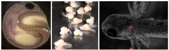
In the Genetics of Cell Behaviour in Development laboratory, at the Department of Genetics, Microbiology and Statistics at the University of Barcelona, we work with the terminal cells (TCs) of the tracheal system of Drosophila melanogaster, which are very interesting cells. During embryonic development, they are able to generate a tube within their cytoplasm, a so-called seamless, subcellular tube. As the larvae hatch and grow these tiny tubes are able to branch inside a single cell to provide oxygen to the various tissues in the animal.
Embryonic and larval tracheal TCs are a powerful model to understand the mechanisms that lead a single-cell to change its shape, and generate a subcellular seamless tube, in a process analogous to capillary blood-vessel sprouting. Cell elongation and subcellular tube formation are dependent on dynamic cytoskeletal reorganization. Microtubules and actin are major players in this active cytoskeletal process. We have been studying the initial events in the formation of this subcellular tube, focusing on what triggers these changes in the cytoskeleton.
When we investigated the role of centrosomes as Microtubule Organizing Centres (MTOCs) in TCs (Ricolo et al., 2016), we became interested in analysing microtubule bundles inside TCs. We tested the tracheal overexpression of several constructs consisting of MT-associated proteins fused with fluorophores. Among them, we used transgenic lines overexpressing the Drosophila spectraplakin Short-stop (Shot).
Shot is a giant multifunctional protein responsible for many aspects of cytoskeleton organization in different tissues. Shot can operate as a single cytoskeleton component, can coordinate cytoskeletal elements such as actin and microtubules, and can also interact with other cellular structures. The functional versatility of Shot is probably reflected by the abundant generation of different isoforms, and by the characteristics of its protein domains. The full-length isoform A (Shot-A) contains an NH2-terminal actin-binding domain (ABD), different central domains (rod-like domain, plakin spectrin-like repeats and an EF-hand), and a C-terminal domain involved in MT interaction (GRD and C-tail domains). Shot-A has been shown to act as an actin-microtubule crosslinker. Isoform C (Shot-C) lacks a proper actin-binding domain and has been described to interact mainly with MTs (Voelzmann et al., 2017).
Since we were interested in visualizing MTs in TCs in live embryos, we tested the tracheal overexpression of Shot-C using the transgenic line UAS-shotC-GFP. This was a clear winner! GFP staining of embryos overexpressing Shot-C-GFP, allowed us to beautifully label MTs; we could clearly see the MT-fibres inside the TCs and they were bright enough that we could image them live, as the embryo develops.
But… (there is always a but). When you want to see a biological structure through the use of an overexpression system in Drosophila embryos, as in other organisms or cell culture systems, you have to be very careful. Often the overexpression of a specific protein can perturb the physiological state of the cell; in other words, it can give rise to a mutant phenotype. So, we decided to analyze if the embryonic overexpression of Shot caused any changes during TC extension and lumen development.
Surprisingly, we found that Shot overexpression induced the generation of an extra-subcellular lumen inside the TC, a very similar phenotype to the one caused by extra centrosome numbers. So, by searching for ways of illuminating MTs, we had serendipitously found another way to induce luminal branching at embryonic stages. Of course, our next question was whether Shot was inducing higher numbers of centrosomes in TCs. Nicely, we were able to genetically prove that Shot acted in a different way to generate the extra-subcellular lumen branching event.
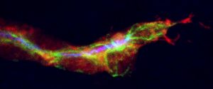
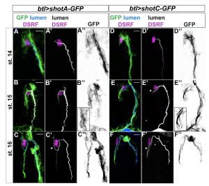
ShotOE induces luminal branching through its microtubule-binding domain. Lateral view of DB tip cells from st.14 to st.16, of btl >shotA-GFP embryos (A–C) and btl >shotCGFP (D–F). Embryos were stained with GFP (green in A-F and grey A’’-F’’) to visualise Shot-GFP, DSRF to mark the TC nuclei (in magenta) and CBP to stain the chitinous lumen (blue in A-F and white A’-F’). Both overexpressing conditions induced Extra subcellular lumina (white stars). Note the GFP was more diffuse in the cytoplasm of the TCs of embryos overexpressing shotA, and more organised in bundles in the TCs overexpressing shotC. Anterior side of embryo is on the left and dorsal side up. Scale bars 5 μm. From Ricolo and Araújo, 2020, eLife.
The results of our experiments told us that the more Shot we expressed in the TC, the more subcellular branching we could generate. So, how does this Shot overexpression generate branched subcellular lumina? One hypothesis was that an excess of MT-stabilization could induce the generation of acentrosomal branching points along pre-existent microtubule bundles along the luminal track. If this was true, the overexpression of other proteins involved in MT stabilization should be also able to induce extra subcellullar lumina. We tested this hypothesis and found that overexpression of the microtubule-associated protein Tau was able to generate the same luminal phenotype.
Analyzing the complete loss-of-function of shot we observed defects in luminal formation and cell elongation. We observed, using rescue experiments and analysis of endogenous Shot localization that this phenotype depended on Shot’s ability to crosslink MTs and actin. Surprisingly, we were able to rescue the shot null phenotype overexpressing Tau indicating a role for Tau in mediating the connection between actin and microtubules in TCs.
With our work, we showed the importance of actin-microtubule crosslinking in the formation of novel subcellular tubes by single-cells (Ricolo and Araújo, 2020). But also, this work has taught us that by doing research you can open new exploratory paths in an unpredictable way. Serendipity plays a role in science. You just need to keep an open mind and follow your curiosity!
The Evolutionary Developmental Biology Lab at the Francis Crick Institute is seeking a laboratory research scientist that will help establish the laboratory, manage its day-to-day operations, and lead the generation of large-scale genomics datasets.
The Evolutionary Developmental Biology Lab will open early 2021. We study how organs originate and how they diversify in form and function across species. We tackle this problem by combining evolutionary and developmental biology with large-scale comparative genomics. We are an interdisciplinary group and highly collaborative.
In our lab we will generate and analyse our own large-scale genomics datasets, including single-cell transcriptomics and epigenomics, whole-genome sequencing, and spatial transcriptomics. The laboratory research scientist will set-up the wet lab and lead the data production of the group. This will be done in close collaboration with several science technology platforms at the Institute (animal facilities, single-cell genomics, sequencing).
Evolutionary Developmental Biology Lab
One of the most fundamental problems in biology is understanding how novelties arise, whether they be new cells, new tissues, or whole new organs. Our group tackles this problem by identifying the genetic and developmental processes responsible for the origin, and subsequent evolution, of vertebrate organs.
Our group uses the placenta as a model to understand how organs originate and evolve. We study the placenta because placentas have evolved independently many times across vertebrates and because they exhibit an extraordinary diversity of forms and functions. We study the placenta within two distinct, but complementary, contexts. In mammals, we work on one of the most fascinating aspects of pregnancy, the mother’s tolerance to the intimate contact between its own cells and those of her foetus. We want to understand how these critical interactions between the mother and the fetal cells have evolved across mammals. In fish, we address the question of how organs are created. We study a family of fish where placentas have evolved independently multiple times. By studying multiple independent inventions of the placenta we try to identify general principles guiding the evolution of new organs at the level of genes, cells and developmental processes.
PhD position available in the lab of Dr Chema Martin (Queen Mary University of London) in collaboration with Oxford Nanopore Technologies, Dr Alex de Mendoza (QMUL) and Dr Paul Hurd (QMUL) to dissect the evolution of 3D genome structure with Pore-C Nanopore sequencing.
Background
The 3D structure of the genome – the set of DNA molecules with the guidelines to build an organism – is fundamental for proper expression of the genetic information contained in it [1]. However, how this 3D structure evolves and is influenced by other features of the genome is still unclear. This is because our understanding of this biological phenomenon is mostly limited to a handful of model organisms and by the fact that current methods to reconstruct the 3D structure of genomes depend on short-read sequencing. In late 2019, Oxford Nanopore Technologies (ONT) released Pore-C, a method to apply long-read sequencing to reconstruct 3D genome structure that overcomes current limitations in chromatin conformation approaches [2]. In parallel, the Martín-Durán lab is establishing a set of marine segmented worms (annelids) as research model systems for comparative evolutionary genomics [3]. What are the genomic features controlling 3D genome structure in annelids? How do these mechanisms generate variability in 3D genome structure among species? Can we develop Pore-C as a widespread approach to reconstruct 3D genome structure?
In this project, you will rigorously answer these questions combining state-of-the-art experimental and computation approaches in a unique academic-industry collaborative environment.
– You will have access to large genomic databases, and in-house live organisms to fuel your investigation.
– You will gain experience of molecular techniques (epigenomics, nanopore long-read sequencing), bioinformatics (pipelines to analyse Pore-C data), and statistics.
– You will be encouraged to develop your own ideas and hypotheses.
Benefits
This is a BBSRC LIDo iCASE PhD fully funded position, including home (UK) tuition fees and a tax-free stipend in the region of £17,285. The student will become part of Queen Mary’s Doctoral College, which provides training and development opportunities and financial support for research. The student will also have access to a Researcher Development Programme designed to help recognise and develop key skills and attributes needed to effectively manage research, and to prepare and plan for the next stages of their career. In addition, the student will enjoy a 6-months placement at Oxford Nanopore Technologies headquarters in Oxford (UK), where the students will integrate within the research and development team.
Skills/Qualifications
In a multidisciplinary project like this, candidates are unlikely to have a background in all disciplines involved. The most important qualification is motivation, enthusiasm and that the project appeals to you. However, previous computational experience would be important. We can envisage strong candidates coming through a variety of routes including:
– practical molecular biology
– developmental and cell biology
– biotechnology
– computational biology
Application
To apply, students should have a 1st class degree or have received a MSc in a relevant field (i.e. molecular biology, genetics and evolutionary genomics, developmental and cell biology, biotechnology, bioinformatics) or are about to finish their MSc.
1. Rowley, M.J., Corces, V.G. (2018) Organizational principles of 3D genome architecture. Nat Rev Genet 19, 789–800. https://doi.org/10.1038/s41576-018-0060-8
2. Netha Ulahannan, et al. (2019) Nanopore sequencing of DNA concatemers reveals higher-order features of chromatin structure. bioRxiv 833590; doi: https://doi.org/10.1101/833590
3. José M. Martín-Durán, et al. (2020) Conservative route to genome compaction in a miniature annelid. Nature Ecology and Evolution. https://doi.org/10.1038/s41559-020-01327-6
Drs. Hillary Maddin and Tetsuto Miyashita are seeking motivated students and fellows to join our Carleton-based team in Ottawa, Canada. Successful candidates will participate in a newly funded project aimed at understanding various aspects of the development and evolution of the vertebrate skeleton. In particular, projects will focus on investigating skull and limb development in model and non-model species of amphibians. Collaborators for these projects include Chris Joslin (Carleton), James Hanken (Harvard), and Ryan Kerney (Gettysburg). MSc, PhD, and postdoctoral positions available.
Carleton University has research partnerships with Canadian Museum of Nature, which houses natural history collections of over 14 million specimens, and the University of Ottawa, which offers an extensive network of biomedical and life science labs and the state-of-the-art next-generation sequencing facility. Ottawa is a vibrant, multicultural, and affordable city embraced in the green belt. Browse some overview and Google Image search summary of life in Canada’s capital.
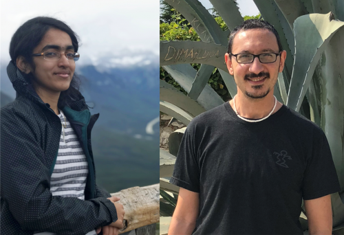
The veins are the vascular networks of plant leaves, functioning as channels for transport of signals and nutrients. A new paper in Development investigates how the spatial regulation of auxin transport contributes to vein patterning in Arabidopsis. We caught up with first author Priyanka Govindaraju and her supervisor Enrico Scarpella, Associate Professor at the Department of Biological Sciences, University of Alberta in Edmonton, Canada, to find out more about the work.
Priyanka (L) and Enrico (R)
Enrico, can you give us your scientific biography and the questions your lab is trying to answer?
ES: I graduated in Biological Sciences at the University of Milan, but I spent my fifth undergraduate year at Leiden University as an exchange student. That’s when I met Annemarie Meijer and Harry Hoge, who would later become my PhD supervisors. Every day I am grateful for the life-changing opportunity they gave me and for how patient they had been – before I could start my PhD with them, I had to go back to Italy, finish my last year of undergraduate studies (university degrees took longer then) and complete a year of civil service (in lieu of the mandatory military service to which I objected).
Early in my PhD – I think it was toward the end of the first year – I fell in love with vascular tissues, especially the mesmerizing networks of veins in the leaves. So after my PhD in Mathematical and Natural Sciences from Leiden University, I moved to Toronto, where Thomas Berleth had generously given me the opportunity to do a postdoc with him.
A little more than a year into my postdoc, Thomas selflessly passed on to me an ad for a position in the Department of Biological Sciences at the University of Alberta. I interviewed there toward the end of my second year of postdoc. Though early in my postdoc, the search committee and the department took a chance on me and offered me an Assistant Professor position, which I took on a year later. I have been an Associate Professor there since 2010, and I have just now applied for Full Professor. The questions my lab tries to address are the same that have been haunting me since my graduate days: how do veins form and how are they assembled into networks?
And Priyanka – how did you come to work in Enrico’s lab and what drives your research today?
PG: During my undergrad, I came to be interested in plant developmental biology. I was looking for labs in which to continue my training as graduate student, and therefore I was doing a lot of background research about the prospective research groups. One of my undergrad supervisors had suggested that I have a look at the leaf vein development research done in the Scarpella lab, and I was very much interested and encouraged by the lab’s research. I also thought it would provide me with the platform to learn techniques such as imaging and molecular biology, and I was very curious and motived to know how veins form: I wanted to solve a small portion of the huge puzzle. I was happy when I saw that there were vacancies in the lab and contacted Enrico showing my interest in the research. The experience that I have gained from Enrico and the lab will certainly help me with my career.
How has your research been affected by the COVID-19 pandemic?
ES: Well, my lab – like all the labs at our university that do not work on COVID-19 – was shut down on March 15, and, until the end of May, we only had permission to maintain essential plant collections – basically, to water plants. Since the beginning of June, we have received permission to resume research part-time – only a maximum of two people can be in the lab at any given time. We currently do not know when we will be able to resume full-time research, but given time my research will recover. Right now, I am more concerned about that of my trainees and how I can minimize the impact this delay in research will have on their career.
Before your paper, how were auxin and its transporter PIN1 thought to regulate vein development?
PG & ES: We thought that, in the epidermis of the leaf, PIN1 would transport auxin toward discrete points. From these ‘convergence points’ of auxin transport in the leaf epidermis, PIN1 would transport auxin into the inner tissues of the leaf, where PIN1-mediated auxin transport would select the cell paths that would later become veins.
Can you give us the key results of the paper in a paragraph?
PG & ES: Contrary to our expectations, we found that reintroducing PIN1 in the epidermis of leaves that lack PIN1 failed to rescue the mutant vein pattern. Furthermore, removing PIN1 from the epidermis of normal leaves, or of leaves that lack PIN1’s most closely related proteins, had no effect on vein patterning. By contrast, reintroducing PIN1 in the inner tissues of leaves that lack PIN1, or PIN1 and its most closely related proteins, rescued the mutant vein patterns. Finally, eliminating PIN1 from the inner tissues of normal leaves led to abnormal vein patterns.
How do you think vascular PIN1 works to promote vein patterning?
PG & ES: We think that the concept at the core of the Canalization Hypothesis formulated by Tsvi Sachs more than 50 years ago still offers the best starting point to understand how PIN1 in the inner tissues of the leaf may promote vein patterning. The Canalization Hypothesis proposes a positive feedback between the auxin that flows through a segment of a cell’s plasma membrane and the capacity of that cell to transport auxin in that same direction. The pre-existing vasculature – at first, the midveins of the cotyledons (i.e. the embryonic leaves) for the first two post-embryonic leaves we analysed in our paper – would act as an auxin sink that orients toward itself auxin transport, mediated by PIN1 and related proteins, in the inner tissues of the developing leaves. Because of the postulated positive feedback of auxin transport on itself, broad domains of auxin transport in the inner tissues of the developing leaf would be gradually refined into narrow paths of auxin transport – the canals the hypothesis refers to. And these narrow paths of auxin transport would define the positions where veins would later form.
How fortunate that we never run out of research questions and opportunities!
But, as usual, the devil is in the details because we still don’t know, for example, how auxin finds, as efficiently as it seems to be doing, a sink located cells away. Nor do we know how a cell senses the auxin that flows through it or how some auxin transport paths end up connecting two sinks to each other. How fortunate that we never run out of research questions and opportunities!
Do you think the epidermis plays any role at all in the process?
PG & ES: Yes, we do, and indeed our results do not rule out a role for the leaf epidermis, or for epidermal auxin, in vein patterning.
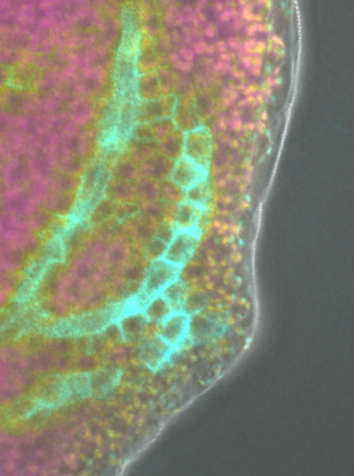
PIN1::cPIN1:GFP expression in a pin1 mutant.
When doing the research, did you have any particular result or eureka moment that has stuck with you?
PG I found that PIN1 expression in the epidermis was not sufficient to rescue the vein pattern defects of pin1 mutants but was sufficient to rescue their inflorescence defects; conversely, PIN1 expression in the inner tissues was sufficient to rescue the vein pattern defects, but not the inflorescence defects. This is the moment that stuck with me: when I found that there is a mechanistic difference in leaf vein patterning and inflorescence morphogenesis.
And what about the flipside: any moments of frustration or despair?
PG: I would not exactly term it as despair, but when I had started the research work, based on previous reports, I was using PIN1 expression in the epidermis as a control. And indeed, PIN1 expression in the epidermis did rescue the inflorescence defects of pin1 mutants. But when I found that the same PIN1 expression in the epidermis did not rescue the vein pattern of pin1 mutants, I suddenly felt that something was wrong as it was not going in the way I predicted it would go.
I understand you’ve since left the Scarpella lab – what are you up to now?
PG: There is a lot of uncertainty due to COVID-19. There were certain exams that I was planning to take and now they have been delayed indefinitely. Right now, I am planning to work as a research fellow and look for labs where I could potentially do a PhD in the near future.
Where will this work take your lab?
ES: We are currently testing hypotheses of the possible role of the leaf epidermis in vein patterning.
Finally, let’s move outside the lab – what do you like to do in your spare time in Alberta?
PG: When I was new to Alberta, in my spare time I preferred to explore the city and walk around the river valley, which was very close to the university. I constantly made a lot of plans, before I finally made it to beautiful lakes and the National Parks in Alberta. Apart from traveling, I enjoy reading books and do gardening during summer.
ES: I love spending time with my wife and my daughter – I particularly enjoy talking, walking and playing with them. I love the ocean, but there isn’t one in Alberta, so I make do with the swimming pool – though, for obvious reasons, I have not been able to see the ocean or its surrogate in a while. And I love listening to and playing music – especially with my daughter – reading, thinking and writing.
 (No Ratings Yet)
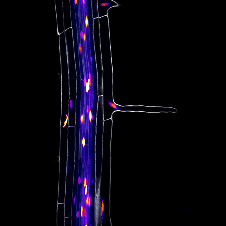
 (1 votes)

 (7 votes)