The Center of Regenerative Medicine at Washington University in St. Louis invites applications for the Rita Levi-Montalcini Postdoctoral Fellows in Regenerative Medicine program. These fellowships honor Rita Levi-Montalcini, whose Nobel-winning discovery of Nerve Growth Factor was among the first regenerative biology research to be conducted at WUSTL.
The Center of Regenerative Medicine (CRM) seeks individuals of outstanding talent with a doctoral degree to provide them with the opportunity to pursue research within a CRM lab. As Rita Levi-Montalcini was herself an international scholar working at WUSTL, we strongly encourage international applicants to apply. Additional information about the Center of Regenerative Medicine can be found at: https://regenerativemedicine.wustl.edu/. Full program details can be found at: https://regenerativemedicine.wustl.edu/about/rlm_fellowship/rlm_programdetails/. We anticipate awarding one fellowship in 2020.
Qualifications
RLM Fellowships are intended for exceptional scientists of great promise who have recently been awarded, or who are about to be awarded, the doctoral degree. Fellows are required to work in the lab of a CRM faculty member on a project that directly focuses on regenerative medicine. Current employees, fellows, and students of Washington University in St. Louis are not eligible. Applicants currently on H-1B visas are not eligible.
Terms of Appointment
RLM Fellowships will be granted for a period of two years.
A Ph.D./D.Sc./M.D. must be awarded and proof furnished to the CRM before the start of the Fellowship.
The RLM Fellowship provides annual compensation of $55,000, as well as fringes and health insurance, research funds, and relocation and travel funds.
Review of applications will begin on August 30, 2020.
Please provide a full current CV, two letters of reference, a brief description of scientific accomplishments and long-term goals, and indicate a potential CRM faculty host (https://regenerativemedicine.wustl.edu/people-page/).
Washington University is an Equal Opportunity Employer. All qualified applicants will receive consideration for employment without regard to race, origin, religion, age, sex, sexual orientation, gender identity or expression, national origin, genetic information, disability, or protected veteran status.
When COVID-19 hit Europe, it quickly became clear that holding scientific conferences was not going to be possible (or responsible) for some time, and that we weren’t going to be able to gather in September for Development’s 4th From Stem Cells to Human Development conference. Having been deeply involved in this meeting series since its conception, I was gutted – I love helping to bring this growing community together, I love the venue we were planning to use, and I love all the exciting science I get to hear about. So the question became – should we cancel/postpone the meeting, or should we try and arrange it in virtual format? To be honest, I wasn’t that excited about a virtual conference – what’s made this meeting so enjoyable in the past has been the relaxed and collaborative atmosphere, and the opportunity to talk science (and life!) over posters, dinner and drinks; how do you recapitulate that online? But then our events team introduced me to Remo – an online conferencing platform that really does give you the opportunity for these kinds of informal interactions (though sadly doesn’t provide the drinks…!).
I don’t want to sound like a sales rep for Remo, but what’s great about it is that you get to sit at a virtual table with other conference attendees and chat with them by video. You can also see who’s sitting at other tables, and move around to find the people you want to meet. So in a similar way as you’d bump into someone in the coffee queue at a conference, you can seek out your friends and collaborators (or hoped-for future collaborators/mentors etc) in the Remo space. You can also find a spot for one-to-one video chat and show someone your latest results by screen-sharing. And of course, you can watch talks, put questions to the speakers and engage in discussion after a presentation.
All this to say that we think (hope!) we’ve found a format for a virtual conference that will make it more than just another set of zoom webinars or Slack chats. For our September meeting (more details in the poster below), we’ve got a fantastic line-up of speakers, slots for short talks selected from abstracts, and we’ll be running poster sessions too (again with video chat). Because of the interactive format, there’s a limit to how many people can attend – we alsohope this will help to reinforce the collegial atmosphere of the meeting, and encourage people to present unpublished results. So if you’re interested in human development and want to meet with a bunch of other people with similar interests, get your application in soon – the deadline is 17 July!
The scuttling of conferences by COVID-19 has deprived scientists of one of their main ways to network*. Gone now are the chance (or not so chance) encounters with people you know only from author lists as you stand in line for coffee between talks, in elevators or bars, at poster sessions and organised socials…This new world has spurredmuch discussion on the future of scientific gatherings, and also spawned a plethora of online meetings and seminar series which may well become part of the new normal post-pandemic. However, many of these online events lack something approaching a networking element.
With this in mind, we’ve decided to conduct an experiment: an online networking event for developmental biology and stem cell researchers across the world. As 2020 marks ten years of the Node, it will also be a kind of birthday party for us, and following literally seconds of thought we’ll be calling it
The Node 10th Birthday Online Networking Event
Wednesday 29 July
4PM BST (should last 1-2h)
The event will take place in the virtual world – more specifically in Remo, a virtual conferencing tool (Development is also planning to use Remo for its upcoming conference on human development – registration is open until 17 July). In the Remo world you join a virtual conference centre and interact with real people at virtual tables via your webcam. We think it’s got a lot of potential for networking to complement your usual Slack channels and Zoom groups.
We’re currently working on the format, and are thinking of a mix of guided and free networking activities. For instance, you might sign up to join a table led by an expert in a certain area of science (e.g. writing papers, starting your first lab, reopening labs after lockdown, diversity and inclusion in science), or be paired with someone at random to talk about your interests, scientific or otherwise.
In the spirit of the Node, we also want community input – what kind of things would you enjoy and benefit from in an online networking event? We’ve set up a survey where you can share your ideas and register to attend (note even if you don’t plan to attend, if you have any opinions or ideas on the format, we’d love to hear them)
We’re looking forward to your feedback, and to meeting you in July in Remo!
* If this word makes you shudder, you could try to see it more as a good way to meet future collaborators, employers, employees, invitees, inviters, allies, friends…you could also try a small dose of it by joining the Node Network, our online database of developmental and stem cell researchers.
Postdoc (f/m/d) – Comparative genomics in planarians –
(Code number 11-20)
Planarians are fascinating animals that can regenerate from tiny pieces, harbor adult pluripotent stem cells, scale their bodies over a wide size range and, as a taxonomic group, display a fascinating spectrum of regenerative abilities, body sizes, reproductive strategies or life expectancies from a few months to seeming immortality. In collaboration with Gene Myers, our group has pioneered planarian high-quality genome assemblies and we have established a large and phenotypically diverse species collection through world-wide field expeditions. Comparative genome mining now promises access to a wealth of intriguing research questions. Current project opportunities include probing of the genomic consequences of asexuality by means of comparisons between the sexually and asexually reproducing strains of S.mediterranea; body size evolution in the giant planarians of Lake Baikal or genomic adaptations to life in the lake’s abyssal zone (~ 1600 m depth); intra-organismal population genomics amongst the many independently replicating pluripotent stem cells or the dynamics and functional relevance of the new class of giant planarian retroelements that we discovered. We have a number of fully funded postdoc positions available for talented individuals to pursue these or other questions.
Your Profile
You have a PhD or equivalent degree in a relevant subject area, e.g., biology, computational biology or computer science and extensive hands-on experience with genomic data.
You have a proven track record in one or more of the following: genome assembly, multi-genome alignments; comparative genomics; structural genome variance; transposon mobility; cancer or evolutionary genomics; phylogenetics and or population genomics.
You are passionate about the scientific endeavor and you are not afraid of pursuing your questions beyond the current scientific frontier.
You are self-motivated and independent and enjoy being part of an international and interdisciplinary work environment.
About us
We are a brand-new department at the Max Planck Institute for Biophysical Chemistry in the historic science town of Goettingen. We represent the organismal end of biophysical chemistry at the institute and investigate the mechanistic and evolutionary underpinnings of planarian regeneration. The department hosts a large zoo of planarian species for comparative analyses and we just established a field station at Lake Baikal in Russia, the Galapagos of planarian biodiversity. We are a thoroughly international and interdisciplinary group of people and based at one of Germany’s premier research campuses. We enjoy generous funding by the Max Planck Society and the proximity to picturesque Goettingen with its bustling student bars.
The Position
The payment and benefits are based on the TVöD guidelines. The Postdoc position are limited to two years with a possibility of extension.
The Max Planck Society is committed to increasing the number of individuals with disabilities in its workforce and therefore encourages applications from such qualified individuals.
Interested? Submit your application including cover letter (explaining background and motivation), CV, transcripts, and publication record preferably via e-mail as one single PDF file to
TB: The joy of seeing an article finally published is always slightly tempered by the long-drawn out process of peer review, re-writing, re-submission, re-review, proof-reading, required to get to that point. But the publication of our article ‘Auxin export from proximal fruit drives arrest in temporally competent inflorescences’ represented the end of a much longer journey. And to be honest, even a third round of peer review didn’t diminish the joy in fulfilling a long held personal ambition. Here, we look back at some of the key moments in the making of this paper.
2003: Inspiration
TB: I don’t know when exactly I first read ‘The fate of inflorescence meristem is controlled by developing fruit in Arabidopsis‘ by Linda Hensel and the late Tony Bleecker. Even 2003 is something of a deduction. What I do remember is that it was one of a bundle of articles photocopied out of the physical journal (those were the days!), and given to me early in my PhD by Jon Booker, one of the post-docs in Ottoline Leyser’s lab. And I remember being immediately struck by the paper upon first reading it. For the uninitiated, Hensel et al (1994) is an absolute masterpiece of experimental plant science, a combination of meticulous observation, meticulous experimentation and nuanced interpretation. It is a bona fide ‘classic’.
Hensel et al focuses on an important but somewhat overlooked developmental phase, namely ‘end-of-flowering’. Flowering itself is the reproductive phase in the most abundant group of land plants, the angiosperms (or ‘flowering plants’). Flowering occurs by the production of specialised shoots (inflorescences), which generate flowers. Each flower typically has male and female organs (staemen and carpels) that produce gametes (pollen and ovules respectively). Pollination of ovules leads to the formation of seeds containing embryos, and conversion of the carpels into a fruit that holds the seeds. Given that the majority of the world’s food supply is derived from the seeds and fruits produced by flowering, it’s a pretty important process. For the plants themselves, it is critical that flowering begins at the right time of year, but also that it ends at the correct time. While we know a lot about the developmental mechanisms that control the start of flowering, we know much less about those that control end-of-flowering.
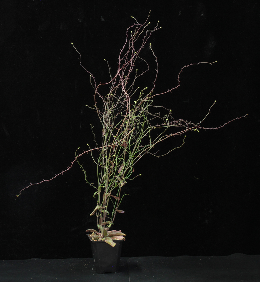
In looking at end-of-flowering, Hensel et al chose to work on what was rapidly becoming the dominant plant model species, the small, short-lived weed Arabidopsis thaliana (or just ‘Arabidopsis’). Arabidopsis reproduction occurs in a single coherent burst, elaborated through a branching inflorescence system. What Hensel et al found was that Arabidopsis flowering seemed to end by the simultaneous developmental arrest of all inflorescences on the plant, in a ‘global proliferative arrest’ (GPA) event (Figure 1). They then showed that this arrest required the presence of fertile fruit, and that removal of fruit could delay arrest, or even re-start flowering if arrest had already happened. Further, they demonstrated that it was very specifically the seed inside the fruit that really seems to drive arrest. Ultimately, they proposed a model in which the cumulative build-up of a fruit-derived signal triggers the simultaneous arrest. I thought this was a stunningly elegant mechanism for coordinating development across the whole plant body, and I knew there and then that I wanted to work on this problem, although not right there and then…this was something I was going to save for the future.
Figure 1: Global proliferative arrest. A wild-type Arabidopsis plant undergoing ‘global proliferative arrest’…except look more closely and it isn’t…
12th March 2016: Inception
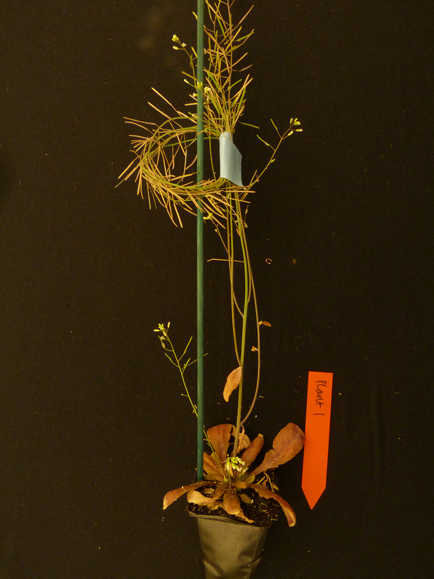
TB: As it turned out, the future was a long time in coming. Originally, I’d thought about this as something to work on towards the end of my PhD, but other tasks took precedence. And then I moved away to post-doc on somewhat different areas of plant development; I never forgot about GPA, but for a long while it was mostly at the back of my mind. Luckily for me, everyone else seemed to forget about GPA for quite a while. There was no follow up from Hensel et al, and no one else picked up the baton in the meantime. Thus, when I finally picked the idea back up in earnest in late 2015, we knew little more about GPA than we had done back in 2003. At the time, I was in the process of applying for academic positions, and GPA was clearly something that I was going to work on when I started my own group. In February 2016, I accepted a position at the University of Leeds to start later that year, and shortly thereafter, I set up my first GPA experiment (the imaginatively titled ‘GPA1’). When I started, I never really doubted that the phenomenon was already defined, and what I was looking for was just the mechanism. But GPA1 didn’t quite go as expected; yes, the plants arrested as expected, but I kept watering the plants just to see what would happen. To my surprise, the plants initiated new inflorescences and started flowering again (Figure 2). Clearly, this wasn’t going to be quite so straightforward as I’d thought…
Figure 2: When is arrest not arrest? “Plant 1” completely misunderstands what it is supposed to do during GPA.
20th January 2017: Proclamation
TB: As the ’new boy’ at Leeds, I was asked to give the ‘keynote’ talk at our annual Centre for Plant Sciences symposium, shortly after I started there. I’d never talked about my plans to work on GPA before, but I decided that’s what I was going to talk about, I think mainly because I was excited by the prospect of getting ‘stuck in’ to the project. I had very little actual data to present, but I pitched the talk on the importance of the phenomenon, and talked mostly about the theoretical background. I wasn’t sure how well it would go down, but as it turned out, it was something of a masterstroke…
CW: I had no idea who Tom was at the start of the symposium (other than the tall man who I occasionally passed in the corridors at work), but by the end of his talk, I had basically decided that I also wanted to contribute to understanding GPA. Tom had outlined some of what he planned to do, but I was particularly interested in the fact that he pointed out it could be relevant in crop species such as oilseed rape – I had an agricultural background, and wanted to do more work with crops. At this stage I had no interest in working with Arabidopsis, and figured I could provide some crop knowledge and avoid Arabidopsis. I wasn’t working for Tom at the time, so I figured any research I did contribute, I would just do in oilseed. Funnily enough, that’s not how things ended up working out.
5th June 2017: Recruitment
CW: Not only did I end up working for Tom, I also ended up spending the majority of my time working on Arabidopsis. With hindsight, my naivety about Arabidopsis was key to me completely buying into the end of flowering being a fascinating process. Very early on, I was carrying out an experiment where I was going to treat plants at floral arrest. During a routine check of the plants, Tom asked why I hadn’t carried out the treatments yet. I was confused by this – clearly the plants hadn’t finished flowering. Tom indicated that the primary inflorescences had all arrested – I countered that many of the lower inflorescences were still vigorously flowering. At this stage, we began really questioning GPA as a concept. If floral arrest was synchronous, how could these plants have some inflorescences that had arrested, and others which were still flowering several days later? The next step was to closely examine the flowering lifetimes of every individual inflorescence of a set of plants, where we clearly showed that floral arrest isn’t synchronous, it follows a basipetal wave, with the upmost inflorescences on the plant arresting first, followed by each inflorescence below it in turn. Each inflorescence class (primary, secondary, tertiary etc) also displayed a characteristic lifetime, where the timing of arrest appeared to be controlled specifically by the time since that inflorescence had initiated. I was hooked on the project from this point on; I needed to know how this was controlled!
1st November 2017: Nottingham
TB: Back in May 2016, a paper on GPA had been published in Plant Physiology, suddenly breaking the radio silence on the subject after 22 years (and giving me that terrible sinking feeling, until I realised that their approach was very different to what I was planning). So I was very much aware that interest in the area was starting to emerge again, but I had very little idea who else might be working on GPA. As it would turn out, at least two other research groups had also recently developed interests in GPA – Cristina Ferrandiz’s group in Valencia, and Zoe Wilson’s group in Nottingham.
It was therefore very serendipitous that my first seminar as a group leader on the ‘academic circuit’ was in Nottingham. I think it came as a surprise to Al Ware (the PhD student working on the project) and Zoe that there was someone else working on GPA, just as it came as surprise to me that my audience was really interested in what I had to say on that subject! Happily, we sat down to talk some more GPA after my seminar, and it rapidly became clear that our data and approaches were completely complementary. We agreed to collaborate there and then, and over the next few months, began to sketch out a plan for a manuscript that combined our data. Collaborating with Al and Zoe has been thoroughly enjoyable, and of massive benefit to both groups, so it really was a genuine stroke of luck to meet them so early in the project!
June 2018: Broken
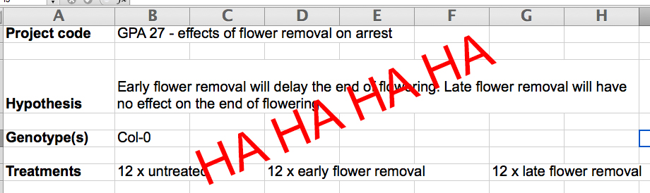
CW: This was an ‘interesting’ period in the development of the GPA research. In each experiment we set up, we were convinced that – this time – we knew what the outcome would be, but each time the results were completely different to what we had expected (Figure 3) and led to multiple new hypotheses and experiments.
Figure 3: Wrong, wrong again. Our hypothesis for GPA27 turns out to be exactly wrong…
Ultimately, this resulted in GPA 27 (we’d come a long way from GPA 3 when I started a year ago) – simultaneously one of my favourite and most-hated experiments to date. The plan was to remove all open flowers from plants daily; I think at this stage we were expecting the plants to arrest at a ‘normal’ time – we were wrong. Not only was floral arrest delayed, but with no fruit production, the plants kept producing more branches, meaning flower production was becoming exponential. What had started out as half an hour each morning removing flowers became the majority of every day pulling flowers off increasingly more gnarled, brittle Arabidopsis (Figure 4). It wasn’t quite as fun as it sounds. I was doggedly insistent that I was going to see this experiment to a close, but was away at a conference for three days, so asked Tom if he’d look after the plants while I was gone – it was only a few days, what could possibly go wrong?
Figure 4: We did this experiment so that you never have to. What happens when you pull every opened flower off an Arabidopsis plant for 5 weeks…
By the second day, Tom had emailed me to tell me that it was probably a good idea to halve the number of plants we were treating; we didn’t need as many as we had, and he’d leave the rest as a ‘recovery’ treatment (traitor). Funnily enough, my horror at this suggestion wasn’t enough to change Tom’s mind, and I returned to the lab to find that the ‘recovery’ plants had produced fruit and…arrested. The treated plants were still flowering on, but the recovery plants had happily arrested and stopped flowering, despite only producing a few fruits per inflorescence. I began to forgive Tom for the ‘recovery’ treatment, and we started questioning how many fruit were actually needed for arrest. Hensel et al. had suggested around 40% seed set was required for arrest, but hadn’t specified whether this was per fruit, per inflorescence… We’d removed hundreds of flowers from these plants, so clearly they hadn’t produced that much seed in the few days of the recovery treatment. We began to question whether it was the position of the fruit, rather than the number, that was crucial to arrest.
After GPA 27 finally finished, I ‘decided’ to take a few weeks of holiday to recover, and swore never to repeat another flower removal experiment. Obviously, when I came back, I’d forgotten the trauma of GPA 27, and carried out another flower removal experiment, only this time we focussed on individual inflorescences. Only a few inflorescences per plant had flowers removed, while the rest of the plant remained untreated. Pleasingly, I found that only the treated inflorescences had a delayed arrest – the rest of the plant arrested when we would expect. Not only had we shown that floral arrest wasn’t synchronous, now we’d also shown that it wasn’t global – it was local, and it was dependent on a small number of fruit.
7th February 2019: Submission
TB: By the end of 2018, we’d managed to collect all the data we needed for the planned manuscript, and we spent the first month of 2019 putting the manuscript together. We’d always planned to submit to Nature Plants; I’d been given some encouragement by one of the editors (Guillaume Tena) at a conference the previous summer, and I felt like our story was of sufficient general interest. Nevertheless, it’s always a tense wait to hear if the journal is going to send the manuscript for review. And often you don’t actually here anything from the journal at all until the reviews land in your inbox. Fortunately, Guillaume let me know fairly quickly that the manuscript was going out for review, making the whole process that bit less stressful!
Summer 2019: Redux
TB: The reviews — all four of them — came back at the start of April. Two of them were very positive, while the other two were positive, but had some serious reservations, particularly with respect to our divergences from the GPA model. But I have to say that, the comments were all very fair, and ultimately very helpful. Overall, this was by far the best experience I’ve had of peer review; I really feel like the reviewers helped us make a better and more coherent paper. We got straight to work on trying to provide new data to address the comments.
CW: Following the reviewers’ comments, we set up several key experiments to really focus in on the numeric and positional fruit requirement for arrest. My primary focus for the paper at this stage involved removing fruits at different timings prior to arrest, and allowing recovery periods to see under what conditions arrest occurred. Through this, we showed that only a small number of fruit are required to bring about arrest – and that the closer they are to the meristem, the faster the arrest; whereas a large number of fruit far from the meristem will not bring about arrest at all.
Interestingly, I accidentally further refined the number of fruit required for arrest while working on a totally different area of my PhD. I was looking at whether removing increasing numbers of flowers along an inflorescence (every other flower, 2 out of every 3, etc.), increased the final size of fruits. It didn’t. But nor did it really affect arrest either. If I removed 4 in every 5 flowers, the inflorescences arrested only a couple of days later than untreated inflorescences, despite only having 20% of the fruit. I particularly love that this experiment, which had a completely different purpose, ended up helping us to answer a different question we were working on. It’s so satisfying to see different areas and ideas link up in unexpected ways, but I hadn’t really experienced that in my own research before this time.
4th April 2020: Acceptance
TB: We finished re-writing the paper just in time for Christmas, and re-submitted on 17th December. I was pretty confident we’d addressed the reviewers comments, but after a second round of peer review (17th February), there were one or two new issues to address, though it was pretty clear publication was going to happen, as long as we addressed those issues. Fortunately, we already had the data, so we resubmitted within a week, and went back out for a final round of review. Then, finally, on 4th April, we received the good news…
CW: Looking back over the lifetime of this project, it’s incredibly satisfying to see how things have progressed so much – not just the research, which I’ve loved, but also generally how our approach and understanding developed as a whole. The first summer I was in the lab was very much a case of trying to get to grips with a new concept and species. The second summer was incredibly productive, but also very draining, both physically and mentally. We got great data out of that time, but it did come at a cost (although I did learn that 30+ consecutive days in the lab really is my limit). By the summer of 2019, we really knew what we were doing. Granted, we still found answers in accidental ways, but our whole approach was much smoother and more refined. Having the manuscript accepted really provides a satisfying point of perspective over how things have developed in the last few years. Now we’re on to the next exciting thing, and I can’t wait to find the answer. It won’t even involve any flower removal. Probably.
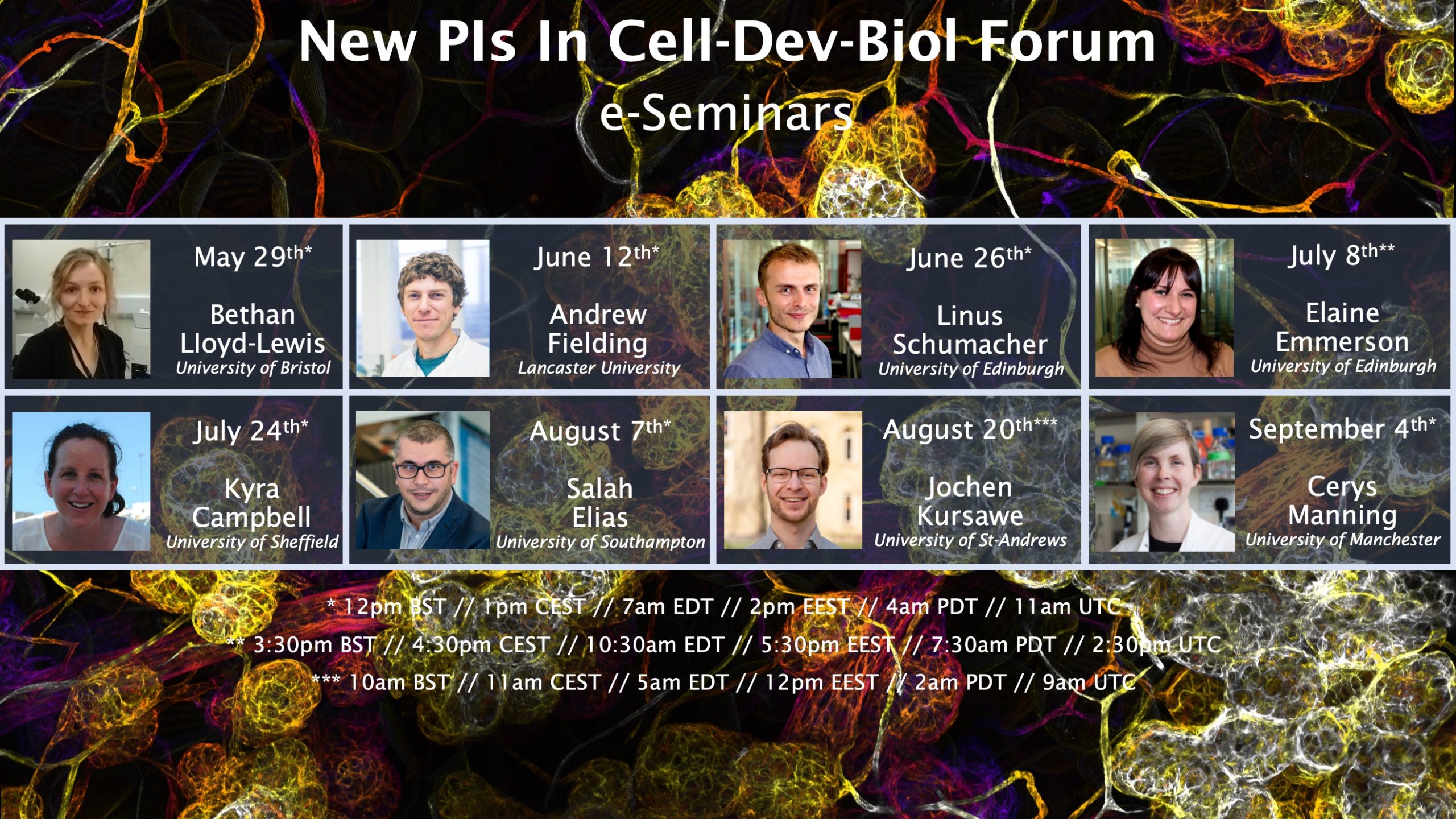
Of the many virtual seminar series that have sprung up in response to the COVID-19 pandemic, one in particular caught our eye – the New PIs in Cell and Developmental Biology Forum (you can follow updates and find information by following #NewPICellDev on Twitter). To find out more about the series we met Salah Elias, new PI at the University of Southampton and organiser of the forum (you can read Salah’s bio at the end of the interview).
How did you come to run your own lab and what are your main research questions?
My move to the University of Southampton in 2017 and appointment to a lectureship enabled me to start my own independent laboratory building on my postdoctoral research. This has involved bringing together expertise from cell biology, physics and mathematics to investigate new microtubule- and actin-mediated mechanisms of oriented cell divisions (OCDs) in the normal mammary epithelium, and determine how this influences stem cell fate and dynamics during development and homeostasis. OCDs represent an essential mechanism during development that ensure proper epithelial integrity and differentiation in several epithelial systems. Yet their functional requirement for mammary epithelial biology remains subject to deliberation. This is a fundamental and important question for my lab, particularly in the mammary gland where tissue turnover is very high. Moreover, given increasing evidence linking dysregulation is OCDs to breast cancer, my lab aims to understand how imbalance in OCDs can lead to the abnormal cell fate and behaviour that contributes to the initiating events of malignant transformation. Our multidisciplinary approach has allowed us to develop a set of cutting-edge tools and novel ideas that open up new research directions and help us establish our own niche.
How has your research been affected by the lock down?
The COVID-19 pandemic has caused unprecedented disruptions within the entire research community. It has also exacerbated existing inequalities, with new PIs representing one of the most affected groups. As a new PI, closing my lab, which I have managed to get up and running after a challenging and time-consuming process, was heart-breaking. After I joined the University of Southampton, I have been able to secure major grants, hire talented PhD students and postdocs and develop excellent collaborations. We have started to work on our ideas and get exciting findings, which we were looking forward to sharing this year at conferences. The pandemic reminded me how vulnerable and precarious my position as a new PI was. In few days we have had to cull several experimental mouse cohorts which we have been preparing several months in advance and thrown away many long-term in vivo imaging experiments which have taken months to prepare. We stopped all our research within a week. Suddenly, I also found myself with childcare responsibilities, which I couldn’t share with my wife who is a key NHS worker. This has considerablydisrupted my productivity and challenged my ability to run my lab. All this would delay time to publish my lab first independent publications, putting at risk the competitiveness and sustainability of my research program.
How did the idea for a young PI forum in cell and developmental biology come up?
One of the immediate consequences of the lockdown was the cancellation of conferences. For my lab, this has taken away many opportunities to showcase and discuss our research. We know how conferences are important particularly for new PIs, by offering excellent networking and career development opportunities. They are also the place where we can make the scientific community aware of our accomplishments and thereby build a reputation that is important for our careers. For me, working from home has also been very isolating and challenging mentally. Many peers around the world have shared their frustrations and struggles through social-media platforms such as Twitter. It was great to see many established PIs relaying actively those concerns and urging the scientific community to protect new PIs during and after the pandemic. However, I thought that new PIs, as a community, could be proactive and help each other. This is how the idea for the New PIs in Cell-Dev-Biol Forum emerged. The forum would include e-seminars, but I also wanted it a platform that maintains connections and fosters peer support. I discussed the idea with Dr. Bethan LIoyd-Lewis, new PI at the University of Bristol, which she endorsed immediately. Then, everything went fast, the first colleagues we have contacted responded positively, and within a week we had our e-seminars with a fantastic fist line-up of speakers. We advertised on Twitter, it was really heartening to see how positively the scientific community has responded and helped us promote the forum. The e-seminars have started already with success.
How does the forum work?
The primary goal of the forum is to be a platform for peer support and collaboration, supported by an e-seminar series where talks are organized every two weeks. Our top priority, particularly during the lockdown, is to offer opportunities for new PIs to give and host talks. Speakers will be encouraged to present unpublished results to promote discussions and collaborations. We advertise through Twitter, where we post a link for each talk. We have also created a Slack (@NewPI_CellDev), which allows us to maintain connections, support each other and exchange ideas about the future of the forum. I am pleased to see how everyone is taking ownership of the forum, which I believe is the best way to maximize contributions and achieve our goals together.
If people are interested in giving a talk, what do they have to do?
Three weeks after its creation, 30 new PIs from 20 UK and European research institutions have joined the forum already. We have a full program of talks until May 2021. Everyone who joins the forum is invited to give and host a talk. As I mentioned above, priority will be given to preliminary results, so colleagues who have just opened their labs are also welcome to share their research, particularly if they are seeking help for their grant applications. Advertising the forum and talks on Twitter allows us to reach the wider scientific community. Generally, when we receive expression of interests from colleagues, we ask them to send us their contact details through direct messaging or e-mail, so we can add them to our mailing list and send them an official invite. Our members are doing a fantastic job in promoting the forum within their own networks and institutions. However, we are also aware that not all our colleagues have a Twitter account. We hope that societies such as the Company of Biologists will help us to be more visible to increase our membership.
Do you expect to keep the forum going when the lockdown ends, whenever that may be?
Our first successes reinforce our ambition to create an international forum for new cell and developmental biologists, which lasts beyond the lockdown. We are aware that the consequences of the COVID-19 pandemic will affect our research and careers for the next few years at least. The forum will offer opportunities for peer support, networking and collaboration, andl represent a powerful mechanism fostering long-term equal partnership with mutual benefits. The forum will also be rewarding on a personal level, and I am looking forward for some really great friendships.
Do you think COVID-19 will change the way scientists share their work with each other?
Unquestionably, the COVID-19 pandemic has brought new opportunities for science communication. It is amazing to witness how proactive and altruist the scientific community is, by turning big conferences virtual, and creating a variety of webinars and e-tutorials and open resources, in such a very short time. We have learned effective ways to make conferences more accessible and more ecologically friendly, while minimizing the costs. I am proud that our forum is taking part of this revolution in science communication. As a community, we need to continue in this vein to bring open science a step closer to reality.
Biography
Salah Elias studied cell biology and Physiology at Rouen University, France. He then did a PhD in neuroscience at INSERM U982, France, where he investigated the mechanisms of secretory vesicles biogenesis and trafficking in neuroendocrine cells, under the co-supervision of Prof Maite Montero-Hadjadje and Prof Youssef Anouar. During his first postdoctoral work in the lab of Dr. Sandrine Humbert at the Curie Institute, France, he showed that huntingtin and kinesin-1 regulate spindle orientation and apical polarity in mammary epithelial cells during development and homeostasis. He then moved to Prof Elizabeth Robertson lab at the Sir William Dunn School of Pathology, University of Oxford, where he identified a new subset of mammary stem cells, expressing Blimp1, that drive gland morphogenesis and homeostasis. In 2017, Salah was appointed Lecturer at the University of Southampton. His lab aims to understand the mechanisms of oriented cell division (OCD) in the normal mammary epithelium to discover cell division defects that are unique to breast cancer cells. Salah is the recipient of an MRC New Investigator Research Grant and Wellcome Trust Seed Award in Science.
Contact details:
School of Biological Sciences, University of Southampton, Highfield Campus, Southampton SO17 1BJ, United Kingdom
Highly motivated postdoctoral candidates are invited to lead several new projects to address fundamental questions on RNA homeostasis (Zhang et al. Molecular Cell 2018; Haeusler et al. Nature 2014) and protein homeostasis (Lu et al. Nature Neuroscience 2019; Liu et al. Genes & Development 2018; Periz et al. PLoS Biology 2015) related to neurodegenerative diseases in the laboratory of Jiou Wang. The position is open to candidates with a wide range of backgrounds including biochemistry, molecular biology, cell biology, and structural biology on all topics of biology. Those with experience in RNA biology, protein biochemistry, and bioinformatics are particularly encouraged to apply.
The Johns Hopkins Medical Institutions provide a stimulating and collaborative environment for biomedical research. Our lab is affiliated with the Department of Biochemistry and Molecular Biology at the Bloomberg School of Public Health and the Department of Neuroscience at the School of Medicine. The Baltimore/Washington D.C. area also offers rich professional and living opportunities.
Candidates should have a doctoral degree and strong research background. Please send a statement of research experience and career goals, a copy of Curriculum Vitae, and contact information of at least one reference to Dr. Jiou Wang at jiouw@jhmi.edu.
A complete listing of PubMed-accessible publications can be accessed at the following URL: http://www.ncbi.nlm.nih.gov/pubmed/?term=Jiou+Wang.
More information available at: http://www.jhu-bmb-phd.org/faculty/jiou-wang. The Johns Hopkins University is an Equal Opportunity Employer.
The ongoing pandemic has resulted in many scientific conferences moving to an online format, and researchers who can no longer attend seminars at their institutes have been organising and attending various virtual seminar series (here on The Node there are currently over 40 online events listed in developmental biology and beyond). Several considerations about virtual events have been brought up (see for example here and here), but a better understanding of participant experience is crucial to inform conference organisers on specific areas that could be improved in future virtual events.
A group of early-career researchers who contribute to preLights have designed a survey and are seeking responses from students, researchers, journal editors, or anyone who has attended a virtual scientific event.
Please take our anonymous survey and feel free to share it with your colleagues.
The survey will be open until 31 July 2020. The results of the survey will be shared on the preLights website.
Study of the molecular mechanism of Endothelial to Mesenchymal Transition during muscle regeneration and crosstalk with the immune system in vivo and in vitro.
A 3 year PhD studentship is available within the Heva Research Group, School of Medicine and Surgery, University of Milano Bicocca, Milan, Italy (https://hevaresearch.unimib.it/), under the supervision of Prof. Silvia Brunelli.
The position is the framework of the H2020 Marie Skłodowska-Curie ITN funded project RENOIR (REcreating the ideal Niche: environmental control Of cell Identity in Regenerating and diseased muscles, https://renoir-itn.eu/).
The PhD project (https://renoir-itn.eu/esr1/) focusses on how the inflammatory and vascular components integrate to coordinate muscle regeneration and how the process of Endothelial to Mesenchymal Transition (EndoMT) contributes to fibrosis in pathological conditions, using in vitro and in vivo models.
The student will be enrolled in the Ph.D. Programme in Translational and Molecular Medicine (DIMET), University of Milano Bicocca (www.dimet.org).
The animation is a result of collaborative work of scientists from the Novo Nordisk Foundation Center for Stem Cell Biology (DanStem) and visual storytellers from the Animation Workshop (VIA), telling the story of a scientific attempt to learn what happen to the liver when damaged and how this knowledge could be translated in the future and help healing liver diseases and improve patients quality of life.
 (No Ratings Yet)
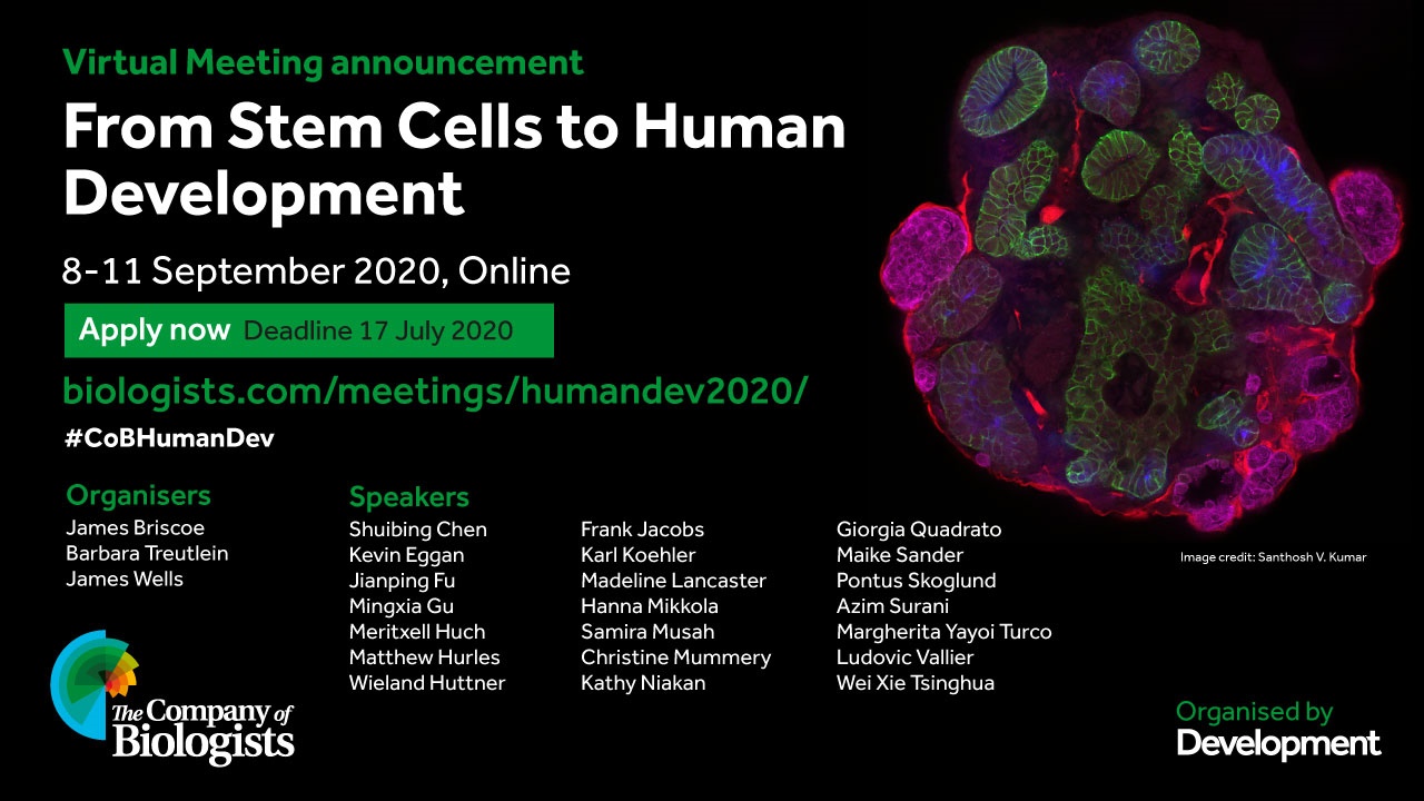
 (3 votes)
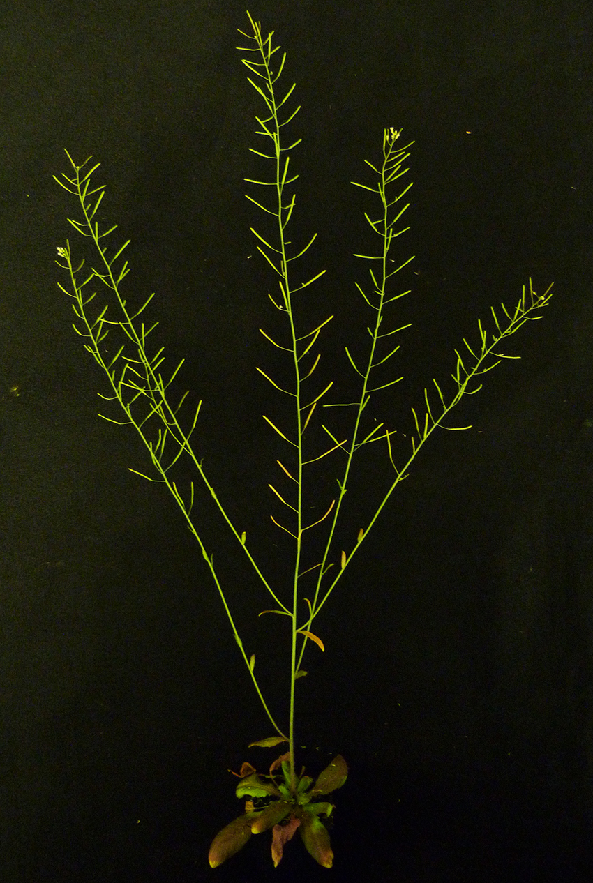

 (6 votes)