We would like to take this opportunity to thank the Node for the chance to write a post about JoVE and how our resources can be beneficial for the research and teaching of developmental biology and multiple other disciplines.
All researchers will be familiar with the challenges of replicating an experiment you’ve read in a paper, or learning a new technique in the lab. Spending hours looking through reference lists or trying to work out exactly what is meant by “shake vigorously” or “aspirate gently”. In an ideal world you ask someone who knows the technique to show you how, but with the global nature of scientific research, time restraints and physical distance means this isn’t always possible. But what if a researcher on the other side of the world could show you how to perform their methodology at any time of day with no travel costs involved? This is where JoVE comes in!
JoVE is dedicated to publishing scientific research in a visual format, capturing the intricate details of life science research and overcoming two of the biggest challenges faced by the scientific community:
1) Poor reproducibility and low transparency of biological experiments
2) The time, labour, and cost intensive nature of learning new experimental techniques
With technology so ubiquitous in our everyday lives, it stands to reason that we should harness its power in scientific research and education.
Since launching in 2006, JoVE Video Journal has published more than 12,000 scientific video demonstrations on experimental methods in 13 discipline specific sections including Developmental Biology. JoVE Video Journal was the first and remains the only peer-reviewed journal of visualised experiments, and today JoVE video articles are viewed by millions of users making scientific research more productive and reproducible.The field of developmental biology employs a multitude of complex and rapidly developing research methodologies. As such, the Developmental Biology section of JoVE Journal, featuring insightful video articles authored by renowned experts in the field, is invaluable for researchers worldwide to learn experimental methods quicker and more precisely.
With more than 500 articles in our developmental biology section, it’s difficult to to highlight just a few examples of the excellent discoveries that have furthered our knowledge in this field. Why not take a look at this selection of some of our more recent additions to the journal that demonstrate the broad range of research:
It’s not just seasoned researchers that can benefit from this video format, we also have visual teaching aids for students at the start of their scientific careers. JoVE’s database of educational videos are designed for educators and students, to better teach and learn key scientific concepts with the aid of animations, and fundamental lab techniques with easy-to-understand video demonstrations. By providing a visual approach to learning basic techniques, JoVE Science Education makes experimentation more accessible to undergraduates in developmental biology classes.
Our Science Education collections cover a range of lab techniques valuable to developmental biologists from very basic skills such as centrifugation, volume measurements, and pipetting, to more advanced developmental biology specific techniques such as culturing embryonic stem cells, explant culture, and genetic engineering of model organisms.
For teaching foundational scientific theory we have JoVE Core Biology, an animated textbook organised into sections including cellular processes, genetics, and human biology. Instead of flicking through hundreds of pages of dry text and confusing diagrams, Core Biology uses concise animated videos to bring the concepts alive.
During the current COVID-19 pandemic, JoVE is aiding universities, colleges and secondary schools in the transition to online teaching by providing free access to all of our educational content until the 15th of June 2020. So now is the perfect time to try it out. Just head to www.jove.com to activate your free trial, and take a look at our faculty resource center for help setting up remote access for your students.
We are constantly adding to our content with new educational resources released all the time, and 150 new journal articles published each month. However, this does not mean that we prize quantity over quality. We believe carefully produced, peer-reviewed scientific videos represent the best way for scientists to share a new technique, and for researchers and students to learn from it. With every video, we aim to drive the next breakthrough in science research and education.
So remember, the next time you are in the lab or want to boost student performance, check JoVE first.
Rebecca Ellerington, Curriculum Specialist UK and Ireland
Like many science enthusiasts, I read the book The Double Helix when I was a student. It’s a dramatic tale of how American geneticist James Watson and British molecular biologist Francis Crick discovered the structure of DNA back in the early 1950s. Of course, being written by Watson himself, it’s no surprise that he’s the dashing hero of the story.
The names of James Watson and Francis Crick are inextricably linked with the discovery of the DNA double helix. And if you’ve been paying attention, you’ll also know that credit is due to Rosalind Franklin, Maurice Wilkins and Ray Gosling too. But what about Elwyn Beighton, Fred Griffith or Rudolf Signer?
In this episode we’re unwinding history to uncover some of the less well-known stories behind the discovery of the structure and function of DNA.
If you enjoy the show, please do rate and review on Apple podcasts and help to spread the word on social media. And you can always send feedback and suggestions for future episodes and guests to podcast@geneticsunzipped.com Follow us on Twitter – @geneticsunzip
This year the Node hits double digits – our first post was published way back on April 1, 2010 (we’re now at over three and a half thousand posts and counting, and get over thirty thousand page views a month). We’ll be celebrating this developmental milestone with a look over some of our favourite content of the decade, but we’re also thinking of ways to move forward and stay relevant to researchers the world over. So we’ve designed a user survey that covers various aspects of how the Node works and what more we could be doing. We are a community site and would love to get a widespread response, so please consider taking the survey and telling your colleagues about it. Here’s the link:
The Southeast Regional Society for Developmental Biology Conference will be held virtually in a Zoom Webinar on May 20-21.
Registration is required for all attendees by May 6, 2020 (limited space). Registration is Free, please use Fee Waiver Code: FREE when completing your registration.
Abstract submission for short talks will be through April 30, with notification to presenters by May 10. Only SDB members are eligible to submit an abstract, links for becoming an SDB member can be found on our website.
Zippering is a striking phenomenon whereby two opposing epithelial tissues become progressively united in one direction over a period of time. Similar, to the travel of a zip fastener, zippering leads to forward progression of a point of fusion, and can occur over significant distances along an organ or tissue, to eventually seal an opening. This is fundamental to establish de novo continuity between two opposing epithelial layers in the embryo, or to repair a gap for example following an injury (Begnaud et al., 2016; Jacinto et al., 2001; Martin and Parkhurst, 2004).
In development, zippering can be first observed during morphogenesis of the neural tube, whereby the flat 3-layered embryo, resulting from gastrulation, undergoes a major transformation to establish the final 3D basic body plan of the future fetus. This remarkable reshaping event is driven by precise coordination between: (i) invagination of the neural plate along the midline, (ii) elevation of the neural folds to meet at the dorsal midline, and (iii) continuous progression of zippering to seal the entire rostro-caudal axis of the developing embryo (reviewed in Nikolopoulou et al., 2017).
Failure of neural tube closure results in the development of severe congenital malformations, collectively known as neural tube defects (NTDs) (Copp et al., 2013; Greene and Copp, 2014; Wallingford et al., 2013). As 0.5 to 2 in every 1000 established pregnancies are affected world-wide, the clinical implications are highly significant to our society. Open spina bifida is the most common NTD observed in new-born babies, caused by failure to complete spinal neural closure by the end of the first month of gestation (Copp et al., 2015).
Alongside the neural tube, several other organs employ epithelial zippering as a mechanism for their development: for example, the optic fissure, palatal shelves, tracheoesophageal foregut and presumptive genitalia. Failure of zippering leaves the organ unsealed, resulting in severe open defects such coloboma, cleft palate, tracheoesophageal fistula and hypospadias.
Our story started with a completely unexpected observation in 2013 which brought us to explore the mechanism behind epithelial zippering during closure of the mouse neural tube. Initially, we aimed to investigate the role of cell-extracellular matrix (ECM) interactions during neural tube closure as a whole. We started examining the structure of the major ECM components including laminins, collagen IV and fibronectin, together with expression of the main integrin subunits that mediate these interactions. However, we noticed that a specific integrin receptor combination, a5b1, localised precisely at the site of zippering. Furthermore, it did not seem a coincidence that this site was also enriched for thick radially-oriented fibrils of fibronectin, which represents the central interacting ligand of a5b1 integrin (Figure 1).
Figure 1. Whole-mount mouse embryo at E9.5 stained for fibronectin (Fn1) (left). Integrin b1 (Itgb1) localisation at the site of zippering in whole-mount view (middle, top) and cross section (middle, bottom). Arrangement of cells in a semi rosette configuration around integrin b1 and radial orientation of fibronectin fibrils (right).
Therefore, we decided to embark on a highly risky and (so it turned out) long experimental route to genetically inactivate the integrin b1 subunit, to assess if the inability to interact with fibronectin at this site would affect propagation of zippering. The integrin b1 knockout mouse dies early in development (Fassler and Meyer, 1995; Stephens et al., 1995) so we had to use conditional genetic approaches. We targeted the neuroepithelium, as the precursor of the neural tube, and we separately targeted the surface ectoderm, that connects to the neuroepithelium at the zippering point. The experiments showed that integrin b1 expression is required on surface ectoderm cells, and its inactivation impaired fusion leading to highly penetrant spina bifida. This discovery was a really exciting and rewarding moment of our research. However, we then had another important question to answer: how do these local cell-ECM interactions, via integrin receptor a5b1, mediate progression of zippering and closure of the neural tube?
It took a long time to find the missing piece of the puzzle to solve the above question. At the beginning, we looked at the cytoskeleton, cellular protrusions, cell death and cell proliferation, which are all downstream of the highly intricate integrin signalling network (Barczyk et al., 2010; Lowell and Mayadas, 2012; Schwartz, 2010). However, we could not find a clear answer by exploring any of these avenues. A very inspiring study was the paper from Edwin Munro’s group (Hashimoto et al., 2015) which involved live imaging of neural tube zippering in the ascidian Ciona intestinalis. The authors described a process of sequential cell junction shortening at the site of zippering. Therefore, we decided to zoom in and look at the cell shapes in the closing mouse neural tube and, similar to Ciona, we noticed that cells around the site of zippering had significantly shorter junctions compared to those that had not yet entered the zippering point. In very elegant geometry, the cells adopted a wedge shaped morphology and formed a semi-rosette configuration which converged precisely to a central fulcrum where the integrins localised (Figure 1). In the targeted mutant embryos, the semi-rosette was severely disrupted which prevented the cell junctions from converging.
At this time, we submitted the paper and received very positive responses. However, we faced a further big challenge: one of the reviewers asked us to live image the process to see if indeed the dynamics of cell shape change and junction remodelling, as predicted by our model, could be observed in vivo. Live imaging of mouse neural tube closure is technically very challenging, as the embryos need to be maintained in culture in a healthy state, with optimal heartbeat, and yet need to be static for confocal imaging. The work from Lee Niswander’s group (Massarwa and Niswander, 2013) had shown that live imaging of mouse embryos is possible, but their images lacked the resolution we needed to visualise individual cell behaviour. We decided to take on the challenge and, after many attempts and long evenings and weekends spent in the confocal room, we finally managed to get images with sufficient cellular resolution. Thanks to the exceptional dissecting skills of Gabriel Galea, we made a small hole through the yolk sac and amnion to expose the closing spinal neural folds, and then kept the embryo steady on an agarose surface using an implanted microsurgical needle. The embryos were very healthy – their hearts beat vigorously – but this meant we had to manually acquire each z-stack and re-centre the region of interest each time!
Despite all the effort, the result was worth it. After a post-imaging surface subtraction, to remove the underlying tissue, we could clearly see for the first time the live behaviour of the cells and how zippering propagates along the body axis. Cells in close proximity to the site of fusion display shorter proximal junctions than cells that have not yet entered the fusion site. Within a 15 minute time-frame, cells close to the zippering point further shorten their proximal junctions and become incorporated into the semi-rosette configuration around a common vertex (Figure 2). This process of junctional remodelling brings cells from the two sides of the neural folds into contact for the first time, converging at the shared site of integrin-mediated anchorage. Once in contact, cells within the semi-rosette establish new junctions with their contralateral counterparts, and then exit the semi-rosette into the region of surface ectoderm that overlies the closed neural tube. In this way, zippering is able to progress, as new cells bordering the open region undergo the same cycle of sequential constriction and semi-rosette formation.
Figure 2. Live imaging of surface ectoderm cells during zippering propagation. Individual cells shorten their proximal junctions over time (a) to form the semi-rosette configuration (c) and then exit the zippering point rostrally (d, e), with further cell elongation.
While the two classical models of epithelial fusion, contraction of an actomyosin cable and protrusion-mediated cell migration, are undoubtedly important in closure, the precise travel of the zipper over long distances, as in neural tube formation, cannot be explained by either of those mechanisms. Genetic or pharmacological disruption of the cytoskeleton does not halt spinal zippering (Escuin et al., 2015) and, while cellular protrusions make the first contacts at the zippering point (Rolo et al., 2016) we found they were intact in embryos lacking integrin b1. Zippering relies instead on small localised cell junctional rearrangements mediated by the action of integrin α5b1. Coordinated adhesion toward a common site of basal anchorage, probably involving fibronectin, brings pairs of contralaterally positioned cells into close proximity (Figure 3). This intermediate state of cell-ECM anchorage is crucial for the subsequent maturation and extension of novel cell-cell junctions between opposing cells, promoting closure and enabling progression of zippering.
Figure 3. Model of semi-rosette formation and zippering propagation. Surface ectoderm cells upregulate integrin 1 at the zippering site (a). Coordinated adhesion to fibronectin (SE = Fn1) causes proximal junctions to shorten forming a semi-rosette (b). Opposing junctions are brought into close proximity, enabling cross-midline junction formation at the site of shared basal adhesion (Se = Fn1 = SE) (c). This propagates zippering forward with novel cell-cell junction formation.
Indeed, a recent whole-exome sequencing study of families affected by NTDs identified variants in the integrin b1-encoding gene, ITGB1, among affected individuals, suggesting ITGB1 as a key predisposing gene in human NTDs (Lemay et al., 2018). The strong similarity between the open lesion in our mouse model and the condition of lumbo-sacral spina bifida in humans, emphasises the possibility that integrin-mediated anchorage may represent a conserved mechanism for neural tube zippering in humans as well as mice. Hence, impaired integrin function could represent a potentially significant risk factor in the aetiology of open spina bifida.
IRIBHM is a research institute of the Medical School of the Free University of Brussels (Université Libre de Bruxelles, ULB). The institute offers an internationally prominent research environment in molecular biology and life sciences, that engage different topics that span receptor pharmacology and new therapeutic targets discovery, early embryonic development, neurobiology, stem cells and cancer. The institute has trained over the years a number of talented young scientists both at the graduate and postdoctoral levels.
In order to expand internationally and keep rising its level of excellence, the IRIBHM (https://iribhm.org/) launches an international graduate programme in order to prepare the future leaders in biomedical sciences. At least 2 PhD scholarships are available. The successful candidates will have the opportunity to work in a warm and stimulating research environment aligned with the highest international standards.
As the administrative center of the European Union, Brussels is a perfect location for an International PhD programme. In addition, the city shows an active cultural life and is hosting most nationalities from all over the world.
We look forward to welcoming you in Brussels!
Requirements:
Diplomas and degrees equivalent to a European Union Master’s degree, which includes project work summarized in a written “small thesis”
Two referees willing to provide letter of recommendations
Excellent knowledge of English
Excellent interpersonal and organisational skills
Due to COVID 19, closing date for CV and letters submission are extended: June 30.
Accepted candidates may start research projects as early as November 2020
We are looking for a POSTDOC to join our EVODEVO-GENOMICS lab in the University of BARCELONA.
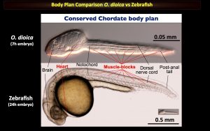
Our lab studies the chordate model Oikopleura dioica to better the impact of gene loss on the evolution of gene regulatory networks. In particular, our research focusses on heart development, long-non-coding RNAs and de novo genes. Click here for a tour “A day in our lab” posted in The Node
Recently, we have also engaged a new EcoEvoDevo line investigating how mechanisms of development in marine embryos respond to climate change, including biotoxins derived from algal blooms. Click here for a tour on this new EcoEvoDevo adventure.
Our approaches include single-cell, RNAseq, Embryo microinjection, RNAi, Confocal-Microscopy, Bioinformatics, population genomics and soon CRISPR
Marie Skłodowska-Curie Individual Fellowships | Postdoc Call ID: H2020-MSCA-IF-2020
Open April 8th – Deadline September 9th 2020. (for University of Barcelona internal procedures, the application forms should be ready by the end of July)
CONTACT: Interested postdoc candidates, please send an email to Cristian Cañestro (canestro@ub.edu) ASAP, including a brief letter of interest, a brief CV, including list of publications with their impact factor and quartile, and technical skills (specially those related with our approaches) all together in ONE single pdf file.
This year I had a unique experience arising from my PhD – 2 weeks giving training in Nigeria, by invitation of Prof. Amos Abolaji from the University of Ibadan. Amos established one the first Drosophila labs in Nigeria in 2014, and in 2017 he co-organized workshop with DrosAfrica – a charity I collaborate with and that you should check out. I met him after we invited him to our EMBL PhD symposium in 2018. He kindly invited me to be a trainer at a Drosophila workshop he was organizing in Abuja in March last year, and to visit his lab in Ibadan too – to do embryo collections and stainings together, but also to prepare science outreach activities and visit two schools. In this article, I am going to share my experience and perhaps convince you to take on a similar adventure too.
Warm weather, warm hearts
When I got off the plane, which arrived to Lagos at 9pm, I didn’t know what to expect. It was hot, really hot! It stayed like that for the two weeks: we had on average 40-degree days, sometimes with a sensation of more. Amos was there to pick me up, dressed in traditional Yoruban clothes. My first impression of Nigeria remained loyal to what I would experience the following two weeks: very warm weather, very colourful attires, very warm and joyful hearts. Also, it is loud, there is always sound…In fact, when I returned home, one of my strongest impressions was that Heidelberg felt extremely quiet even though I live in the centre, which can be quite noisy. The Yoruba language sounds very musical – I learned a bit but wish I had learned more. I stayed for 9 days at the University of Ibadan (UI) and 5 days in Bingham, near Abuja. It is important to note that Nigeria is very big and multiculturally diverse, so what I share mostly reflects what I witnessed more closely in the South West region. I was the only visitor for a week, and, on my 7th day there, Dr. Alex Whitworth joined. Alex is a group leader at the MRC Mitochondrial Biology Unit in the University of Cambridge. It was great to experience both ways: all the new things you discover on your own, fully immersed, but also having someone from a (more) similar background to share experiences with. Alex was an incredible travel buddy to whom I am super grateful, and it wouldn’t have been the same without him; he also showed me that PIs can be cool travel buddies too! Finally, I had heard before going that food was spicy. Well, food is really spicy, and portions are very generous.
No excuses
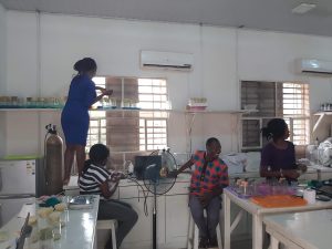
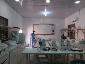
Abolaji lab members in the lab, Bukola (left) is taking care of the flies
The Nigerian scientists I met had a constant mindset of “no excuses”, which comes from trying to overcome lack of resources with perseverance and creativity. This was not entirely new to me. In Portugal – where I come from and where I started working the bench – there is also this attitude. We had to plan seriously and we would coordinate with neighbouring labs to make sure resources were used to the maximum. At EMBL the conditions are very different, as we are quite wealthy and I would say a lot of people (including myself) are spoiled. There are a lot of things we don’t have to worry about such as preparing buffers, animal diet, washing our glassware etc.
We have the tendency to believe that science is objective and therefore democratic. But science is made by humans, and the socioeconomic context plays a huge role in the opportunities people have to realize their potential. If I felt a resource gap after travelling 2000 km between Portugal and Germany, this was even greater with the 6700 km between Heidelberg and Ibadan. I am not talking about not having fancy equipment. I am talking about, for example, not having constant power supply. What does that imply? It implies that you need to spend so much more time caring for the animals – flies in this case – which would be happier less 10-15 degrees Celsius of heat. It implies that you might have to run experiments until 5am because electricity is more stable during the evening. I met students who literally did this. And these students often face overscrutinity of their research credentials by recruiters in US/EU institutions, without taking into account the context and resources in place. Excellent people who dream about great experiments to their bold hypotheses, but sometimes cannot perform these tests with what is available to them. But still, they say “no excuses” – they are very creative to always find a way to improvise. This was true both for the Abolaji lab in UI and also the Bingham workshop participants I met in Abuja. I had heard Amos talk about the lack of power supply when he visited EMBL, but these are things you have to see for yourself to really grasp it. It is important, for the less aware, not to confound excellence gaps with resource gaps. And importantly, in Amos’ lab they share a lot of their equipment with other labs in the department. Since they have a KVA inverter to make up for power shortages, they even share power sockets: I often saw visitors coming in just to use their laptops and plug them in or charge their phones. This contrasted a lot to me with what I have seen in some Western labs, sometimes in the same institute, not wanting to share machines or reagents.
Adapting to your context
The culture I am used to doing science & science outreach in (from my experience in Portugal, UK, Denmark and Germany) is different from the culture of Nigeria. Again, this was not my first cultural shock, as I experienced it by moving abroad, first to the UK and then to Germany. Even after almost 3 years in Germany, I still struggle to adapt to some things. I also had been to Africa before, but it had been 10 years since I was there last, and this time it was for work, not vacation, and on my own, not with my family.
I love making plans and scheduling things to the detail. I am not super punctual, but with African time, there was never a dull moment. Even after some days of realizing that the pace of life was different, our brains are so wired in habits that Nigeria found ways to surprise me everyday. And I think that is great! In EMBL, if a seminar is at 1pm, it starts latest at 1.05pm. In Portugal, the lights of the amphitheatre would probably still be off at 1.05pm, and the audience joins around a quarter past. I felt that in Nigeria it was even more flexible. But this also needs to be put into context. Sometimes I was surprised by some breaks during the middle of the day, but it made sense because we experienced such hot weather (hot even for Nigerians themselves!), that it made more sense to continue going after it cooled a bit off in the evening, and we could stay until late. We made it to a domestic flight after arriving to the airport 15 minutes before departure, which was quite an adventure for me, but the friends I made there assured me that there usually is no “African time” for flights! In terms of planning, I would just say that no amount of planning is too much!
Amos in my first day in Ibadan, when he showed me his lab
I was used to certain health and safety measures from Europe, for example for dealing with chemicals. Guess what, if you don’t have many resources, it is hard to handle chemicals with such care. For me that was initially quite a shock, but eventually that shock grew into respect of the scientists I met who have to make much more sacrifices, including health and safety ones, to carry on with their experiments.
For me, the biggest lesson was that it takes more than ‘adapting’ to fully learn and grow from such an experience. You can go along new circumstances but still be making judgements on your mind, rejecting the new reality because it is done different from what you are used to. It is the jump from that to accepting and embracing it that makes all the difference. If I could go back in time, I wish I had resisted change less in my mind and had learned to accept it quicker, because without judgement you can live better in the present moment and make the most of it. Still, my new friends and fellow students there say that I adapted quite well! They would say – and this is a phrase everyone uses all the time, you just need to make eye contact with someone and you will hear it – “well done”.
Adapting to your audience
I was also faced with cultural difference in how to do outreach. And I remembered the motto “Adapt to your audience”. Instead of resisting different ways of doing it, I realized that the local way is probably the most effective, because the audience is local too. And I believe this goes beyond science communication. If we want to contribute to scientific capacity building, we cannot think we can turn up somewhere and just impose the way we are used to doing it. Again, the contexts are so different, how could there be a one size fits all approach? Regarding the school visits, it was great that these were made as team with the Abolaji lab, who taught me a lot about how to use flexibility and improvisation and also about approachability skills with kids. As curiosity, I was surprised that every single lab member out of 6-7 that came for these visits were natural talents with kids. How come not even one was an odd one out like me? I love doing outreach but I feel very shy. In the Nigerian context, besides having big families of 3+ siblings, the big religious influence means most are used to dealing with kids and to passing on (other types of) information to them. In fact, their deep religious practice makes them great storytellers, which is a key skill for science communication. This was also a great lesson, since we can antagonize science and religion without looking into the full picture.
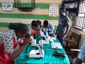
Kenny and Ona-ara with primary school pupils from Ibadan
Outreach for development
Being involved with DrosAfrica – which wants to use the power of Drosophila to empower scientists all across Africa and has plans underway to build a big new institute – and looking up initiatives of scientific capacity building, I didn’t quite realize until being there how relevant outreach and science communication is in this context too. I always thought that there was so much to build for the research practice per se, that I didn’t realize I was putting outreach in a “bonus” category. But the way outreach is intricately involved in research is truly universal and outreach shouldn’t be neglected by the pressure of building core resources in labs. After all, we should spark interest in the young and spread science to society to help strengthen the developing scientific structures. So, I would like to encourage professional science communicators to pay it forward and embark on similar exchanges and visits too. Take your expertise where it can be so valuable and I guarantee you will walk home having learned a lot of new things too. I wrote specifically about my school visit experience in the EMBL School Ambassador website and you can check the EMBL-Nigeria pen pal project I started with my colleague Rafael Galupa in our blog.
Combating imposter syndrome
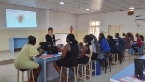
Me teaching in the Bingham workshop
As a PhD student, I strongly felt imposter syndrome when I was invited to train and teach, and that it was a big responsibility and investment for the little amount of knowledge I felt I could pass on. But I realized two things:
Even if you have less experience than a professor, you have a relatability factor that has great power speaking to young people like you.
I didn’t realize how many tools I have gathered from different experiences, and I was surprised to be able to go back and pick them up to share – such as a model used for a school visit or a draft structure a friend once sent me for a motivation letter I had to write. For me this underlined the interconnection of my life experiences and reminded me the hidden value of subtle things or experiences you don’t think about often anymore.
I also learned that the most effective way to contribute to scientific capacity building is to collaborate – meaning visiting, sharing expertise, sharing grants together and sharing or exchanging students. Finally, even if I am shy to admit it, I must say that I left Germany with the mindset that I was going there to teach. I was excited about this new experience, but I only realized after I arrived, that I was also going to learn A TON. And, for sure, I brought more with me than I left there. I swear that this did not come from arrogance but from naivety. If I had focused earlier on how much I would also learn, maybe I would have worried less about my imposter syndrome.
A final message
“Nothing in life is to be feared, it is only to be understood. Now is the time to understand more, so that we may fear less.” Marie Curie
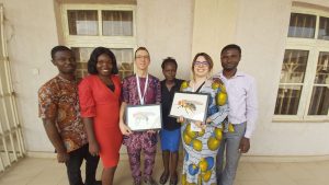
Kenny, Bolaji, Alex, Adeola, me & Ona-ara, at our last day in the workshop in Bingham, Abuja.
Oda-aro in Yoruba means good night, but in my heart it means seen you soon. I miss the warm friends I made there, I miss their uncontained joy, their spontaneity and generosity, their wise and serene takes on life (which actually brought me closer to religion), I miss the colourful attires in the street and the feeling like I time travelled to summer in February. I miss the hawks which flew around the UI campus and the hazy sky. I try to make it up with listening to Nigerian music almost every day and staying in touch.
When I told people around me I was going to Nigeria, a country where the travel security advice is quite grim, I was met with so much resistance and doubt – it cannot be overstated. You hear not only about petty crime or terrorism but also about regular kidnappings, and the advice not to travel by road or be out in the dark makes one realize security things we take for granted, at least I did in my quiet Heidelberg home. And yes, I was hesitant and apprehensive, but I trusted my guts and my values: I deeply believe that science is global and has to be shared. And I put my actions where my words were: I went. It was crucial for me that I had the support of DrosAfrica and that I trusted my host – who was incredible – and this is something you should take into account too. Everything went fine, but I lived a very controlled life: I never left campus or walked around after dark without company; for the outreach, we visited gated schools with security; our accommodation was chosen strategically; between Ibadan and Abuja, we took a plane instead of the road. Indeed, I heard of the precautions people take in their daily life in regards to which cabs they get into; I heard a testimony in a church service about a kidnapping that fortunately ended well. But the people I met live regular lives which didn’t seem to me to be overwhelmed by these issues. So although we had no issues, I also confirmed that indeed, the travel advice is based on threats that do exist. This for me presents an interesting paradox: if we haven’t been somewhere, we can only rely on the pictures the news gives us. But if you take precautions, that shouldn’t be a reason to stay home, because there is so much life behind the news, and even I speak humbly from a miniscule experience of 2 weeks. So much warmth, so much culture, so much diversity, news ways of living and thinking and looking at science and life and on and on…
If you read this and you are torn, please go. Prepare, take cautions, but go!
Nature in UI
This visit had the support of DrosAfrica, for which I am very grateful. In particular, thanks to Isa Palacios for always being there. ELLS and EMBL Comms supported the school visits – thanks Eva and Verena! I am grateful to my supervisor Justin Crocker for being supportive and to several colleagues (in and out EMBL) who shared material, insight, and especially those who shared encouragement.
A postdoctoral position is available for an initial period of 2 years at the “Stem Cell and Brain Research Institute” (www.sbri.fr) in the lab of Dr Olivier Raineteau.
The project follows two recent studies from the team demonstrating transcriptional heterogeneity of postnatal neural progenitors (Donega et al., Cell Report, 2018), and their amenability to pharmacological manipulation (Azim et al., Plos Biology, 2017). It aims at using transcriptomic, as well as fate-mapping approaches to study neural progenitors (endogenous or induced) and their capacity to produce define cell types, including distinct neuronal subtypes. The project involves close collaborations with the laboratories of Denis Jabaudon (CMU, Geneva) and Christophe Heinrich (within the SBRI institute).
We are seeking highly motivated candidates with a PhD in biological sciences with a strong interest in developmental neurosciences. The candidate will have an expertise in fate mapping approaches. Previous experience with bio-informatics analysis (e.g. single cell RNA-Sequencing analysis, R Studio, Seurat) is also required.
Environment: The team has a long-standing expertise in the field of postnatal forebrain neurogenesis and CNS repair. It is part of the Stem cell and Brain Research Institute (SBRI), and exciting international multidisciplinary research centre. The working languages are French and English.
Job information
Closing date: 15/06/2020
Employment start date: 01/09/2020
Institution: Inserm U1208
Department: Stem cell and Brain Research Institute
Contact Information
Dr Olivier Raineteau Stem Cell and Brain Research Institute
INSERM U1208
Applicants should send their CV, a motivation letter, 3 references (name, e-mail, address, phone no.) before the 15th of June to: olivier.raineteau@inserm.fr
Selected publications :
Donega V, Marcy G, Lo Giudice Q, Zweifel S, Angonin D, Fiorelli R, Abrous DN, Rival Gervier S, Koehl M, Jabaudon D, Raineteau O (2018) Transcriptional Dysregulation in Postnatal Glutamatergic Progenitors Contributes to Closure of the Cortical Neurogenic Period. Cell Rep. 22(10):2567-2574
Azim K, Angonin D, Marcy G, Pieropan F, Rivera A, Donega V, Cantu C, Williams G, Berninger B, Butt AM, Raineteau O (2017). Pharmacogenomic identification of small molecules for lineage specific manipulation of subventricular zone germinal activity. Plos Biology; 15(3):e2000698.
Azim K, Hurtado-Chong A, Fischer B, Kumar N, Zweifel S, Taylor V, Raineteau O (2015) Transcriptional Hallmarks of Heterogeneous Neural Stem Cell Niches of the Subventricular Zone. Stem Cells. 33(7):2232-42
Social media gets a hard time these days, and in some instances rightly so. For the scientific community however, it’s a powerful platform for informal science communication and fruitful collaborations. On Twitter for example, the developmental biology community shares news, discusses research, celebrates publications and commiserates grant rejections. It can be a supportive, informative and fun place that highlights life at and beyond the bench. With 13.9k and 15.8k followers respectively, Development and the Node use Twitter to interact with this community, promoting their content as well as engaging with authors and readers.
While Twitter is our go-to, it isn’t used everywhere and The Company of Biologists (the not-for-profit publisher of Development) has been focusing its efforts on audiences that can’t be reached through the Twittersphere. One such audience is in China, where different social media platforms are used, and manuscript submissions and readership figures have been steadily increasing. So, last summer we launched a Chinese WeChat channel.
Social media in China
WeChat was developed and released by Tencent in 2011 and has since become one the most popular social media applications in China. Want to search for news articles? Use WeChat. Talk to friends? Use WeChat. Order a takeaway? Use WeChat. Pay for a meal before you even arrive at the restaurant? Use WeChat. Figures from the end of 2019 indicate that WeChat’s monthly users have increased to a whopping one billion.
(L) The Company of Biologists’ WeChat feed; (R) The first post on the account included introductions from the company’s Board of Directors.
Science on WeChat
Much like on Twitter, a lot of scientific discussion happens on WeChat, and many Western scientific organisations already have WeChat channels to promote their work. Since the launch of our WeChat channel, we’ve accumulated a following edging towards 600 and this number is continually growing. Given our relatively low profile and lack of a physical presence in China, we’re pleased with this growth and are excited to build a community of engaged Chinese researchers.
We publish articles on the channel once a week that highlight some of the many thing The Company of Biologists does, be it the latest research, interviews, posts from the Node, Company announcements, grant information, Workshops, Meetings – there’s a lot to share. We’re also running a series providing advice to support Chinese authors in their publishing experience with us. Subjects in this series covered so far include a video about how to frame a research question, the process of submitting to all five of The Company of Biologists’ journals and top tips on promoting research after publication.
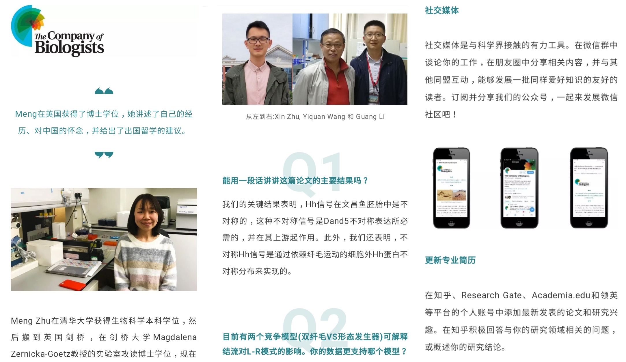
Screenshots from three articles published on WeChat: (L) an interview with a preLighter; (middle) a Q&A with first authors; (R) tips on how to promote research.
We’re fortunate to work with a supportive agency who assist us with the translations of our articles. Once the articles are published in local time, users can share content that they find interesting in WeChat groups or tap the ‘Wow’ button to promote the article in their own networks.
The future
Our main aim this year is to establish a working group of Chinese early-career researchers. We’d invite members of this group to work on the channel with us, which could be in the form of guest posts, checking the translations of our content, or providing insights into relevant topics. If you or anyone you know would be interested, please do get in touch – simply email wechat@biologists.com.
 (2 votes)
 (No Ratings Yet)
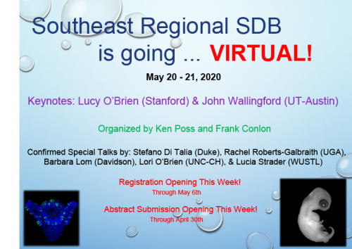


 (13 votes)
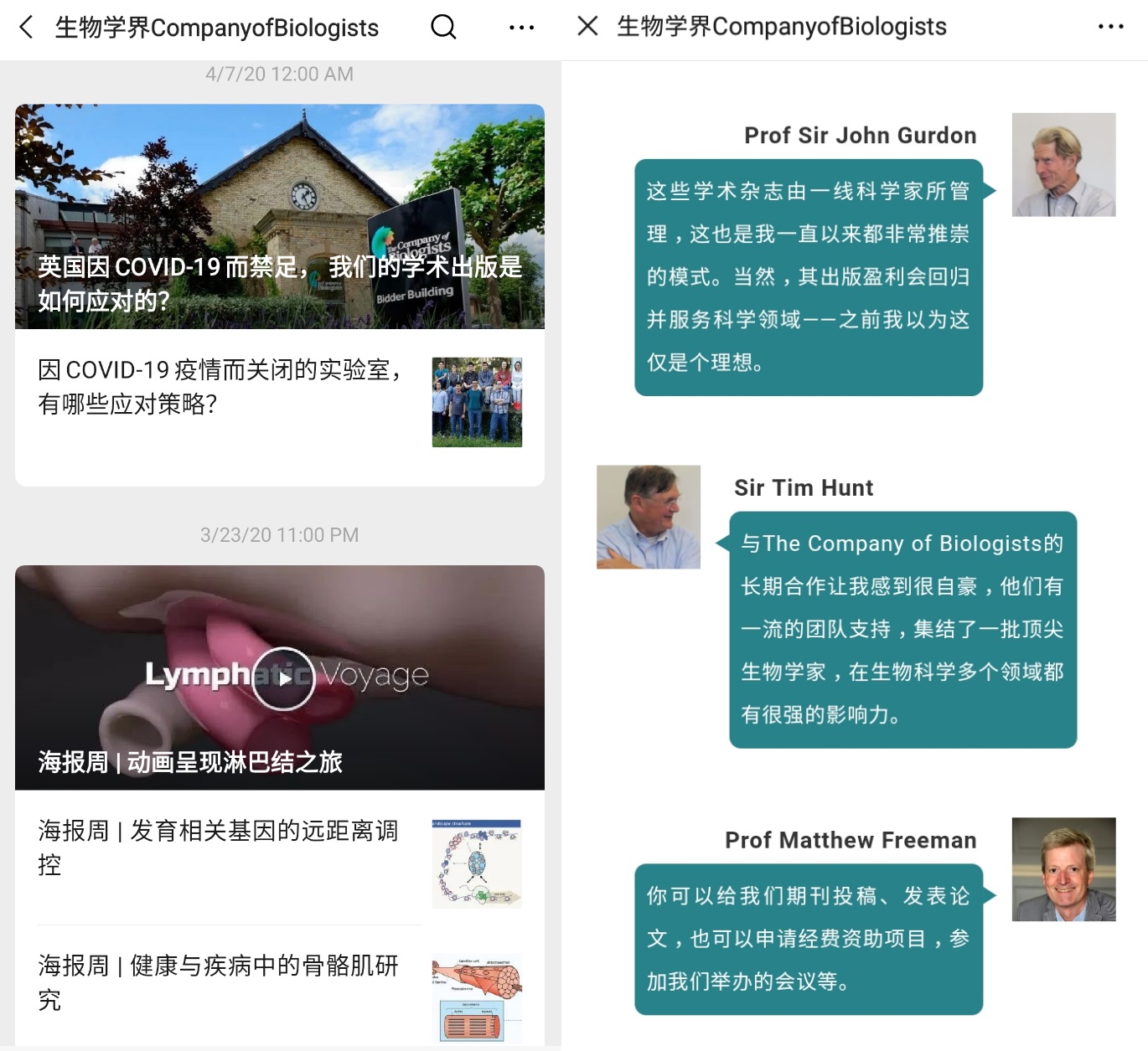
 If you have WeChat, give us a follow!