We are looking for highly motivated individuals who share our passion for science and would like to work in a friendly and collaborative environment.
Fully funded PhD student and Postdoc positions are available in the laboratory of Dr. Peng Huang in the Department of Biochemistry and Molecular Biologyat the University of Calgary, Canada.We use zebrafish as a model system to understand how tissue patterning is achieved and how tissue integrity is maintained. We study the spinal cord patterning to understand how different cell signaling pathways (Hedgehog and Notch signaling) interact during cell fate specification. We also study how non-muscle cells (e.g., tendon fibroblasts and muscle progenitor cells) contribute to muscle development, degeneration and regeneration. For more information about the lab and our recent publications, please visit: https://people.ucalgary.ca/~huangp/index.html
PhD student candidates should have a BS or MSc in Molecular Biology, Genetics, Developmental Biology or a related discipline, a strong academic background, good English skills and an enthusiasm for research. Previous lab experience with genetic model organisms is preferred but not required. Excellent written and verbal communication skills are critical.
Postdoc candidates should have a PhD in Developmental Genetics or a related discipline, excellent molecular biology skills, and a strong interest in developmental biology. Excellent written and verbal communication skills are critical. The candidate must have a track record of academic success as evidenced by peer-reviewed publications, awards and scholarships.
To apply, please send a cover letter summarizing previous research experiences and future goals and the CV with names of 2-3 references to Peng Huang, peng.huang@gmail.com with the subject line “PhD Student Position” or “Postdoc Position”.
Calgary, Canada’s fastest growing major city, is vibrant and multicultural with a population of more than 1.2 million. Situated near the Rocky Mountains, Banff National Park and Lake Louise, Calgary offers great quality of life and outstanding recreational activities.
A postdoctoral position is available in the group of Elke Ober at DanStem. The team studies fundamental mechanisms of tissue morphogenesis in the liver. The aim is to understand how different cell types communicate with each other and how the resulting cell behaviours establish an organ with a functional tissue architecture and of the correct size. To achieve this, the group utilises zebrafish genetic tools and state-of the-art molecular and cell biology techniques, such as mRNA sequencing, genome editing, confocal and light-sheet microscopy. This research project will employ a multi-disciplinary approach to understand how liver size is controlled combining zebrafish models of liver growth with human in vitro models. Starting date for this position is 1 May 2020, or upon agreement with the chosen candidate.
Institute:
The Novo Nordisk Foundation Center for Stem Cell Biology – DanStem is located at the University of Copenhagen. DanStem addresses basic research questions in stem cell and developmental biology and has activities focused on the translation of promising basic research results into new strategies and targets for the development of new therapies for cancer and chronic diseases such as diabetes and liver failure. Find more information about the Center at http://danstem.ku.dk/.
Job description:
We are seeking a highly motivated and ambitious postdoctoral candidate with a strong background in cell, developmental and/or stem cell biology to join our team. The candidate will investigate the molecular and cellular mechanisms controlling start and termination of organ growth in unique zebrafish liver mutants and human in vitro models of hepatic stem cell differentiation using e.g. advanced live cell imaging and transcriptomic approaches. The position is for 2 years with a possible extension.
Qualifications:
Candidates must hold a PhD degree in cell/developmental/stem cell biology or similar, with a track-record of successful scientific work.
Candidates should have a strong background in zebrafish genetics, cell biology and/or live cell imaging and advanced quantitative image analysis.
Previous experience in bioinformatics, next-generation sequencing and/or human stem cell differentiation are considered an advantage.
Good English communication skills, both oral and written, are prerequisite for the successful candidate.
Terms of employment:
The employment is planned to start 1 May 2020 or upon agreement with the chosen candidate. The full-time position is for 2 years with possiblity of extension. The terms of employment are set according to the Agreement between the Ministry of Finance and The Danish Confederation of Professional Associations or other relevant professional organization. The position will be at the level of postdoctoral fellow and the basic salary according to seniority. Currently, the salary starts at 34.360 DKK/approx. 4,.590 Euro (October 2019-level). A supplement could be negotiated, dependent on the candidate´s experiences and qualifications. In addition a monthly contribution of 17.1% of the salary is paid into a pension fund.
Non-Danish and Danish applicants may be eligible for tax reductions, if they hold a PhD degree and have not lived in Denmark the last 10 years.
The position is covered by the “Memorandum on the Job Structure of Academic Staff at Universities” of 11 December 2019.
Questions:
For further information contact Associate Professor Elke Ober, elke.ober@sund.ku.dk.
Application Instruction:
The application must be submitted in English, by clicking on “Apply online” below. Only online applications will be accepted.
The application must include:
Cover letter detailing the basis on which the applicant scientific qualifications meet the requirements for this position.
Curriculum vitae.
List of references (full address, incl. email and phone number)
Diplomas – all relevant certificates.
List of publications
Deadline for applications: 5 March 2020, 23.59pm.
The further process:
After the expiry of the deadline for applications, the authorized recruitment manager selects applicants for assessment on the advice of the Appointments Committee. All applicants are then immediately notified whether their application has been passed for assessment by an expert assessment committee. Selected applicants are notified of the composition of the committee and each applicant has the opportunity to comment on his/her assessment. You may read about the recruitment process at http://employment.ku.dk.
The applicant will be assessed according to the Ministerial Order no. 242 of 13 March 2012 on the Appointment of Academic Staff at Universities.
University of Copenhagen wish to reflect the diversity of society and welcome applications from all qualified candidates regardless of age, disability, gender, nationality, race, religion or sexual orientation. Appointment will be based on merit alone.
We are looking for a curious, enthusiastic and creative team member for new exciting projects in our lab (postdoc or staff scientist). The Fuhrmann Laboratory investigates the role of signaling pathways (Wnt, TGFbeta, Hippo, Hedgehog) during ocular development and regeneration in mouse. We investigate the cellular and molecular mechanisms regulating early eye patterning, RPE development and closure of the optic fissure during formation of the optic cup. We determine how signaling pathways control regeneration of the retinal pigment epithelium (RPE) in the adult eye. The lab is located in the Vanderbilt Eye Institute and is affiliated with the Cell and Developmental Biology Dept. at Vanderbilt University in Nashville, TN (https://www.vumc.org/fuhrmannlab/).
We are seeking candidates with a strong motivation in applying advanced imaging techniques and functional physiology techniques, and a solid background in cell biology, biochemistry and tissue culture paradigms to study aspects of ocular development and/or regeneration in mouse. Successful candidates must have a recent PhD in Life Sciences or equivalent with at least one first-author publication from their graduate work. We look forward to a talented and highly motivated team member. Candidates with less than 2 years of postgraduate work are specifically encouraged to apply. To apply, email a brief cover letter describing research accomplishments and future research goals, current CV with list of publications, and contact information for 3 professional references to:
Welcome to our monthly trawl for developmental biology (and related) preprints.
This month’s trawl includes a veritable farm, with developmental studies of potatoes, beetroot, tomato, maize, wheat, pigs, cows and goats, plus many of the usual suspects. They were hosted on bioRxivandarXiv. Let us know if we missed anything. Use these links to get to the section you want:
Differences in mitochondrial activity trigger cell competition during early mouse development
Ana Lima, Gabriele Lubatti, Jörg Burgstaller, Di Hu, Alistair Green, Aida Di Gregorio, Tamzin Zawadzki, Barbara Pernaute, Elmir Mahammadov, Marian Dore, Juan Miguel Sanchez, Sarah Bowling, Margarida Sancho, Mohammad Karimi, David Carling, Nick Jones, Shankar Srinivas, Antonio Scialdone, Tristan A. Rodriguez
Local retinoic acid directs emergence of the extraocular muscle functional unit
Glenda Comai, Marketa Tesarova, Valerie Dupé, Muriel Rhinn, Pedro Vallecillo Garcia, Fabio da Silva, Betty Feret, Katherine Exelby, Pascal Dollé, Leif Carlsson, Brian Pryce, Francois Spitz, Sigmar Stricker, Tomas Zikmund, Jozef Kaiser, James Briscoe, Andreas Schedl, Norbert B. Ghyselinck, Ronen Schweitzer, Shahragim Tajbakhsh
Cell-type specific impact of glucocorticoid receptor activation on the developing brain
Cristiana Cruceanu, Leander Dony, Anthi C. Krontira, David S. Fischer, Simone Roeh, Rossella Di Giaimo, Christina Kyrousi, Janine Arloth, Darina Czamara, Silvia Martinelli, Stefanie Wehner, Michael S. Breen, Maik Koedel, Susann Sauer, Monika Rex-Haffner, Silvia Cappello, Fabian J. Theis, Elisabeth B. Binder
Hearts and kidneys from Cott-Solomon and Kuruvilla
Regulation of human development by ubiquitin chain editing of chromatin remodelers
David B. Beck, Mohammed A. Basar, Anthony J. Asmar, Joyce Thompson, Hirotsugu Oda, Daniela T. Uehara, Ken Saida, Precilla D’Souza, Joann Bodurtha, Weiyi Mu, Kristin W. Barañano, Noriko Miyake, Raymond Wang, Marlies Kempers, Yutaka Nishimura, Satoshi Okada, Tomoki Kosho, Ryan Dale, Apratim Mitra, Ellen Macnamara, Undiagnosed Diseases Network, Naomichi Matsumoto, Johi Inazawa, Magdalena Walkiewicz, Cynthia J. Tifft, Ivona Aksentijevich, Daniel L. Kastner, Pedro P. Rocha, Achim Werner
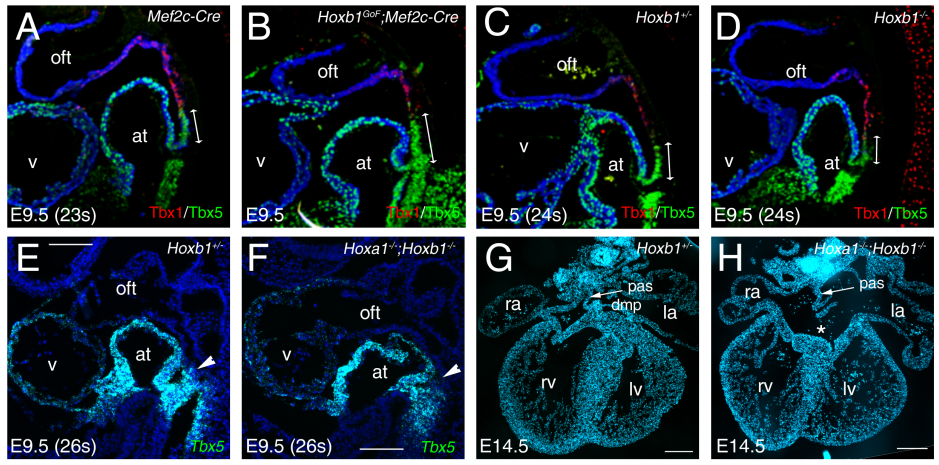
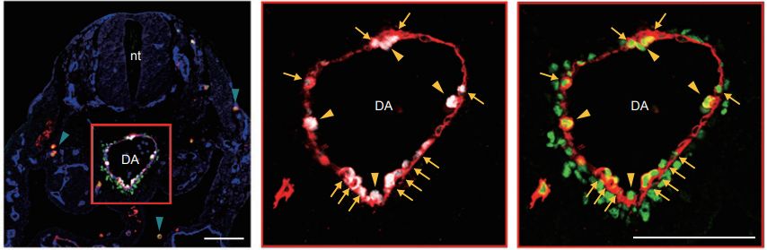
Hox-dependent coordination of cardiac progenitor cell patterning and differentiation
Sonia Stefanovic, Brigitte Laforest, Jean-Pierre Desvignes, Fabienne Lescroart, Laurent Argiro, Corinne Maurel-Zaffran, David Salgado, Christopher de Bono, Kristijan Pazur, Magali Théveniau-Ruissy, Christophe Béroud, Michel Pucéat, Anthony Gavalas, Robert G. Kelly, Stéphane Zaffran
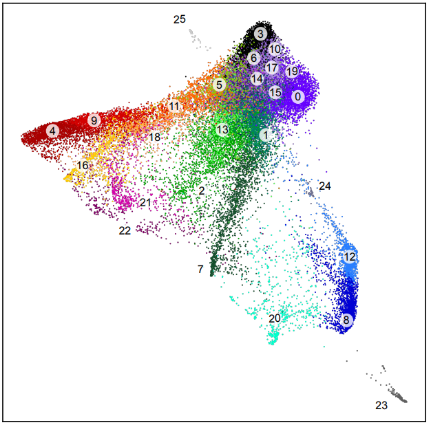
Single cell epigenomic atlas of the developing human brain and organoids
Ryan S. Ziffra, Chang N. Kim, Amy Wilfert, Tychele N. Turner, Maximilian Haeussler, Alex M. Casella, Pawel F. Przytycki, Anat Kreimer, Katherine S. Pollard, Seth A. Ament, Evan E. Eichler, Nadav Ahituv, Tomasz J. Nowakowski
Molecular and genetic regulation of pig pancreatic islet cell development
Seokho Kim, Robert L. Whitener, Heshan Peiris, Xueying Gu, Charles A. Chang, Jonathan Y. Lam, Joan Camunas-Soler, Insung Park, Romina J. Bevacqua, Krissie Tellez, Stephen R. Quake, Jonathan R. T. Lakey, Rita Bottino, Pablo J. Ross, Seung K. Kim
Sox2 modulation increases naïve pluripotency plasticity
Kathryn Tremble, Giuliano G. Stirparo, Lawrence E. Bates, Katsiaryna Maskalenka, Hannah T. Stuart, Kenneth Jones, Amanda Andersson-Rolf, Aliaksandra Radzisheuskaya, Bon-Kyoung Koo, Paul Bertone, José C. R. Silva
Neural stem cells traffic functional mitochondria via extracellular vesicles to correct mitochondrial dysfunction in target cells
Luca Peruzzotti-Jametti, Joshua D Bernstock, Giulia Manferrari, Rebecca Rogall, Erika Fernandez-Vizarra, James C Williamson, Alice Braga, Aletta Van den Bosch, Tommaso Leonardi, Agnes Kittel, Cristiane Beninca, Nunzio Vicario, Sisareuth Tan, Carlos Bastos, Iacopo Bicci, Nunzio Iraci, Jayden A Smith, Paul J Lehner, Edit I Buzas, Nuno Faria, Massimo Zeviani, Christian Frezza, Alain Brisson, Nicholas J Matheson, Carlo Viscomi, Stefano Pluchino
ATR expands embryonic stem cell fate potential in response to replication stress
Sina Atashpaz, Sara Samadi Shams, Javier Martin Gonzalez, Endre Sebestyén, Negar Arghavanifard, Andrea Gnocchi, Eliene Albers, Simone Minardi, Giovanni Faga, Paolo Soffientini, Elisa Allievi, Valeria Cancila, Angela Bachi, Oscar Fernandez-Capetillo, Claudio Tripodo, Francesco Ferrari, Andrés Joaquin López-Contreras, Vincenzo Costanzo
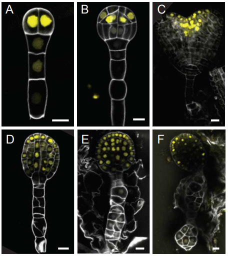
Maternal genome dominance in early plant embryogenesis
Jaime Alaniz-Fabián, Daoquan Xiang, Gerardo Del Toro-De León, Axel Orozco-Nieto, Peng Gao, Andrew Sharpe, Leon V. Kochian, Gopalan Selvaraj, Nathan Springer, Cei Abreu-Goodger, Raju Datla, C. Stewart Gillmor
COP1 destabilizes DELLA proteins in Arabidopsis
Noel Blanco-Touriñán, Martina Legris, Eugenio G. Minguet, Cecilia Costigliolo-Rojas, María A. Nohales, Elisa Iniesto, Marta García-León, Manuel Pacín, Nicole Heucken, Tim Blomeier, Antonella Locascio, Martin Černý, David Esteve-Bruna, Mónica Díez-Díaz, Břetislav Brzobohatý, Henning Frerigmann, Matías D. Zurbriggen, Steve A. Kay, Vicente Rubio, Miguel A. Blázquez, Jorge J. Casal, David Alabadí
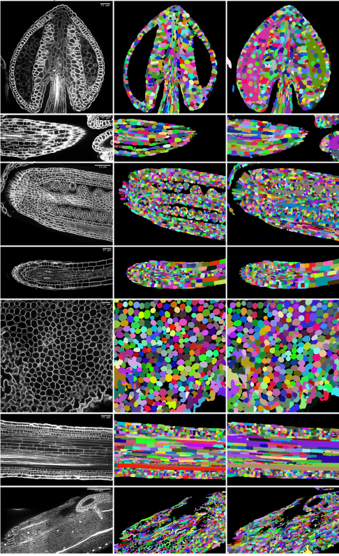
Accurate and Versatile 3D Segmentation of Plant Tissues at Cellular Resolution
Adrian Wolny, Lorenzo Cerrone, Athul Vijayan, Rachele Tofanelli, Amaya Vilches Barro, Marion Louveaux, Christian Wenzl, Susanne Steigleder, Constantin Pape, Alberto Bailoni, Salva Duran-Nebreda, George Bassel, Jan U. Lohmann, Fred A. Hamprecht, Kay Schneitz, Alexis Maizel, Anna Kreshuk
On the origin and evolution of RNA editing in metazoans
Qiye Li, Pei Zhang, Ji Li, Hao Yu, Xiaoyu Zhan, Yuanzhen Zhu, Qunfei Guo, Huishuang Tan, Nina Lundholm, Lydia Garcia, Michael D. Martin, Meritxell Antó Subirats, Yi-Hsien Su, Iñaki Ruiz-Trillo, Mark Q. Martindale, Jr-Kai Yu, M. Thomas P. Gilbert, Guojie Zhang
A chemical tool for improved culture of human pluripotent stem cells
Laurence Silpa, Maximilian Schuessler, Gu Liu, Marcus Olivecrona, Lucia Groizard-Payeras, Elizabeth Couper, Carole J. R. Bataille, Mark Stevenson, Len W. Seymour, Stephen G. Davies, William S. James, Sally A. Cowley, Angela J. Russell
CRISPR-Cas13d induces efficient mRNA knock-down in animal embryos
Gopal Kushawah, Joaquin Abugattas-Nuñez del Prado, Juan R. Martinez-Morales, Michelle DeVore, Javier R. Guelfo, Emry O. Brannan, Wei Wang, Timothy J. Corbin, Andrea M. Moran, Alejandro Sánchez Alvarado, Edward Málaga-Trillo, Carter M. Takacs, Ariel A. Bazzini, Miguel A. Moreno-Mateos
The Developing Human Connectome Project: typical and disrupted perinatal functional connectivity
Michael Eyre, Sean P Fitzgibbon, Judit Ciarrusta, Lucilio Cordero-Grande, Anthony N Price, Tanya Poppe, Andreas Schuh, Emer Hughes, Camilla O’Keeffe, Jakki Brandon, Daniel Cromb, Katy Vecchiato, Jesper Andersson, Eugene P Duff, Serena J Counsell, Stephen M Smith, Daniel Rueckert, Joseph V Hajnal, Tomoki Arichi, Jonathan O’Muircheartaigh, Dafnis Batalle, A David Edwards
Curious about novel lymphatic cell types in the meninges?
Excited to explore links between non-neuronal cells, brain activity, and sleep using zebrafish as a model system?
Then consider the Research Associate Position NOW available in the Rihel lab at University College London (Deadline Feb 22, 2020).
Details: Full Time, Grade 7.
Salary (inclusive of London allowance) £35,965 – £43,470 per annum
Duties and Responsibilities: The post-holder will research the genetic and cellular mechanisms of zebrafish sleep in the laboratory of Dr. Jason Rihel. They will join a team of researchers working on various aspects of sleep behaviours in zebrafish, with a focus on the interaction between the brain and a novel lymphatic cell type in the meninges. This position is funded for a period of 24 months in the first instance and will start immediately.
Key Requirements: Applicants must have a PhD in Neurobiology, Genetics, or a related field, together with significant experience in genetics, molecular biology, in vivo imaging, and behavioral neuroscience of a model organism such as zebrafish.
IRIBHM is a research institute of the Medical School of the Free University of Brussels (Université Libre de Bruxelles, ULB). The institute offers an internationally prominent research environment in molecular biology and life sciences, that engage different topics that span receptor pharmacology and new therapeutic targets discovery, early embryonic development, neurobiology, stem cells and cancer. The institute has trained over the years a number of talented young scientists both at the graduate and postdoctoral levels.
In order to expand internationally and keep rising its level of excellence, the IRIBHM (https://iribhm.org/) launches an international graduate programme in order to prepare the future leaders in biomedical sciences. At least 2 PhD scholarships are available. The successful candidates will have the opportunity to work in a warm and stimulating research environment aligned with the highest international standards.
As the administrative center of the European Union, Brussels is a perfect location for an International PhD programme. In addition, the city shows an active cultural life and is hosting most nationalities from all over the world.
We look forward to welcoming you in Brussels!
Requirements:
Diplomas and degrees equivalent to a European Union Master’s degree, which includes project work summarized in a written “small thesis”
Two referees willing to provide letter of recommendations
Excellent knowledge of English
Excellent interpersonal and organisational skills
Closing date for CV and letters submission: June 30.
Accepted candidates may start research projects as early as November 2020
The Lindstrom Laboratory in the Broad CIRM Center and Department of Stem Cell Biology and Regenerative Medicine, Keck School of Medicine of USC, has an opening for a postdoctoral scholar to study the transcriptional regulation underpinning the formation of functional cell-types in the kidney from stem and progenitor populations in the developing embryo. Applicants are expected to have a Ph.D. in a discipline of biological sciences or expect to complete it by the date of hire. Preference will be given to candidates with research experiences in developmental biology and genetics, stem cells and tissue engineering. However, all interested candidates are encouraged to apply.
The Lindstrom lab focuses on understanding how cells coordinate their behavior during development in order to form complex tissues that perform critical physiological functions. This enables us to make important insights into how birth defects occur, form strategies to replicate developmental process in stem-cell based systems, and to allows us to develop new regenerative therapies. Our main focus is on the kidney, where we apply state-of-the-art technologies such as single-cell omics, confocal imaging, and organoid models to delineate mechanisms controlling its formation and function. Additional information can be found at https://lindstromlab.usc.edu.
We are seeking a highly motivated and passionate individual wishing to take a leading role in developing their research projects while also contributing and gaining experience in writing grants and fellowship applications. The successful candidate will be expected to also oversee the work of other lab personnel and coordinate their research with fellow lab members. The candidate should anticipate engaging in highly collaborative and cross disciplinary research.
The Broad CIRM Center and Department of Stem Cell Biology and Regenerative Medicine, Keck School of Medicine of USC (http://keck.usc.edu/stemcell/) offers an exciting and nurturing research environment with great opportunities to conduct cutting edge science, use of latest equipment, and postdoctoral training and support. A weekly seminar series invites leading scientists in the stem / regenerative / synthetic / and developmental sciences (https://stemcell.keck.usc.edu/events/) and at regular intervals postdoctoral scientists present their data to the department.
When applying, please submit the required documentation online. A cover letter should highlight why you are interested in joining the lab, projects that you may wish to develop, and how your existing experience will synergize with our goals (2 pages max). In addition, please provide the names of a minimum of two references.
The Seifert lab in the Department of Biology at the University of Kentucky (UK) is seeking an exceptionally creative postdoctoral researcher to join our group. The successful candidate will lead an effort to isolate stable embryonic stem cell lines and/or derive iPSC lines for genomic modification and subsequent generation of transgenic spiny mice. This project will provide freedom to work on additional projects related to early embryonic development in spiny mice or projects investigating the cellular and molecular basis for complex tissue regeneration in mammals. To pursue these lines of research we maintain an active breeding colony of spiny mice (Acomys cahirinus) at UK.
Ideal candidates will have a strong background in developmental biology, experience working with ESCs and iPSCs and mouse transgenesis, experience working with model or non-model organisms, expertise in microscopy and comfortability with bioinformatics. While this is a funded position, postdocs in the Seifert lab are strongly encouraged to develop their own projects and external funding portfolios as a pathway toward independence. Salary follows NIH guidelines for postdoctoral researchers. Informal inquiries by email are strongly encouraged. For additional information visit: http://www.ashleyseifert.com/opportunities.html
Review of applications will begin on a rolling basis and will continue until the position has been filled. Ideal start date is Spring 2020. Candidates will have completed their Ph.D. prior to starting the position but need not have defended their dissertation prior to applying. Applicants should submit their application materials through the UK Jobs site (upload under Specific Request 1) at http://ukjobs.uky.edu/postings/251962. In addition, applicants should send a single pdf document to Ashley Seifert (awseifert@uky.edu) that includes their CV, names of three references, and a 1-2-page synopsis of their current research interests and how these complement our overall research program.
The Department of Biology houses a strong group of research labs interested in regenerative and stem cell biology using a diverse array of animal models (e.g., spiny mice, salamanders, planarians, lampreys, zebrafish) and in vitro systems. Together, these labs create a vibrant atmosphere to pursue interdisciplinary projects across comparative genomics, developmental, regenerative and evolutionary biology.
The University of Kentucky is an Equal Opportunity Employer and encourages applications from veterans, individuals with disabilities, women, African Americans, and all minorities.
The Department of Biology at the University of Kentucky (UK) is seeking applications for the Thomas Hunt Morgan Postdoctoral Fellowship in the Seifert Lab. This fellowship provides exceptional PhDs an opportunity to conduct independent research in any aspect of regenerative biology. The Fellows are funded for two years through the College of Arts and Sciences and provided support to develop their own projects while they build external funding portfolios as a pathway toward independence. The Seifert lab uses an array of animal models to study complex tissue regeneration and maintains large, active breeding colonies of spiny mice (Acomys cahirinus) and salamanders at UK.
The Department of Biology houses a strong group of research labs interested in regenerative and stem cell biology using a diverse array of animal models (e.g., spiny mice, salamanders, planarians, lampreys, zebrafish) and in vitro systems. Together, these labs create a vibrant atmosphere to pursue interdisciplinary projects incorporating cutting edge science in regenerative, developmental, and evolutionary biology.
Review of applications will begin on a rolling basis and will continue until a candidate is selected. Candidates will have completed their Ph.D. prior to starting the position but need not have defended their dissertation prior to applying.
Interested applicants should apply online at http://ukjobs.uky.edu/postings/251988. Applicants should submit their application materials through the UK Jobs site (upload under Specific Request 1) as well as send a single pdf document to Ashley Seifert (awseifert@uky.edu). Application materials submitted through UK Jobs as well as submitted to Dr. Seifert should include a CV, names of three references, and a 2-page statement of proposed research interest. Informal inquiries by email are strongly encouraged. More information is available here: http://www.ashleyseifert.com/opportunities.html
In this episode exploring great ideas in genetics, we’re discovering our inner fish – finding out whether we really do go through a fishy phase in the womb, and looking at the legacy of Tiktaalik, the first fish to walk on land.
Born in 1834, Ernst Haeckel was a German zoologist with a flair for illustration – and a knack for creating incredibly detailed and widely shared scientific images. But do his infamous embryo drawings really show the true picture of early development?
Haeckel thought that we went through a ‘fish’ stage in the womb because our embryos appear to have gills during early development. Although his theory that ‘ontogeny recapitulates phylogeny’ has subsequently been shown to be incorrect, we now know there is a close connection between development and evolution, or ‘evo-devo’ as it’s sometimes known.
In short, our evolutionary history is written in our developmental genes, and it’s a history that we can trace right the way back to the very first vertebrates. The best example of this is Tiktaalik – our oldest ‘fishapod’ ancestor that forms the missing link between fish and land-dwelling tetrapods.
If you enjoy the show, please do rate and review on Apple podcasts and help to spread the word on social media. And you can always send feedback and suggestions for future episodes and guests to podcast@geneticsunzipped.com Follow us on Twitter – @geneticsunzip
 (No Ratings Yet)
 (1 votes)


 In this episode exploring great ideas in genetics, we’re discovering our inner fish – finding out whether we really do go through a fishy phase in the womb, and looking at the legacy of Tiktaalik, the first fish to walk on land.