Roles of metabolism in the developmental origins of health and longevity
A postdoctoral research position funded by the Wellcome Trust is available in the laboratory of Dr. Alex Gould at the Francis Crick Institute in London. The lab works on the mechanisms by which dietary nutrients during development can have profound long-term effects upon adult metabolism and lifespan. We are looking for a highly motivated researcher with experience in molecular biology and/or metabolism. The successful applicant will be able to choose from several Drosophila and mouse models that have been established in our lab (PMID: 21816278, PMID: 26451484, PMID: 29123106, PMID: 29515102 and unpublished). They will be exposed to a range of techniques including genetics, molecular biology, confocal microscopy, biochemistry, metabolomics as well as mass spectrometry imaging (PMID: 22246326, PMID: 26451484). Access will be provided to state-of-the-art facilities in advanced light and electron microscopy, metabolomics and single-cell sequencing. Examples of other projects ongoing in the lab can be seen at: www.agouldlab.com www.crick.ac.uk/research/labs/alex-gould
Applications are now open for this year’s Gene Regulatory Networks for Development at The Marine Biological Laboratory in Woods Hole, USA from October 13-26. The application deadline is July 17th. The course is for graduate students, postdoctoral researchers, staff scientists and faculty members. It focuses on using experimental data and computational modeling to analyze gene regulatory networks controlling development.
This unique course is an intense and always interesting experience and has had great reviews in all of the previous years. Students will meet with renowned experts in the field for an in-depth treatment of experimental and computational approaches to GRN science. Through lectures, highly interactive discussions, and group projects we will explore the GRN concept and how it can be applied to solve developmental mechanisms in various systems and contexts. Topics include structural and functional properties of networks, GRN evolution, cis-regulatory logic, experimental analysis of GRNs, examples of solved GRNs in a variety of developmental contexts, and the computational analysis of network behaviour by continuous and discrete modelling approaches.
Scott Barolo – U. of Michigan (co-director)
James Briscoe – Francis Crick Institute
Martha Bulyk – Harvard U.
Ken Cho – UC Irvine
Doug Erwin – Smithsonian Institution
Robb Krumlauf – Stowers Institute
Arthur Lander – UC Irvine
Bill Longabaugh – Institute for Systems Biology
Lee Niswander – U. of Colorado
Isabelle Peter – Caltech (co-director)
Alexander Stark – IMP Vienna
Zeba Wunderlich – UC Irvine
We are looking to appoint a Research Technician who will provide support to a one-year Neuroblastoma UK-funded project entitled “Establishment of an in vitro model of neuroblastoma initiation using pluripotent stem cell differentiation”. The project aims to dissect the cellular and molecular basis of neuroblastoma initiation using human pluripotent stem cell (hPSCs) differentiation and hPSC lines engineered to ectopically overexpress common neuroblastoma-associated oncogenes.
You will join a research team under the guidance of Dr Anestis Tsakiridis, providing support for routine hPSC culture and differentiation, preparation of samples/analysis and carrying out molecular cloning/genetic modification of hPSCs. Appropriate training will be provided. This is an excellent opportunity to gain hands-on laboratory experience and to be part of a leading research team. Our group’s research aims to define the molecular basis of cell fate decisions during human embryonic development and determine how “altered” embryonic multipotent states drive tumourigenesis (https://www.tsakiridislab.com/).
Applicants must have a good honours degree or equivalent experience in a developmental/stem cell biology-related subject along with previous experience of working in a research laboratory. Familiarity with some/all of the following techniques is desirable: hPSC culture; RNA isolation; molecular cloning; mammalian cell transfection; quantitative real time PCR; immunostaining; fluorescence microscopy; flow cytometry. Applicants should also have an interest in stem cell and developmental biology.
To apply: visit the University of Sheffield job portal (https://www.sheffield.ac.uk/jobs/index)
Closing date: 7th August 2019
Expected start date: 1st September 2019
For more details/questions contact: a.tsakiridis@sheffield.ac.uk
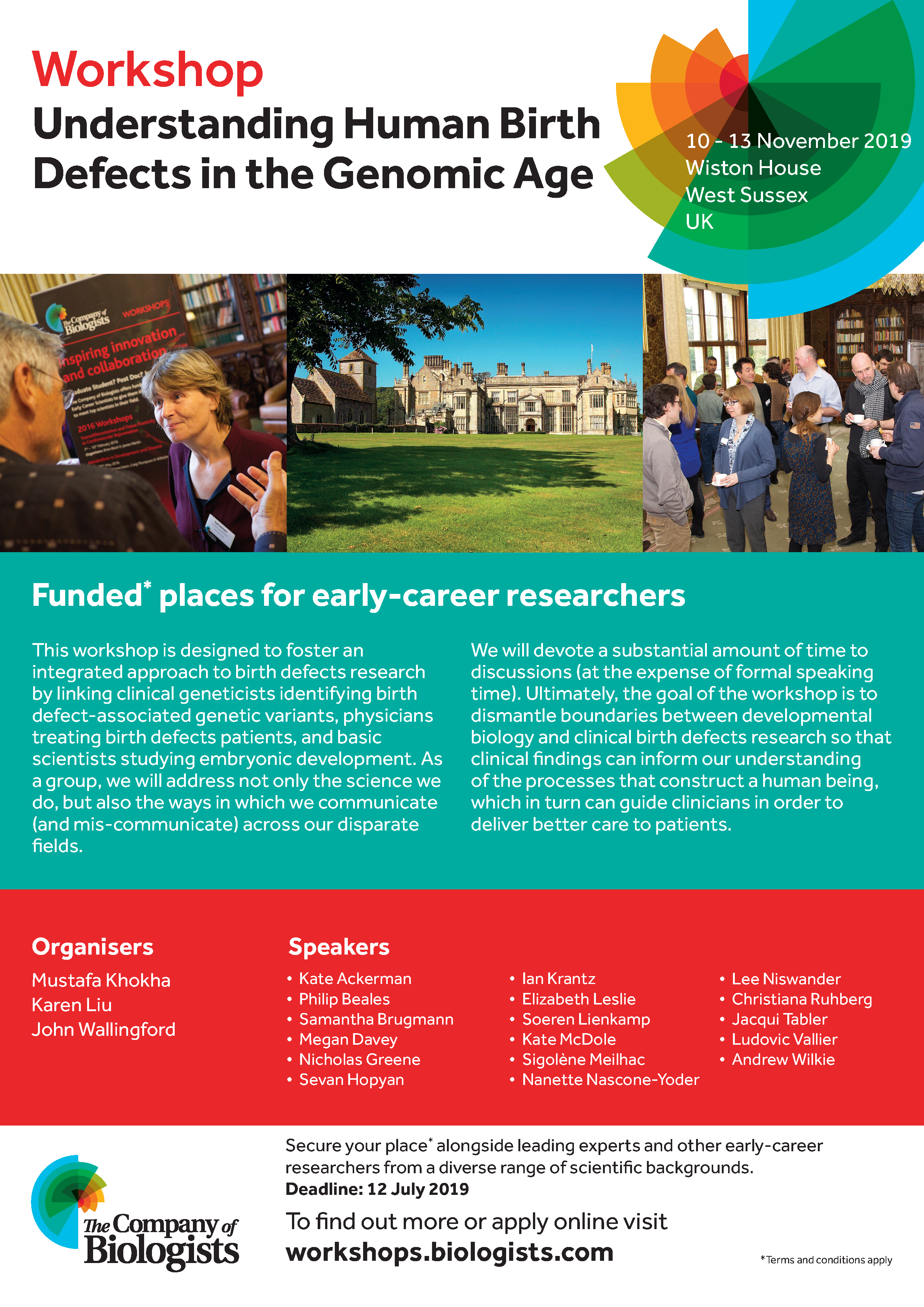
The Company of Biologists Workshops provide leading experts and early-career researchers from a diverse range of scientific backgrounds with a stimulating environment for the cross-fertilisation of interdisciplinary ideas. In November, experts will gather in the beautiful surroundings of Wiston House in West Sussex with the aim of ‘Understanding Human Birth Defects in the Genomic Age‘. Organised by Mustafa Khokha, Karen Liu and John Wallingford, the Workshop is an amazing opportunity to explore applied developmental biology.
There are around 10 funded places for early-career researchers available – a fantastic opportunity to share your research with leading scientists in an intimate setting.
Transcriptional autoregulation occurs when transcription factors bind their own cis-regulatory sequences, ensuring their own continuous expression along with expression of other targets. During development, continued expression of identity-specifying transcription factors can be achieved by autoregulation, but until now formal evidence for a developmental requirement of autoregulation has been lacking. A new paper in Development provides this proof with the help of CRISPR/Cas9 gene editing in the C. elegans nervous system. We caught up with the paper’s two authors: postdoc Eduardo Leyva-Díaz and his supervisor Oliver Hobert, Professor of Biological Sciences and HHMI Investigator at Columbia University, New York, to find out more about the work.
Oliver Hobert (L) and Eduardo Leyva-Díaz (R).
Oliver, can you give us your scientific biography and the questions your lab is trying to answer?
OH I started out investigating signal transduction for my PhD with Axel Ullrich and Gerhard Krauss in Germany, and then moved to the USA for my postdoc with Gary Ruvkun. In Gary’s lab, I started working with C. elegans on transcription factor regulation and specification of neuronal fates. In my own lab, we have continued to pursue our interest in understanding the molecular mechanisms that control the generation of diverse cell types in the nervous system. More recently, we are also becoming more and more interested in understanding how neuronal identity features are modulated by certain factors, such as environmental conditions or sexual identity.
And Eduardo, how did you come to work in the Hobert lab, and what drives your research today?
EL-D My fascination with science began in biology laboratory classes in high school, with a very dedicated and passionate teacher. Since then, I’ve been always attracted to genetics and molecular biology, and my first research experience as an undergraduate student was in Prof. Jose Luis Micol’s lab working on Arabidopsis thalianagenetics. Towards the time of my graduation, I became interested in the nervous system, specifically in learning and memory, although I have never really worked on that field. The one thing I was not interested in at all at that time was developmental neurobiology, but funnily enough, after my rotation in different labs at the Instituto de Neurociencias de Alicante, I was totally captivated by it, and devoted my next 6 years to studying mouse brain development in Guillermina Lopez-Bendito’s lab. After my thesis defense, I stayed for a few months in the lab and worked on a new research line aimed at reprogramming endogenous astrocytes into different projection neurons. With this experience in identity reprogramming and transcriptional regulation, I developed a deep interest in neuronal identity specification, particularly regarding the maintenance of neuronal features. The Hobert lab was then a clear perfect match, with C. elegans representing an excellent model system to study neuronal identity specification and maintenance.
When did you first become interested in transcriptional autoregulation? And given it has been known about for decades, why do you think it has taken so long to formally test its functional requirement?
OH & EL-D A key characteristic of several terminal selectors, identity-specifying transcription factors, is their role in the maintenance of neuronal identity, which is thought to be achieved by transcriptional autoregulation. However we, as well as others, had only inferred transcriptional autoregulation from the presence of binding sites of a transcription factor in its own genomic locus, and from genetic loss-of-function studies in which the activity of a transcription factor is removed and a loss of transcription of this locus is consequently observed. Formal proof for the functional relevance of autoregulation has been sparse, however. The advent of CRISPR/Cas9 technologies has been key to providing formal proof for this requirement, because it enabled us to disrupt autoregulation, but not other functions of a specific transcription factor. We could therefore precisely ask what it is that autoregulation actually does – and we came up with a surprise that we had not anticipated.
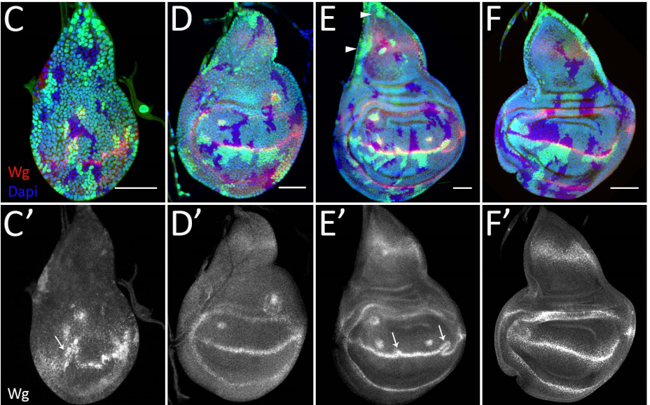
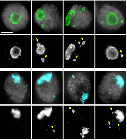
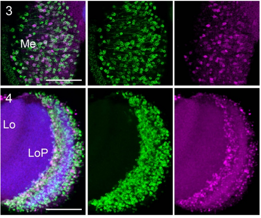
C. elegans embryo in which the che-1 locus has been tagged with gfp through CRISPR/Cas9 genome engineering. che-1::gfp expression can be observed in the bilaterally symmetric ASE neuron pair (ASEL + ASER) and their sister cells, which are in the process of undergoing apoptotic cell death.
Can you give us the key results of the paper in a paragraph?
OH & EL-D In this paper, we use CRISPR/Cas9 to remove a cis-regulatory motif from a cell identity-specifying transcription factor, showing that the disruption of transcriptional autoregulation leads to a failure to maintain the differentiated state of the cell. Upon regulatory motif mutation, we observe a gradual decrease in neuronal function and cell identity marker expression. This was an expected result that provided formal proof for the importance of identity-triggering transcription factors in maintaining the identity state of a cell. However, we also found that transcriptional autoregulation is not only required to maintain a specific cellular state, but is also required during development to amplify the expression levels of the autoregulating transcription factor to a critical threshold level in order to allow it to initiate expression of its target genes, which will define the differentiated state of the cell.
Do you think the early function in initiation of che-1expression is likely to be a general feature of autoregulation?
OH & EL-D In general, we think that if a gene can autoregulate it makes sense that this autoregulation is also used early in development. However, we have found in the literature examples of other autoregulating transcription factors for which maintenance relies on autoregulation, while the initial amplification is achieved by different means. Interestingly, this dual role of autoregulation, early amplification/late maintenance, seems to be modular and context dependent, since in some cases the autoregulation of other factors is only important early in development. Nonetheless, it does not seem far-fetched to propose that the functional duality of transcriptional autoregulation constitutes a widely used gene regulatory principle during animal development.
It does not seem far-fetched to propose that the functional duality of transcriptional autoregulation constitutes a widely used gene regulatory principle during animal development
When doing the research, did you have any particular result or eureka moment that has stuck with you?
EL-D For me, the eureka moment was when we realized about the function of transcriptional autoregulation in early development. We were very satisfied with the close correlation between che-1 expression and neuronal functional performance through the different developmental stages. But when we looked earlier, we were at first surprised by finding already low levels of che-1 expression in the embryo. Then we realized that it would only make sense if autoregulation also contributed to transcription factor initial amplification and, consequently, acquisition of the differentiated state.
And what about the flipside: any moments of frustration or despair?
EL-D Without any doubt, the moments of frustration and despair were at the very beginning of the project. Generating precise motif mutations in the che-1promoter was key for this story, and obtaining some of the cis-regulatory mutations took longer than expected. The application of CRISPR/Cas9 engineering to different projects was just becoming established in the lab at that point, and we were at the initial phase of standardization and protocol set up. Of course, we got our mutants, and the road was mostly paved after that.
So what next for you after this paper?
EL-D I am intensively working on a second project, where we are trying to understand how the expression of pan-neuronal genes is controlled. Neuronal identity is determined by the expression of neuron-type specific genes and pan-neuronal genes, which are shared by all neurons in the nervous system. We now know several examples about neuron-type specific gene regulation, but not that much about pan-neuronal genes. Previous work form the Hobert lab has shed some light into the how, and now I am trying to find the who, identifying key factors controlling pan-neuronal gene expression. And then, job hunting.
Where will this work take the Hobert lab?
OH This work will hopefully not present the endpoint of studying transcriptional autoregulation. While there’s plenty of evidence to suggest that positive autoregulation is a widespread phenomenon, we also know that some identity-specifying terminal selectors do not autoregulate, even though their expression is maintained throughout the life of a neuron. How does this work? In at least one other case, we also have reason to believe that there is negative autoregulation, in which a terminal selector dims down its own expression. We would love to understand how and why this is.
Finally, let’s move outside the lab – what do you like to do in your spare time in New York?
EL-D New York is an amazing place and I love to explore the city and its surroundings with my wife and friends. I especially enjoy discovering all the culinary options, and I try to take advantage of the different cultural activities that the city has to offer. I also like to stay active, running and playing different sports. Finally, I love to travel when possible, to discover new places or back to Spain to enjoy the weather, food, family and friends.
OH I don’t have much to add to this. New York is an amazing, dynamic and constantly changing place that leaves new things to discover even if one has lived in the city for a while.
In this episode we’re celebrating the actual birthday of the society – founded on the 25th June, 100 years ago – with past president, Nobel laureate and winner of the Genetics Society’s first centenary medal, Sir Paul Nurse.
To mark this auspicious day, the Genetics Society held a very special birthday party at the John Innes Centre in Norwich. First we were treated to a wonderful exhibition of artefacts from the society’s history, including co-founder William Bateson’s original microscope and some fascinating photos. Then past president of the society and Nobel prize-winner Sir Paul Nurse unveiled two blue plaques dedicated to each of the founders, followed by the first ever Centenary medal lecture.
If you enjoy the show, please do rate and review and spread the word. And you can always send feedback and suggestions for future episodes and guests to podcast@geneticsunzipped.com
Our thoughts are with Suzanne’s family, friends and colleagues.
Suzanne Eaton, the molecular and developmental biologist based at the MPI-CBG in Dresden, is currently missing on the island of Crete (see the MPI’s statementand recent NBC news story for details).
The search team has set up a fundraising page, as “additional costs are anticipated for added search and rescue teams with dogs for land and specialized equipment for sea. A donation account via PayPal has been set up. Unused funds will be donated to organizations who have generously volunteered their time and resources during our search”
The ‘Searching for Suzanne’ page has been posting regular updates and requests – including a request to manually check surveillance footage from airplanes, drones and security cameras
Applications are invited from highly motivated individuals interested in fundamental mechanisms of neurodevelopment and disease.The focus of the project is to understand neural developmental and behavioural phenotypes in mouse models of Tuberous Sclerosis Complex (TSC1 conditional mouse mutant). The fellowship is in the laboratories of Sara Wilson at the Department of Integrative Medical Biology (IMB), and Leif Carlsson at UCMM both laboratories at Umeå University, Sweden. The facilities provide an interactive modern environment with easy access to good core facilities. The fellowship is administratively placed at IMB, which is an interdisciplinary department focusing on questions in basic and medical sciences. The fellowship is funded for two years and is available immediately. The working ‘day to day’ language in the laboratories is English.
Background of the candidate:
Scientists with a keen interest in developmental neuroscience are encouraged to apply!
Technical experience with mouse genetics and handling, developmental biology, neuroscience, molecular and/or cell biology. Experience with rodent behaviour analysis is an advantage but not required.Technical experience with imaging, molecular biology, immunohistochemistry, in situ hybridisation, vertebrate embryonic model systems is advantageous. Technical experience with embryo electroporation and /or neurite outgrowth/migration assays will also be positively evaluated. Full training will be given!
Qualifications of the candidate:
The successful candidate will have or about to receive a Ph.D. in a relevant discipline, have good communication skills and be proficient in written and spoken English. The most successful scientist will have a high level of motivation, be organised rigorous and have the ability to work both independently and within a team.
Application:
Please submit your application (reference 2019SW7) by 20thAugust 2019 to sara.wilson@umu.se by sending the following documents as a single pdf file:
A short cover letter (not more than 1 page) to include a description of your research experience and suitability for the position.
Curriculum Vitae including: publication list, technical expertise, names and contact information for three referees.
Welcome to our monthly trawl for developmental biology (and related) preprints.
Another big haul this month covering everything from great ape cerebral organoids to collectively contracting choanoflagellates, the genes that control iris development to the signals and forces making boundaries in the hindbrain. The preprints were hosted on bioRxiv, PeerJ, andarXiv. Let us know if we missed anything, and use these links to get to the section you want:
Genomic architecture of Shh dependent cochlear morphogenesis
Victor Muthu, Alex. M. Rohacek, Yao Yao, Staci M. Rakowiecki, Alexander S. Brown, Ying-Tao Zhao, James Meyers, Kyoung-Jae Won, Shweta Ramdas, Christopher D. Brown, Kevin A. Peterson, Douglas J. Epstein
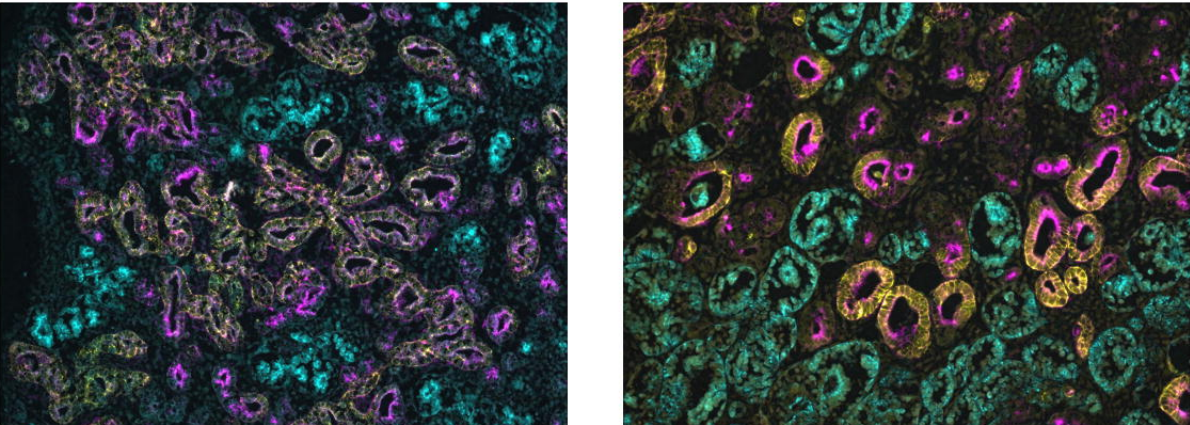
Kidney organoids from Hildebrandt, et al.’s preprint
Control iPSC lines with clinically annotated genetic variants for versatile multi-lineage differentiation
Matthew R Hildebrandt, Miriam S Reuter, Wei Wei, Naeimeh Tayebi, Jiajie Liu, Sazia Sharmin, Jaap Mulder, L Stephen Lesperance, Patrick M Brauer, Caroline Kinnear, Alina Piekna, Asli Romm, Jennifer Howe, Peter Pasceri, Rebecca S Mok, Guoliang Meng, Matthew Rozycki, Deivid de Carvalho Rodrigues, Elisa C Martinez, Michael J Szego, Juan Carlos Zúñiga-Pflücker, Michele K Anderson, Steven A Prescott, Norman D Rosenblum, Binita M Kamath, Seema Mital, Stephen W Scherer, James Ellis
Early Stem Cell Aging in the Mature Brain
Albina Ibrayeva, Maxwell Bay, Elbert Pu, David Jörg, Lei Peng, Heechul Jun, Naibo Zhang, Daniel Aaron, Congrui Lin, Galen Resler, Mi-Hyeon Jang, Benjamin D. Simons, Michael A. Bonaguidi
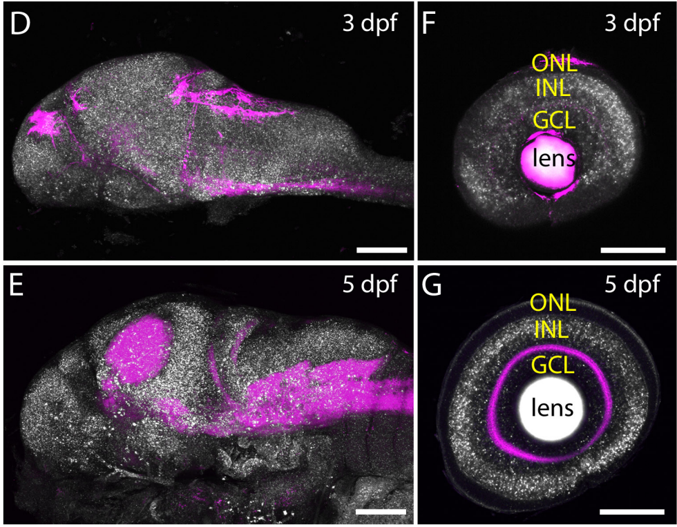
Zebrafish Dscaml1 is Essential for Retinal Patterning and Function of Oculomotor Subcircuits
Manxiu Ma, Alexandro D. Ramirez, Tong Wang, Rachel L. Roberts, Katherine E. Harmon, David Schoppik, Avirale Sharma, Christopher Kuang, Stephanie L. Goei, James A. Gagnon, Steve Zimmerman, Shengdar Q. Tsai, Deepak Reyon, J. Keith Joung, Emre R. F. Aksay, Alexander F. Schier, Y. Albert Pan
Multiparametric phenotyping of compound effects on patient derived organoids
Johannes Betge, Niklas Rindtorff, Jan Sauer, Benedikt Rauscher, Clara Dingert, Haristi Gaitantzi, Frank Herweck, Thilo Miersch, Erica Valentini, Veronika Hauber, Tobias Gutting, Larissa Frank, Sebastian Belle, Timo Gaiser, Inga Buchholz, Ralf Jesenofsky, Nicolai Härtel, Tianzuo Zhan, Bernd Fischer, Katja Breitkopf-Heinlein, Elke Burgermeister, Matthias P. Ebert, Michael Boutros
Negligible-Cost and Weekend-Free Chemically Defined Human iPSC Culture
Hui-Hsuan Kuo, Xiaozhi Gao, Jean-Marc DeKeyser, K. Ashley Fetterman, Emily A. Pinheiro, Carly J. Weddle, Michael V. Orman, Marisol Romero-Tejeda, Mariam Jouni, Malorie Blancard, Tarek Magdy, Conrad Epting, Alfred L. George Jr., Paul W. Burridge
CLIJ: Enabling GPU-accelerated image processing in Fiji
Robert Haase, Loic A. Royer, Peter Steinbach, Deborah Schmidt, Alexandr Dibrov, Uwe Schmidt, Martin Weigert, Nicola Maghelli, Pavel Tomancak, Florian Jug, Eugene W. Myers
Expanding the CRISPR Toolbox with ErCas12a in Zebrafish and Human Cells
Wesley A. Wierson, Brandon W. Simone, Zachary WareJoncas, Carla Mann, Jordan M. Welker, Bibekananda Kar, William A. C. Gendron, Michael A. Barry, Karl J. Clark, Drena L. Dobbs, Maura A. McGrail, Stephen C. Ekker, Jeffrey J. Essner
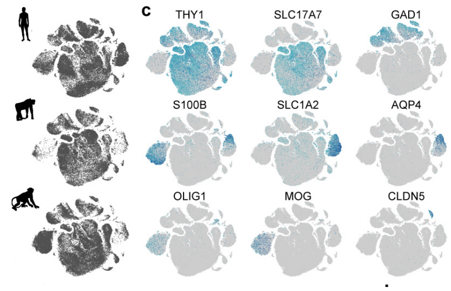
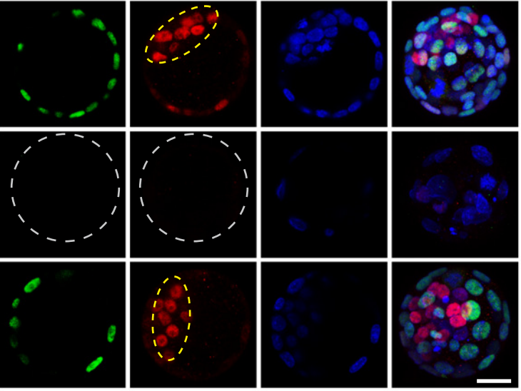
Cross-species blastocyst chimerism between nonhuman primates using iPSCs
Morteza Roodgar, Fabian P. Suchy, Vivek Bajpai, Jose G. Viches-Moure, Joydeep Bhadury, Angelos Oikonomopoulos, Joseph C. Wu, Joseph L. Mankowski, Kyle M. Loh, Hiromitsu Nakauchi, Catherine A. VandeVoort, Michael P. Snyder
Purified Aequorea proteins from Lambert, et al.’s preprint
Aequorea victoria’s secrets
Gerard G. Lambert, Hadrien Depernet, Guillaume Gotthard, Darrin T. Schultz, Isabelle Navizet, Talley Lambert, Daphne S. Bindels, Vincent Levesque, Jennifer N. Moffatt, Anya Salih, Antoine Royant, Nathan C. Shaner
You can also read the Research Highlight for this press released article.
The capacity of the human central nervous system to regenerate after injury or illness is limited, and the resulting functional impairments carry a vast societal and personal burden. In glaucoma, degeneration of retinal ganglion cells (RGCs) – the axons of which form the optic nerve connecting the retina to the brain – leads to permanent blindness; there is currently no effective treatment for RGC degeneration. Now, University of Nebraska Medical Center researcher Iqbal Ahmad and colleagues show that human RGCs can be regenerated in an in vitro setting helped by lessons learned in rodent models. The discovery is detailed in the journal Development.
“This finding could lead to new methods of screening for drugs and genes impacted by glaucoma to help treat and possibly reverse vision loss in people suffering from the disease,” said Dr. Ahmad, a professor in the department of ophthalmology and visual sciences at UNMC.
RGCs are key in sending messages to the brain through a series of synapses and connections that tell us what the eye sees. In people who suffer from glaucoma, it’s the degeneration of these cells that lead to loss of sight, Dr. Ahmad said.
Dr. Ahmad and his team of investigators found that when the mTOR signaling pathway, present in all cell types and essential for cell survival, is activated in RGCs the cells begin to regenerate and thrive. The researchers used a microfluidic chamber system to see how axons regenerated after axotomy.
Dr. Ahmad has spent 25 years studying the stem cell approach to understand and treat glaucoma, which is called a silent robber of vision because it strikes without warning or any noticeable symptoms. Glaucoma is the second leading cause of irreversible blindness and affects more than 3 million people in the United States and 60 million people worldwide.
The significance of this work, Dr. Ahmad said, is that it is done using human adult pluripotent stem cells, whereas previous work was done only in rats and mice. While those animal models provided insight into better understanding the disease progression of glaucoma, research using human RGCs will translate more readily when it comes to potential drug and gene therapies, he said. His lab has already applied for a patent on the technology that shows how RGCs can be regenerated.
“We are hopeful this process will bring us one step closer to recapturing sight in those patients who suffer from vision loss because of glaucoma,” he said.
 (No Ratings Yet)
 (1 votes)