A postdoc position is available in the Laird lab at the University of California, San Francisco to study cell heterogeneity during development using quantitative imaging, lineage tracing and single cell approaches in mouse and ESC models. Our collaborative team focuses on the basis of heterogeneity in the developing germline and its consequences for gamete function. The position is in the UCSF Edythe and Eli Broad Center for Regeneration Medicine and Stem Cell Research located at the UCSF Parnassus Heights campus, in the heart of San Francisco. UCSF offers an outstanding developmental biology community, access to cutting edge technologies and a supportive working environment. Candidates with a Ph.D. degree in a biological science, publications, demonstrated creativity and research experience in a relevant field such as genetics, biochemistry, live imaging or basic bioinformatics should submit a C.V. and names of at least 2 references via email to diana.laird@ucsf.edu.(more…)
Fully funded postdoctoral positions are presently available in the Conlon Lab, whose studies focus on identifying the molecular networks that are essential for early heart development and how alterations in these networks lead to congenital heart disease and sexual dimorphism. For these studies, we use a highly integrated approach that incorporates developmental, genetic, proteomic, biochemical and molecular-based studies in mouse, Xenopus and stem cells.
Recent advances and projects of interest in the Conlon lab include studies that define the cellular and molecular events that lead to cardiac septation, those that explore cardiac interaction networks as determinants of transcriptional specificity, the mechanism and function of cardiac transcriptional repression networks, and the regulatory networks of cardiac sexual dimorphism.
Candidates should have recently obtained or be about to obtain a Ph.D. or M.D. in a field of biological science and should have a strong publication record. Outstanding and highly motivated candidates should apply by email to Dr. Frank L. Conlon (frank_conlon@med.unc.edu) and include a CV/resume, three references and description of your specific interest in our research programs.
In the mid-1900s, Conrad Hal Waddington introduced the idea of development as a series of branching decisions taken under the control of genes1. In mammals, the first of these decisions takes place before the implantation of the embryo in the maternal uterus and leads to the distinction between the trophectoderm (TE, future placenta) and the inner cell mass (ICM, future embryo and yolk sac). In our recent study, we dissected the role of the Notch signalling pathway during early preimplantation development and found that it promotes the gradual loss of potency prior to the first lineage choice.
This project started back in 2014, after our previous work led by Teresa Rayon in which we characterised a regulatory element upstream of Cdx2, key gene in the specification of the trophectoderm2. Cdx2 was known to act downstream of the Hippo pathway, but we found that this enhancer was activated by the convergence of two pathways: Hippo and Notch. I had recently started my PhD in Miguel Manzanares’ lab at the Centro Nacional de Investigaciones Cardiovasculares (CNIC) in Madrid, and we decided to explore in more detail what Notch was doing in these early stages of mouse development.
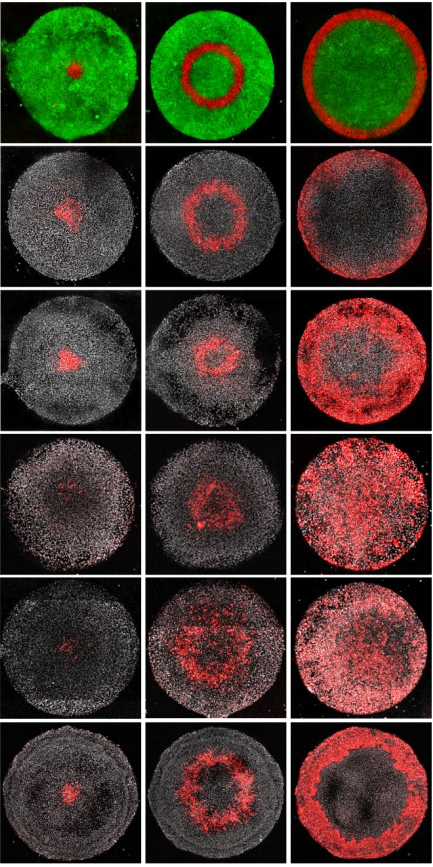
Different labs had been studying the role of the Hippo pathway during preimplantation development for a few years. So, we recapitulated all the information to try to find a cue of how Notch could be cooperating with it. Hippo is known to act as a readout of cell polarity and thus, only polarised cells (facing the outside) allow YAP to bind the transcription factor TEAD4 and activate Cdx2. However, initial expression of Cdx2 in the early compacted morula occurs in both inner and outer cells. Also, in the original work by Hiroshi Sasaki’s lab3, the authors claimed that the drop of Cdx2 expression in Tead4 mutant embryos was more dramatic in the blastocyst, when Cdx2 is only in the polarised TE, than in the morula. Therefore, our first hypothesis was that Notch could be acting in the early phases of Cdx2 expression, when this expression could not be completely explained by the role of YAP in polarised cells. We checked CDX2 in Rbpj and Notch1 mutant morulae (transcription factor and receptor of the Notch pathway respectively) and interestingly, we saw that CDX2 was strongly diminished specifically in the early morulae (<16 cells). After that, CDX2 levels were recovered, presumably because of the action of the Hippo pathway. Only double mutants for both Rbpj and Tead4 completely lacked CDX2 expression in the morula (and they did not reach the blastocyst stage). Cdx2 was responding to both pathways, but it seemed to behave differently depending on the stage. To be able to modulate the action of each pathway and verify if they were acting differently in these time windows, we used pharmacological inhibitors that allowed us to block them in a time-controlled manner. In the morula stage, only the blockade of the Notch pathway affected the expression of Cdx2. In contrast, the inhibition of TEAD/YAP was the one reducing Cdx2 from morula to blastocyst. Thus, we confirmed that although both pathways converged to regulate the same target gene, they did not act the same way.
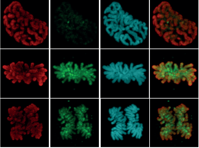
CDX2 expression in wildtype and Rbpj mutant morulae. Reduction of CDX2 is evident in the early stages of Rbpj mutant embryos (left panel, 8-16 cells).
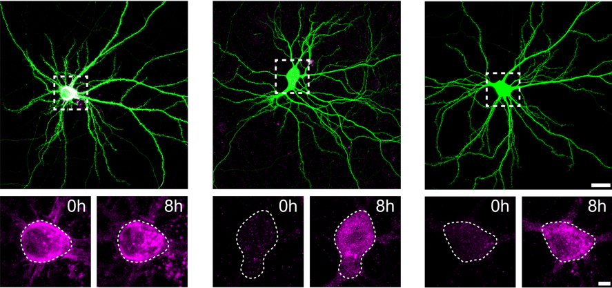
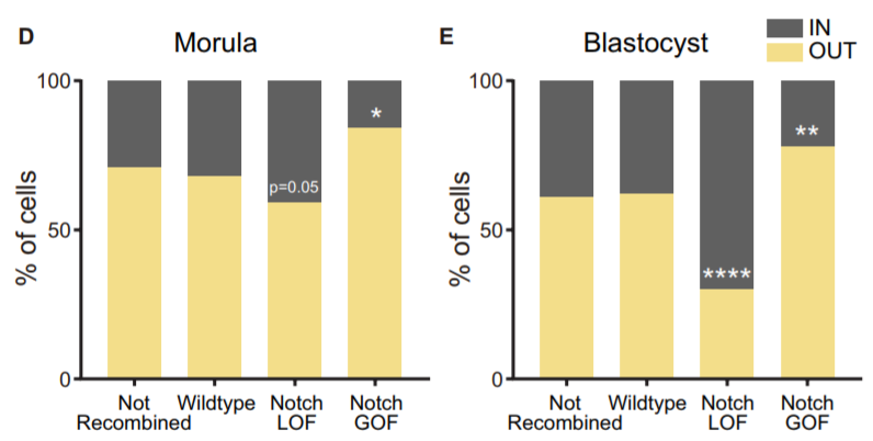
Another aspect we wanted to study was how Notch behaved during the morula to blastocyst transition, when its activity gets gradually restricted to the TE. In 2015, I was awarded a short stay fellowship from the Spanish Government that allowed me to visit the lab of Kat Hadjantonakis at the Sloan-Kettering Institute in New York for a few months, to continue with our collaboration and perform live imaging in embryos from the Notch activity reporter line (CBF1-VENUS)4. After some struggle, and with the help of Min Kang, we finally got movies where the embryos stayed in focus during the whole time-lapse (~24 hours). But the tougher part appeared later, back in Madrid. How could we manage all the information from those movies? The cell tracking for each embryo took a while… a long while. And once it was done, we still needed to consider many aspects. Fortunately, Antonio Lopez-Izquierdo, a biomedical engineer student, joined the lab to carry out the final project for his degree. He could programme in Matlab, so he developed a tool to 3D-reconstruct the embryos in each time frame and analyse the behaviour of the intensity of the reporter according to the position of the blastomeres within the embryo. The results indicated that there were already differences in the reporter intensity levels between outer and inner blastomeres in the morula, and that there was some correlation between these intensity levels and the position that cells occupied within the embryo which required further study. By then, the laboratory of Rui Benedito (our neighbours next door) had generated a transgenic line to produce mosaic cell populations with different Notch activity levels upon specific LoxP/CRE recombination5. We used this system to confront wildtype blastomeres with Notch loss of function (LOF) and gain of function (GOF) blastomeres. Beautifully, we saw how these differences affected the positioning of the cells within the embryo: Notch GOF blastomeres were more prone to occupy outer positions at the expenses of Notch LOF blastomeres which preferentially occupied inner locations.
Percentage of not recombined cells or recombined for each cassette from the iChr-Notch-Mosaic line (wildtype, Notch LOF or Notch GOF) that are in an inner or outer position at the morula and blastocyst stage.
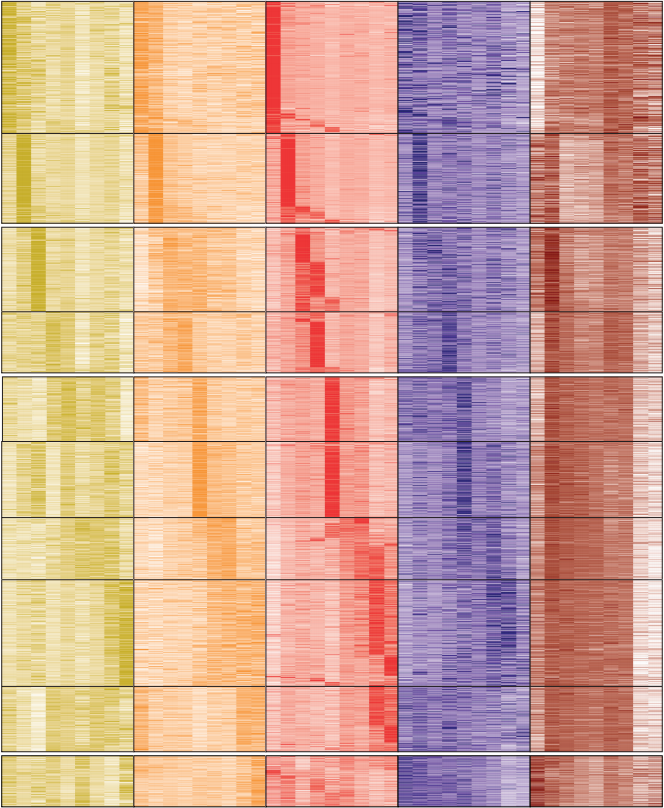
At that point, we knew more details about the role of Notch regulating Cdx2 and favouring the positioning of blastomeres in the embryo. Nevertheless, we had the feeling that Notch was doing something else and we wished to gain a broader view on how it was working. Transcriptome profiling using low amounts of RNA was emerging, so that was the way to go. We decided to carry out single embryo RNA-seq in wildtype and Rbpj mutant morulae. Two issues arose: each sample consisted of only a dozen cells so we had to test and fine-tune the protocol to make sure that it would work with the limited material; and we could only genotype after the sequencing so we did not know how many samples of each genotype we had. Once we had it, the analysis showed that most of the genes (~70%) were downregulated; these were not only TE-related genes, but also some pluripotency genes. So, the role of Notch did not seem to be just the specification of the TE.
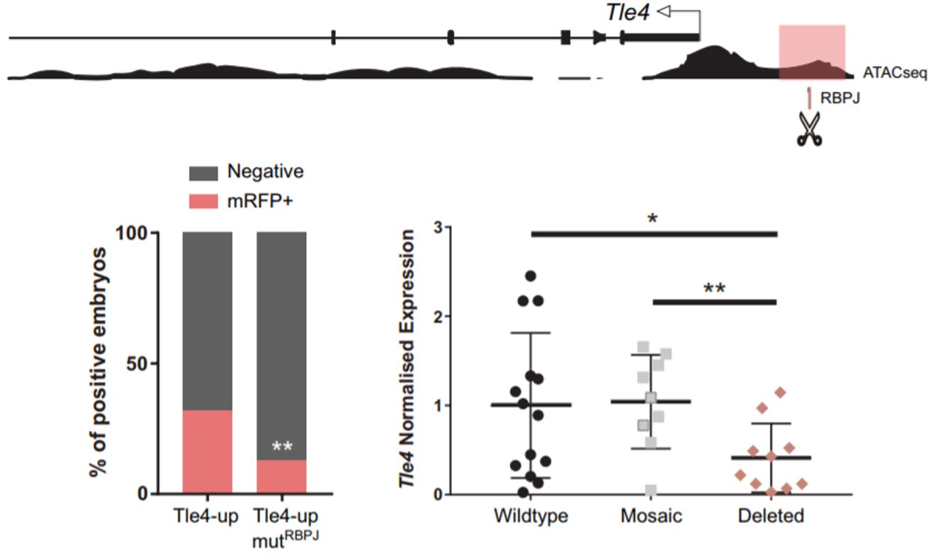
Remarkably, among the genes that were upregulated, we found two naïve pluripotency genes: Prdm14 and Dppa3. When we obtained this result, we had already seen that Notch was active very early, when the embryo consists of only four cells. Interestingly, Prdm14 is known to be heterogeneously expressed at the 4-cell stage and its expression fades away to be re-expressed in the ICM of the blastocyst6. That is the opposite pattern of Notch activity. Hence, we hypothesised that Notch could be blocking the expression of those naïve pluripotency genes in the early embryo, to boost the first differentiation programs. We did not think that this could be a direct target of Notch, given that when Notch is active it mainly activates gene expression. It is when Notch is not active that RBPJ can act as a repressor. We wondered if we could identify any gene that could be activated by Notch to repress the naïve pluripotency genes. To do that, we combined the analysis of our RNA-seq with predicted RBPJ binding motifs and data of ATAC-seq in 8-cell embryos7. In the resulting list (which was much more bearable), we found two candidates that had been described to block naïve pluripotency markers in mouse ES cells: Tle4 and Tbx3; and we decided to study them in parallel. With the help of Isabel Rollan in the lab, we identified two enhancers in the genomic landscapes of those genes that included an RBPJ motif in their sequences (Tle4-up and Tbx3-i7). To see if these sites were important for the regulation of the enhancers, we mutated these motifs and only the activity of the Tle4 enhancer was reduced. Finally, in order to see if these sites also affected the endogenous expression of Tle4 and Tbx3 respectively, we deleted them using the CRSPR/Cas9 system and we observed that Tle4 expression was affected in the edited embryos, but Tbx3 levels remained normal. Thus, RBPJ was important for the activity of the Tle4-up enhancer and for proper Tle4 expression in the morula.
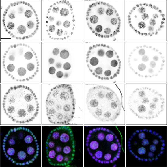
An RBPJ binding motif upstream of Tle4 is important for proper activity of the enhancer in which is included (highlighted in pink). Deletion of this site by CRISPR/Cas9 decreases Tle4 endogenous expression (bottom right).
We concluded that Notch was regulating the transition in the embryo towards the first lineage decision. First, promoting a differentiating scenario by blocking naïve pluripotency genes (possibly through TLE4) and then, inducing the expression of Cdx2 to specify the trophectoderm in cooperation with Hippo.
It is curious to think how the role of Notch during preimplantation development was ruled out more than a decade ago because single mutants were not lethal until postimplantation stages, and how convergence with other inputs can give robustness to embryonic development albeit masking important roles.
I want to finish thanking other lab members who were also involved in this exciting story: Mariajo Andreu, Julio Sainz de Aja and Javier Adan.
Sergio Menchero, Isabel Rollan, Antonio Lopez-Izquierdo, Maria Jose Andreu, Julio Sainz de Aja, Minjung Kang, Javier Adan, Rui Benedito, Teresa Rayon, Anna-Katerina Hadjantonakis, Miguel Manzanares
eLife 2019;8:e42930 DOI: 10.7554/eLife.42930
References:
Slack, J. M. W. Conrad Hal Waddington: the last Renaissance biologist? Nat. Rev. Genet.3, 889–895 (2002).
Rayon, T. et al. Notch and hippo converge on Cdx2 to specify the trophectoderm lineage in the mouse blastocyst. Dev Cell30, 410–422 (2014).
Nishioka, N. et al. Tead4 is required for specification of trophectoderm in pre-implantation mouse embryos. Mech Dev125, 270–283 (2008).
Nowotschin, S., Xenopoulos, P., Schrode, N. & Hadjantonakis, A. K. A bright single-cell resolution live imaging reporter of Notch signaling in the mouse. BMC Dev Biol13, 15 (2013).
Pontes-Quero, S. et al. Dual ifgMosaic: A Versatile Method for Multispectral and Combinatorial Mosaic Gene-Function Analysis. Cell170, 800–814.e18 (2017).
Burton, A. et al. Single-Cell Profiling of Epigenetic Modifiers Identifies PRDM14 as an Inducer of Cell Fate in the Mammalian Embryo. Cell Rep.5, 687–701 (2013).
Wu, J. et al. The landscape of accessible chromatin in mammalian preimplantation embryos. Nature534, 652–657 (2016).
3-year fixed term postdoctoral position funded by the MRC
About the position:
The Percharde lab at the MRC London Institute of Medical Science (MRC LMS) is looking to recruit 1-2 talented and highly-motivated postdocs to join our group. The Chromatin & Development group is a recently-established team focused on understanding the molecular events surrounding cell fate choices during early development.
Our recent research indicates that Transposable Elements (TEs) have important, undiscovered roles during development, and projects in the lab include uncovering how TE regulation is important during embryogenesis, as well as how unrestrained TE expression may contribute to cases of disease. To answer these questions, we routinely use mouse and human embryonic stem cells (ESCs) and mouse embryos as models, adopting a combination of genome-wide techniques, CRISPR/Cas9 technology, high-resolution microscopy, biochemistry and molecular embryology approaches to probe gene and TE function. Visit www.perchardelab.com and https://lms.mrc.ac.uk/research-group/chromatin-and-development/
for more information. Closing date 16th May, interviews on 3-5th June 2019.
Candidate specifications:
Candidates should have a PhD or be in the final stages of completing one, and have a strong background in epigenetics, developmental biology and/or gene or transposon regulation. They should also have one or more first-author publications published or being submitted to a peer-reviewed, internationally recognised journal. Experience working with mouse and/or human ESCs/iPSCs or mouse embryos is highly desirable, as well as an interest in transposon biology.
About the institute:
This is an MRC Postdoctoral Scientist position providing training and development supporting post-doctoral scientists in early or changed career paths helping establish them as successful research scientists in their chosen field.
The MRC London Institute of Medical Sciences is a research institute within UK Research and Innovation. UK Research and Innovation is a new entity that brings together nine partners to create an independent organisation with a strong voice for research and innovation, more information can be found at www.ukri.org
Apply:
For full details of this post and to complete an online application, visit: https://mrc.tal.net/vx/lang-en-GB/appcentre-1/candidate/postings/1195 and upload your CV, the names and contacts of two scientific references, along with a cover letter stating why you are applying for this post (providing evidence against the requirements as per the Job Description and Person Specification). Please quote reference number LMS – 1071.
Journal of Cell Science and its publisher, The Company of Biologists, are seeking to appoint a Community Manager, based in Cambridge, UK, to run a new microscopy resource website.
An extensive consultation told us that the microscopy community would welcome a trusted, curated and centralised site to connect people, resources and information. Our exciting new initiative is therefore intended to create a platform for the microscopy community to share news and techniques, discuss issues relevant to the field and read about the latest research and events.
We are now looking for an enthusiastic and motivated person to join us to develop, launch and maintain this site, which will be hosted by Journal of Cell Science but will be relevant to all of the Company of Biologists’ journals.
Core responsibilities of the position include:
Creating and commissioning content, including writing posts and soliciting content from the academic community, societies, companies and other organisations.
Providing user support and ensuring site functionality on a day-to-day basis.
Providing creative and practical input into the design and development of the site.
Maintaining and developing the site’s presence on social networking sites such as Facebook and Twitter.
Developing and managing sponsorship, ads and commercial relationships.
Representing Journal of Cell Science and the resource site at international conferences.
Applicants should have a PhD with research experience in a relevant scientific field and extensive imaging experience – ideally involving the application or development of new methods for microscopy or image analysis. The successful candidate will have proven social media skills and a clear understanding of the online environment as it applies to scientists. Applicants should have excellent writing and communication skills, and strong interpersonal and networking abilities – both online and in person. Experience with additional media, such as video or podcasting, and an eye for aesthetics, would be an advantage. We are looking for an individual with fresh ideas and a willingness to learn new skills, and who will contribute broadly to the Company’s activities.
This is an exciting opportunity to develop a hub for the microscopy community – in a similar vein to the Company’s established community site for developmental biologists, the Node – and to engage with relevant people at all levels: academics, developers, facilities, institutes and companies. The Community Manager will work alongside an experienced in-house team, including the Executive Editor of Journal of Cell Science, as well as with the journal’s international team of academic editors. Additional responsibilities may be provided for the right candidate. The Company of Biologists is based in attractive modern offices on the outskirts of Cambridge, UK.
The Company of Biologists (biologists.com) exists to support biologists and inspire advances in biology. At the heart of what we do are our five specialist journals – Development, Journal of Cell Science, Journal of Experimental Biology, Disease Models & Mechanisms and Biology Open – two of them fully open access. All are edited by expert researchers in the field, and all articles are subjected to rigorous peer review. We take great pride in the experience of our editorial team and the quality of the work we publish. We believe that the profits from publishing the hard work of biologists should support scientific discovery and help develop future scientists. Our grants help support societies, meetings and individuals. Our workshops and meetings give the opportunity to network and collaborate.
Applicants should send a CV along with a covering letter that summarises their relevant experience, and in particular their specific microscopy/imaging expertise, any links to online activities), current salary, and why they are enthusiastic about this opportunity.
Applicants should send a CV along with a covering letter that summarises their relevant experience, and in particular their specific microscopy/imaging expertise, any links to online activities), current salary, and why they are enthusiastic about this opportunity.
Applications and informal queries should be sent by email to hr@biologists.com.
We may request written tests in advance of any interview.
Journal of Cell Science and its publisher, The Company of Biologists, are seeking to appoint a Community Manager, based in Cambridge, UK, to run a new microscopy resource website.
An extensive consultation told us that the microscopy community would welcome a trusted, curated and centralised site to connect people, resources and information. Our exciting new initiative is therefore intended to create a platform for the microscopy community to share news and techniques, discuss issues relevant to the field and read about the latest research and events.
We are now looking for an enthusiastic and motivated person to join us to develop, launch and maintain this site, which will be hosted by Journal of Cell Science but will be relevant to all of the Company of Biologists’ journals.
Core responsibilities of the position include:
Creating and commissioning content, including writing posts and soliciting content from the academic community, societies, companies and other organisations.
Providing user support and ensuring site functionality on a day-to-day basis.
Providing creative and practical input into the design and development of the site.
Maintaining and developing the site’s presence on social networking sites such as Facebook and Twitter.
Developing and managing sponsorship, ads and commercial relationships.
Representing Journal of Cell Science and the resource site at international conferences.
Applicants should have a PhD with research experience in a relevant scientific field and extensive imaging experience – ideally involving the application or development of new methods for microscopy or image analysis. The successful candidate will have proven social media skills and a clear understanding of the online environment as it applies to scientists. Applicants should have excellent writing and communication skills, and strong interpersonal and networking abilities – both online and in person. Experience with additional media, such as video or podcasting, and an eye for aesthetics, would be an advantage. We are looking for an individual with fresh ideas and a willingness to learn new skills, and who will contribute broadly to the Company’s activities.
This is an exciting opportunity to develop a hub for the microscopy community – in a similar vein to the Company’s established community site for developmental biologists, the Node – and to engage with relevant people at all levels: academics, developers, facilities, institutes and companies. The Community Manager will work alongside an experienced in-house team, including the Executive Editor of Journal of Cell Science, as well as with the journal’s international team of academic editors. Additional responsibilities may be provided for the right candidate. The Company of Biologists is based in attractive modern offices on the outskirts of Cambridge, UK.
The Company of Biologists (biologists.com) exists to support biologists and inspire advances in biology. At the heart of what we do are our five specialist journals – Development, Journal of Cell Science, Journal of Experimental Biology, Disease Models & Mechanisms and Biology Open – two of them fully open access. All are edited by expert researchers in the field, and all articles are subjected to rigorous peer review. We take great pride in the experience of our editorial team and the quality of the work we publish. We believe that the profits from publishing the hard work of biologists should support scientific discovery and help develop future scientists. Our grants help support societies, meetings and individuals. Our workshops and meetings give the opportunity to network and collaborate.
Applicants should send a CV along with a covering letter that summarises their relevant experience, and in particular their specific microscopy/imaging expertise, any links to online activities), current salary, and why they are enthusiastic about this opportunity.
Applications and informal queries should be sent by email to hr@biologists.com.
We may request written tests in advance of any interview.
Job Title: Post-doctoral fellow. Position is currently recruiting. Minimum of 2-year commitment is required.
Job Description
The Liao Laboratory at the Center for Regenerative Medicine at Massachusetts General Hospital in affiliation with the Division of Plastic Surgery and the Harvard Stem Cell Institute is seeking a highly motivated research fellow interested in craniofacial developmental biology and genetics. We are currently recruiting 2 post-doctoral fellows to join our team of 10 investigators. One post-doc position is seeking a candidate with experience in iPSC derivation and cell model work. The second post-doc position is seeking a candidate with experience in zebrafish or mouse models. We have a pipeline of studying human subjects with rare craniofacial conditions, where we apply human iPSC, mouse or zebrafish models to dissect the developmental genetic basis of craniofacial malformations.
The fellow will join a collaborative group of scientists who study the basic and translational biology of facial morphogenesis, cranial neural crest specification and development. In this instance, the fellow’s work will have a direct bearing on discovery of drugs to mitigate malformation phenotypes and functional genomics of human candidate genes implicated in orofacial clefts. We are inherently interested in the basic biology of neural crest cell specification, migration, differentiation and potential for regeneration. Integrated approaches of classical developmental and modern stem cell biology are used to explore human tissue regeneration and disease. We apply the latest technology in zebrafish genetics with routine use of CRISPR, iPSC, single cell RNAseq, Cut&Run transcriptional analysis, cell transplants, FACS, live confocal labeling and imaging, transgenics, chemical screening, and many other experimental approaches. Our laboratory has a dedicated zebrafish facility outfitted with the latest imaging, injection and cell manipulation instruments. Researchers work in open format laboratory space that foster interactions with other stellar researchers working in the Center, with ample opportunities to participate in scientific seminars and professional development.
The current lab members include 3 post-doc fellows, 3 graduate students, 3 technicians, 1 undergraduate and 1 high school student. The laboratory has stable funding with 4 extramural grants. There will be ample opportunity for eligible candidate to compete for independent funding.
Job Requirements
The candidate should have PhD or an MD/PhD in a relevant field or equivalent training. A strong experimental foundation in iPSC derivation, iPSC analysis, cell culture, animal models are mandatory. Additional experience with mouse genetics, development, cell, developmental, are desirable. An ancillary knowledge of genomics or systems biology is welcome. Outstanding oral and written communication skills are required.
Please email your inquiry, current CV, statement of interest and career goals, to Eric Liao, cliao@partners.org
Employing Department and Institution:Center for Regenerative Medicine, Massachusetts General Hospital.
Welcome to our monthly trawl for developmental biology (and related) preprints.
This month was notable for a preponderance of plant development preprints, many molecular maps (supported by single cell sequencing), a hearty helping of human development and a multiplicity of (Drosophila) melanogaster mechanics.
The preprints were hosted on bioRxiv, PeerJ, andarXiv. Let us know if we missed anything, and use these links to get to the section you want:
Osteocytes remodel bone by TGF-β-induced YAP/TAZ signaling
Christopher D. Kegelman, Jennifer C. Coulombe, Kelsey M. Jordan, Daniel J. Horan, Ling Qin, Alexander G. Robling, Virginia. L Ferguson, Teresita M. Bellido, Joel D. Boerckel
Early human embryos from Meistermann, et al.’s preprint
Spatio-temporal analysis of human preimplantation development reveals dynamics of epiblast and trophectoderm
Dimitri Meistermann, Sophie Loubersac, Arnaud Reignier, Julie Firmin, Valentin Francois Campion, Stéphanie Kilens, Yohann Lelièvre, Jenna Lammers, Magalie Feyeux, Phillipe Hulin, Steven Nedellec, Betty Bretin, Simon Covin, Gael Castel, Audrey Bihouée, Magali Soumillon, Tarjei Mikkelsen, Paul Barrière, Jérémie Bourdon, Thomas Fréour, Laurent David
Control of glioblastoma differentiated-to-stem cell reprogramming by IRE1α/XBP1s signaling.
Dimitrios Doultsinos, Mari McMahon, Konstantinos Voutetakis, Joanna Obacz, Raphael Pineau, Florence Jouan, Pierre-Jean Le Reste, Akram Obiedat, Juhi Samal, John B Patterson, Quinping Zheng, Afshin Samali, Abhay Pandit, Boaz Tirosh, Aristotelis Chatziioannou, Eric Chevet, Tony Avril
Redox potential defines functional states of adult hippocampal stem cells
Vijay S Adusumilli, Tara L Walker, Rupert W Overall, Gesa M Klatt, Salma A Zeidan, Tim J Fischer, Sara Zocher, Alex M Sykes, Susanne Reinhardt, Andreas Dahl, Dilyana G Kirova, Jörg Mansfeld, Annette E Rünker, Gerd Kempermann
ELIMÄKI locus is required for mechanosensing and proprioception in birch trees
Juan Alonso-Serra, Xueping Shi, Alexis Peaucelle, Pasi Rastas, Matthieu Bourdon, Juha Immanen, Junko Takahashi, Hanna Koivula, Gugan Eswaran, Sampo Muranen, Hanna Help-Rinta-Rahko, Olli-Pekka Smolander, Chang Su, Omid Safronov, Lorenz Gerber, Jarkko Salojärvi, Risto Hagqvist, Ari-Pekka Mähonen, Kaisa Nieminen, Ykä Helariutta
Brassica fruits from Stephenson, et al.’s preprint
Mapping and dynamics of regulatory DNA in maturing seeds
Alessandra M Sullivan, Andrej A Arsovski, Agnieszka Thompson, Richard Sandstrom, Robert E Thurman, Shane Neph, Audra K Johnson, Shawn T Sullivan, Peter J Sabo, Fidencio V Neri III, Molly Weaver, Morgan Diegel, Jennifer L Nemhauser, John A Stamatoyannopoulos, Kerry L Bubb, Christine Queitsch
Fog signaling has diverse roles in epithelial morphogenesis in insects
Nadine Frey, Matthew A. Benton, Rodrigo Nunes da Fonseca, Cornelia von Levetzow, Dominik Stappert, Muhammad Salim Hakeemi, Kai H. Conrads, Matthias Pechmann, Kristen A. Panfilio, Jeremy A. Lynch, Siegfried Roth
Two Y chromosome-encoded genes determine sex in kiwifruit
Takashi Akagi, Sarah M. Pilkington, Erika Varkonyi-Gasic, Isabelle M. Henry, Shigeo S. Sugano, Minori Sonoda, Alana Firl, Mark A. McNeilage, Mikaela J. Douglas, Tianchi Wang, Ria Rebstock, Charlotte Voogd, Paul Datson, Andrew C. Allan, Kenji Beppu, Ikuo Kataoka, Ryutaro Tao
Enabling large-scale genome editing by reducing DNA nicking
Cory J. Smith, Oscar Castanon, Khaled Said, Verena Volf, Parastoo Khoshakhlagh, Amanda Hornick, Raphael Ferreira, Chun-Ting Wu, Marc Güell, Shilpa Garg, Hannu Myllykallio, George M. Church
A simple and efficient CRISPR technique for protein tagging
Fanning Zeng, Valerie Beck, Sven Schuierer, Isabelle Garnier, Carole Manneville, Claudia Agarinis, Lapo Morelli, Lisa Quinn, Judith Knehr, Guglielmo Roma, Frederic Bassilana, Mark Nash
A Pandas complex adapted for piRNA-guided transposon silencing
Kang Zhao, Sha Cheng, Na Miao, Ping Xu, Xiaohua Lu, Ming Wang, Yuhan Zhang, Xun Yuan, Weiwei Liu, Xin Lu, Xuan Ouyang, Peng Zhou, Jiaqi Gu, Yiqun Zhang, Ding Qiu, Shan Wang, Zhaohui Jin, Youzhong Wan, Jinbiao Ma, Ying Huang, Yang Yu
A reference map of the human protein interactome
Katja Luck, Dae-Kyum Kim, Luke Lambourne, Kerstin Spirohn, Bridget E. Begg, Wenting Bian, Ruth Brignall, Tiziana Cafarelli, Francisco J. Campos-Laborie, Benoit Charloteaux, Dongsic Choi, Atina G. Cote, Meaghan Daley, Steven Deimling, Alice Desbuleux, Amélie Dricot, Marinella Gebbia, Madeleine F. Hardy, Nishka Kishore, Jennifer J. Knapp, István A. Kovács, Irma Lemmens, Miles W. Mee, Joseph C. Mellor, Carl Pollis, Carles Pons, Aaron D. Richardson, Sadie Schlabach, Bridget Teeking, Anupama Yadav, Mariana Babor, Dawit Balcha, Omer Basha, Christian Bowman-Colin, Suet-Feung Chin, Soon Gang Choi, Claudia Colabella, Georges Coppin, Cassandra D’Amata, David De Ridder, Steffi De Rouck, Miquel Duran-Frigola, Hanane Ennajdaoui, Florian Goebels, Liana Goehring, Anjali Gopal, Ghazal Haddad, Elodie Hatchi, Mohamed Helmy, Yves Jacob, Yoseph Kassa, Serena Landini, Roujia Li, Natascha van Lieshout, Andrew MacWilliams, Dylan Markey, Joseph N. Paulson, Sudharshan Rangarajan, John Rasla, Ashyad Rayhan, Thomas Rolland, Adriana San-Miguel, Yun Shen, Dayag Sheykhkarimli, Gloria M. Sheynkman, Eyal Simonovsky, Murat Taşan, Alexander Tejeda, Jean-Claude Twizere, Yang Wang, Robert J. Weatheritt, Jochen Weile, Yu Xia, Xinping Yang, Esti Yeger-Lotem, Quan Zhong, Patrick Aloy, Gary D. Bader, Javier De Las Rivas, Suzanne Gaudet, Tong Hao, Janusz Rak, Jan Tavernier, Vincent Tropepe, David E. Hill, Marc Vidal, Frederick P. Roth, Michael A. Calderwood
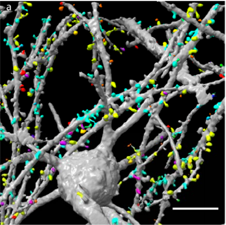
Dendritic spines from Chakraborty, et al.’s preprint
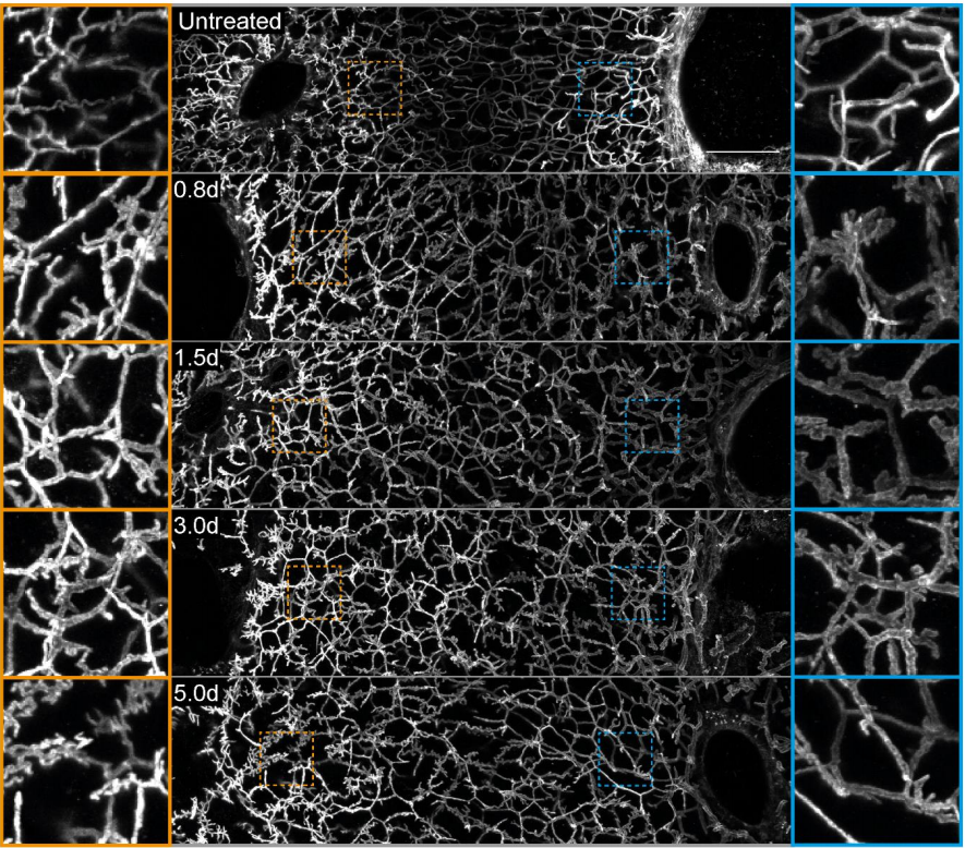
Light-sheet microscopy with isotropic, sub-micron resolution and solvent-independent large-scale imaging
Tonmoy Chakraborty, Meghan Driscoll, Malea Murphy, Philippe Roudot, Bo-Jui Chang, Saumya Vora, Wen Mai Wong, Cara Nielson, Hua Zhang, Vladimir Zhemkov, Chitkale Hiremath, Estanislao Daniel De La Cruz, Ilya Bezprozvanny, Hu Zhao, Raju Tomer, Rainer Heintzmann, Julian Meeks, Denise Marciano, Sean Morrison, Gaudenz Danuser, Kevin M. Dean, Reto Fiolka
3D Multicolor Nanoscopy at 10,000 Cells a Day
Andrew E S Barentine, Yu Lin, Miao Liu, Phylicia Kidd, Leonhard Balduf, Michael R Grace, Siyuan Wang, Joerg Bewersdorf, David Baddeley
The ELIXIR Core Data Resources: fundamental infrastructure for the life sciences
Rachel Drysdale, Charles E Cook, Robert Petryszak, Vivienne Baillie Gerritsen, Mary Barlow, Elisabeth Gasteiger, Franziska Gruhl, Juergen Haas, Jeremy Lanfear, Rodrigo Lopez, Nicole Redaschi, Heinz Stockinger, Daniel Teixeira, Aravind Venkatesan, ELIXIR Core Data Resource Forum, Niklas Blomberg, Christine Durinx, Jo McEntyre
We are seeking an outstanding colleague to join collaborative research and provide bioinformatics expertise. Candidate will collaborate with members of the Center of Regenerative Medicine to analyze and interpret primary sequencing data (ChIP-seq, RNA-seq, genome/exome sequencing) by utilizing state of the art bioinformatics tools. This position assists in developing and conducting research projects, including experimental design, data analysis, and documentation of experiment results.
PRIMARY DUTIES AND RESPONSIBILITIES:
Designs, develops, and implements:
Algorithms and computer software for analyzing high-throughput, massively parallel genomic data sets.
Relational databases (SQL/NoSQL) and web server maintenance.
Familiar with biological data storage and format, including genomics data search, query, verification, and submission.
Independent research projects, including design of research protocols and development of procedures for the collection, verification, and management of data.
Performs integrative analysis of high-throughput biological data and writes interpretative reports.
Trains other researchers on the daily use of bioinformatics software and biology databases.
Assists with grant preparation and reporting of methods, data, and results.
REQUIRED QUALIFICATIONS:
Bachelor’s degree in computer science, bioinformatics, biostatistics, or related field plus 4 years of experience; OR master’s degree in computer science, bioinformatics, biostatistics, or related field plus 2 years of experience; OR combined education and experience of 9 years.
PREFERRED QUALIFICATIONS:
Master’s degree or Ph.D. in computer science or related field highly preferred.
Knowledge of developmental biology and/or regenerative medicine would also be a plus.
Experience with single cell sequencing and epigenomics highly preferred.
Proficiency with Linux system, common programing languages (e.g., R, Python, Perl, PHP), and bioinformatics analysis tools.
Excellent writing, communication and interpersonal skills.
The hiring range for this position is $53,830 – $72,675 annually. This position is eligible for full-time benefits. Please visit our website at http://hr.wustl.edu to view a summary of benefits.
VISIT https://jobs.wustl.edu and search job ID 43692 to apply.
We seek highly motivated post-doctoral Fellows interested in cell signaling at the intersection of development, stem cells and cancer. Candidates will join the group of Dr. Andres Lebensohn, an Earl Stadtman Principal Investigator in the Laboratory of Cellular and Molecular Biology (LCMB) at the Center for Cancer Research (CCR) of the National Cancer Institute (NCI), National Institutes of Health (NIH). We combine functional genomics, CRISPR/Cas9-mediated genome editing, cell biology and biochemistry to study how signaling pathways are used, reused and repurposed to drive the myriad different cellular processes that give rise to tissues and organs during embryonic development, and maintain them in adult life. A major focus of the lab is on WNT/R-spondin signaling, a fundamental pathway that orchestrates embryonic patterning and morphogenesis, and promotes stem cell self-renewal and tissue regeneration. We recently discovered a new mode of signaling by R-spondins that does not require LGRs, thought to be the main R-spondin receptors (https://elifesciences.org/articles/33126). We seek a Fellow who will use mouse genetics to elucidate the physiological functions of LGR-independent signaling by R-spondins during embryonic development and in stem cells. Descriptions of this and other research projects in the lab can be found at https://ccr.cancer.gov/Laboratory-of-Cellular-and-Molecular-Biology/andres-m-lebensohn.
About the Center for Cancer Research and the Laboratory of Cellular and Molecular Biology:
The CCR is the largest division of the NCI intramural research program, comprising nearly 250 basic and clinical research groups. In the words of Dr. Tom Misteli, director of the CCR, “Our scientists enjoy complete intellectual freedom and are expected to creatively and innovatively explore the most important questions in the field of cancer research and treatment.” The LCMB (https://ccr.cancer.gov/Laboratory-of-Cellular-and-Molecular-Biology) is located at the NIH Bethesda campus within a vibrant biomedical research community. We have access to state-of-the art research facilities and close links to the NIH Clinical Center, one of the leading clinical research hospitals in the world. The open lab space, collegial atmosphere and joint weekly seminars shared by the seven groups in the LCMB give Fellows a strong support network and plenty of opportunities to present and discuss their research with close peers. A short metro ride to Washington D.C. and the surrounding areas provides Fellows and their families access to numerous free museums (https://washington.org/free-things-to-do), excellent culinary choices and many outdoor activities.
Qualifications:
Candidates must have completed a Ph.D. and should be recent graduates or have less than 3 years of post-doctoral experience by the desired start date. Candidates with backgrounds in developmental, stem cell or cancer biology are encouraged to apply. Experience in mouse genetics is required for this project, and experience with organoid models is welcome but not required. Fellows will have exceptional stipend support for the entire duration of their post-doctorate, funds for travel to scientific conferences, and full healthcare benefits.
How to Apply:
Please send a cover letter describing your research accomplishments and future interests, including your specific interest in my lab, a CV with bibliography, and contact information for three references to andres.lebensohn@nih.gov. Please use the following email subject: “Application for post-doc position in WNT/R-spondin signaling”
 San Francisco to study cell heterogeneity during development using quantitative imaging, lineage tracing and single cell approaches in mouse and ESC models. Our collaborative team focuses on the basis of heterogeneity in the developing germline and its consequences for gamete function. The position is in the UCSF Edythe and Eli Broad Center for Regeneration Medicine and Stem Cell Research located at the UCSF Parnassus Heights campus, in the heart of San Francisco. UCSF offers an outstanding developmental biology community, access to cutting edge technologies and a supportive working environment. Candidates with a Ph.D. degree in a biological science, publications, demonstrated creativity and research experience in a relevant field such as genetics, biochemistry, live imaging or basic bioinformatics should submit a C.V. and names of at least 2 references via email to diana.laird@ucsf.edu.
 (No Ratings Yet)
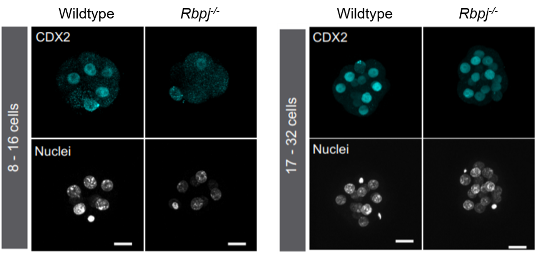
 (1 votes)
 Our
Our