The community site for and by
developmental and stem cell biologists

Organise a Company of Biologists workshop in 2020!

Posted by , on 30 April 2018

As well as publishing Development and four other journals, and supporting scientists through travelling fellowships and meeting grants, The Company of Biologists also runs a successful series of Workshops.

The Workshops  bring leading experts and early career scientists from a diverse range of scientific backgrounds to focus on one topic together. Topics are often interdisciplinary and cover some of the most exciting current biology, as you can see in the archive.


The Workshop Committee are currently seeking proposals for four Workshops to be held during 2020.  They are particularly keen to receive proposals from postdocs for one of the Workshops. We at the Node would also encourage applicants from developmental biology to think about applying!

The deadline date for applications is 25 May 2018


Find out more here:

biologists.com/workshops/propose-new-workshop


Thumbs up (1 votes)
Loading...

Tags:
Categories: Events

Navigate the archive

Use our Advanced Search tool to search and filter posts by date, category, tags and authors.

full-time Research Assistant and Research Associate positions

Posted by , on 27 April 2018

Closing Date: 15 March 2021

The Marine Biological Laboratory is seeking applicants for full-time Research Assistant and Research Associate positions with the Josephine Bay Paul Center for Comparative Molecular Biology and Evolution (http://www.mbl.edu/jbpc/).


To develop CRISPR in rotifers:

We seek a motivated, creative and innovative Research Assistant or Research Associate to join the laboratories of Kristin Gribble and David Mark Welch.  Our research combines comparative genomics, biochemistry, and life history to study aging, maternal effects, and DNA damage prevention and repair using rotifers, a novel aquatic invertebrate model system for studies of aging, neurobiology, genome evolution, and ecology.  The successful candidate will develop genome editing techniques in rotifers, including CRISPR/Cas9, as part of a broad initiative at the MBL to advance new aquatic and marine models for biological discovery.  We invite individuals with experience in genome editing in other animals to join this expanding program.

https://mbl.simplehire.com/postings/3977


To study the biochemistry of DNA repair in bdelloids:

We seek a Research Assistant to join the laboratory of David Mark Welch. Our research combines comparative genomics, biochemistry, and life history to study the evolution of bdelloid rotifers, extraordinarily resilient animals adapted to highly stressful environmental conditions.  The successful candidate will contribute to an ongoing project to clone, express, purify, and assay a variety of rotifer proteins involved in DNA damage prevention and repair.  There is considerable opportunity for motivated, self-directed individuals to participate in technique development, manuscript development and publication. Position level and salary will depend on education and experience.

https://mbl.simplehire.com/postings/3972


To sequence microbial communities:

We seek applicants for a full-time Research Assistant position with the Keck Sequencing Facility of the Josephine Bay Paul Center. The successful applicant will contribute to projects that will explore diversity of microbes in various communities and the influence of changing environments on microbial population structures. Responsibilities include, but are not limited to, laboratory management, preparation and massively high-throughput next-generation sequencing of marker gene and metagenomic libraries from environmental genomic DNA, and computational analysis of such datasets.

https://mbl.simplehire.com/postings/3978


Thumbs up (No Ratings Yet)
Loading...

Categories: Jobs

Postdoctoral Position in Neuroscience: Brain Development in Zebrafish

Posted by , on 26 April 2018

Closing Date: 15 March 2021

A neuroscience Postdoctoral Research Associate position is available in the Robles Lab at Purdue University (www.robleslab.com). Our laboratory applies advanced genetic and microscopic imaging techniques to understand how the nervous system develops in the larval zebrafish. We use confocal laser scanning microscopy to image the structural development of genetically targeted neurons within the intact developing brain. Our major area of emphasis is developing novel, in vivo imaging assays to examine the cellular and circuit-level abnormalities underlying complex neurological disorders such as autism and epilepsy.

Candidates should have a Ph.D. in neuroscience, cell biology, genetics, or a related field. The ideal candidate will have expertise in microscopy, image analysis, and molecular biology techniques. Special consideration will be given to applicants with expertise in developing and implementing computational strategies for data/image analysis.

Please send your CV, a cover letter stating your research interests and professional goals, and the contact information for three (3) references to:

Estuardo Robles

Email: roblese@purdue.edu

Assistant Professor

Department of Biological Sciences

Purdue University

915 W State Street, LILY B-129

West Lafayette, IN, 47907

(765)494-8413

www.robleslab.com

Thumbs up (No Ratings Yet)
Loading...

Categories: Jobs

First impressions of the BSDB Spring Meeting

Posted by , on 24 April 2018

This year, is the BSDB’s 70th anniversary, and this was clearly reflected at our Spring Meeting, 15-18 April 2018 in Warwick! Apart from an outstanding speaker list, and the award of most BSDB medals & prizes of 2018, we saw a history session which looked back at the past, to then project into the future of Developmental Biology. Other specials were a very engaging PhD and postdoc social, the weaving of pedigree networks (with little labels, pins and threads of wool), and the live presentations of DB raps composed and sung by Jerry aka Gerald H Thomsen PhD and produced and mixed by Philip Larsen. See below a few image impressions of the meeting (taken from your many tweets!) and/or listen to the two raps! A detailed meeting report will follow.


Rap 1: BSDB History (part I & II)


Rap 2: Morphogen Mix


Gerald Thomsen, Josh Brickman and Philip Larsen


Only a tiny selection of participants


Emilia receives the Beddington medal from Simon Bullock


Nick Hopwood


The panel discussion


The PhD and postdoc social


PhD and postdoc social


Gearing up for the conference dinner


Weaving pedigree networks: Eric Wieschaus, David Ish-Horowicz, Claudio Stern, Austin Smith


Concluding lecture by Sir John Gurdon

Thumbs up (3 votes)
Loading...

Tags: , ,
Categories: Events, Highlights, Societies

Postdoctoral position: Timing vertebrate limb development

Posted by , on 24 April 2018

Closing Date: 15 March 2021

A Wellcome Trust funded Postdoctoral Research Associate position is available in the lab of Dr. Matt Towers at the University of Sheffield, UK, to work on vertebrate limb development. How embryonic development is timed and scaled remains enigmatic. Understanding the underlying mechanisms remains a central challenge in biology, since these mechanisms coordinate the growth and patterning of all tissues and organs, and when disrupted are likely to form the basis of many congenital disorders. Our recent work on the embryonic chick wing demonstrates a pivotal role for intrinsic timers in limb development (Chinnaiya et al, Nat Comms 2014, Saiz-Lopez et al, Nat Comms 2015; Saiz-Lopez et al, Development 2017, Pickering et al, Biorxiv 2018). The aim of this project is to use a combination of classical grafting techniques and modern genomic analyses in chick embryos to understand the molecular mechanisms of how cells measure time in vivo.

Closing date: 23rd May 2018

See https://towerslab.weebly.com/ for more details of our research.

http://www.jobs.ac.uk/job/BJH713/research-associate-in-cell-and-developmental-biology/

Thumbs up (1 votes)
Loading...

Categories: Jobs

Postdoc Position – Taneyhill Lab, University of Maryland (near Washington DC)

Posted by , on 24 April 2018

Closing Date: 15 March 2021

A position (#123768) is available immediately for a Postdoctoral Scholar to contribute to our multidisciplinary studies aimed at elucidating the molecular basis of chick neural crest and placode cell development. The postdoc will conduct independent research and assist in the training of students in the laboratory of Dr. Lisa Taneyhill at the University of Maryland.

Laboratory skills: Molecular biology and biochemical assays (e.g., recombinant DNA/cloning; DNA, RNA, and/or protein blotting); immunohistochemistry; and/or in situ hybridization. Experience with microscopy and spectroscopy, chick embryology (including microdissections and electroporation), and tissue culture is desirable. For more information on the lab, please see http://www.ansc.umd.edu/people/lisa-taneyhill.

Qualifications: An advanced degree (Ph.D.) in Developmental, Molecular and/or Cell Biology is required. Fluency in spoken and written English is required. Compensation: Salaries are highly competitive, negotiable and commensurate with qualifications. Fringe benefits offered. Applicants must apply through eTerp at https://ejobs.umd.edu. Applications will be accepted until a suitable candidate is identified.

Thumbs up (No Ratings Yet)
Loading...

Categories: Jobs, Research

Medaka fish sheds light on the evolutionary origin of vertebrate pair appendages

Posted by , on 23 April 2018

Link to the paper: https://www.nature.com/articles/s41588-018-0080-5


The evolutionary history of vertebrate appendages

Have you ever wondered how our hands and feet evolved? This question, which commonly evokes fish crawling from sea to land, has long been a subject of interest, both for palaeontologists and developmental biologists. Appendages are an important part of the tool kit of anatomical innovations that emerged in vertebrates. Limbs, wings and fins certainly contributed to the evolutionary success of the whole group, as they are a fertile substrate for all kinds of ecological adaptations. However, the evolutionary history of vertebrate appendages is complex and far from being completely understood, particularly as we go back into the past to the very origin of the vertebrate lineage. It is believed that our chordate ancestors already had median fin folds. Then, both fin rays and endoskeletal elements appeared in the median fins of the first jawless vertebrates, and were maintained in modern members of this group, such as lampreys and hagfish. Later in evolution, paired appendages emerged around 450 million years ago in the first jawed vertebrates (i.e. stem gnathostomes), and were therefore inherited by modern sharks and teleost fish. Finally, when vertebrates conquered land, paired fins evolved to limbs in tetrapods. We know very little about these different steps, particularly about the early ones (1). So, for example, the evolutionary origin of paired fins is still debated and several theories have been around for a while. Most popular hypotheses include a common origin for gills and paired fins, the emergence of the paired appendages from an ancestral lateral fin fold, and, more recently, the exaptation of their developmental program from the pre-existing median fins in jawless vertebrates.


ZRS function in teleost fish

Neil Shubin (University of Chicago), who is well-known for his participation in the discovery of the Tiktaalik roseae (2), is certainly among the scientists most captivated by the evolutionary history of vertebrate appendages. From his long interest in the topic, and through his scientific interaction with an expert in zebrafish epigenomics, José Luis Gómez-Skarmeta (CABD, Seville), stemmed a project that flourished as more people got involved. Igor Schneider (University of Pará, Brasil), a former member of Shubin’s group interested in understanding the fin to limb transition, also joined the venture. Together they focused their attention on investigating the evolutionary origin of a very famous enhancer, the sonic hedgehog (Shh) limb enhancer, ZRS* (i.e. Zone of polarizing activity Regulatory Sequence). Located one mega-base away from the gene promoter in the mouse genome, this cis-regulatory element has been described as solely responsible for directing Shh expression to the posterior limb bud. Gain-of-function point mutations in the ZRS sequence result in polydactyly in human patients; conversely, loss-of-function mutations in mice result in severe distal truncations of the extremities (3). It was known that the ZRS sequence is deeply conserved in the genome of all jawed vertebrates, so the team conducted classical transgenesis assays in mice and zebrafish to ask if the ZRS regulatory code was also functionally conserved. In parallel, they used 4C-seq to determine the 3D architecture of the locus in mice and fish, and thus to evaluate if there was conservation also at this level. All these experiments were positive, and in March 2015 Igor Schneider visited Seville to coordinate the work and discuss the results. It was during that visit that the possibility and the necessity of completing the study by generating a deletion of the ZRS element in fish were contemplated.

In principle, generating a chromosomal deletion in a well-established model organism wouldn’t seem an impossible task in the CRISPR era. However, in the zebrafish genome, as in all other ostariophysan fish (including ≈7900 teleost species), the shh locus was duplicated after the teleost-specific whole genome duplication event. The presence of the paralogs shha and shhb was in this case experimentally very challenging. After some discussion involving some reference literature (3) and logistics, Igor and José simply walked the 5 metres between José Luis’ office and mine. In contrast to ostariophysans, the second shh paralog was specifically lost in the percomorph lineage during evolution, restoring the genomic complexity to a single gene and a single ZRS element. Percomorpha is the largest group of vertebrates, including almost 16000 living species (i.e. one in every four vertebrates), among them the Japanese rice fish known as medaka (Oryzias latipes). Despite its numerous experimental advantages and the increasing size of the community in the last years, still there are not that many groups working with medaka as a genetic tool outside Japan. Luckily, ours is one of them.


More regulatory complexity than anticipated

Joaquín Letelier, a Chilean postdoctoral researcher in my laboratory, immediately set up the CRISPR-Cas9 experiments necessary to ablate a 400 bp region of the ZRS in medaka. Before the end of 2015 we had the first clear and yet disappointing results. In contrast to the situation in mouse, the deletion of the enhancer in medaka was not sufficient to eliminate shh expression in the pectoral fin buds. Although reduced to 50%, the gene was still expressed in the fin primordia, and consequently, the bone architecture of the mature fin was only mildly affected in the ZRS mutant fish. At that point, I must confess, I was very close to dropping the project. However, José Luis and Joaquín never surrendered. They argued that the chromosomal deletion was perhaps too narrow (i.e. it was initially designed to delete specifically critical ETS sites), and convinced me to keep on with a second CRISPR-Cas9 round, this time deleting the entire conserved ZRS region of approximately 950 bp. Months later, my fears were confirmed when the second deletion resulted in an almost identical phenotype. Again a disappointing 50% reduction of sonic expression and very mild endoskeletal defects in adult fins. How was this possible? The simplest explanation may come from the existence of alternative enhancers controlling shh expression in the fin buds. Even when a single ZRS enhancer had been described in mice, for many other genes there was growing evidence for the existence of totally or partially redundant enhancers, also known as shadow enhancers (4): a pervasive theme in living organisms that ensures transcriptional robustness. We then used all information on epigenetic marks that we could gather from published resources, as well as from our own data in medaka and zebrafish, to identify promising regions in the shh locus that may act as alternative enhancers. Several elements were tested in transgenesis assays and we were lucky to identify a region located a few kilobases away from ZRS, both in the fish and human genomes, that was indeed able to drive reporter expression in the developing pectoral fins. The expression pattern confined to the posterior region of the bud was very similar if not identical to ZRS. We named this region as shadow ZRS (sZRS). This was by itself a very important observation showing that the simple regulatory logic observed in mice was not the general rule, and that additional enhancers played a role in different vertebrate species.


Hot news from the medaka front

The discovery of the new shadow enhancer kind of satisfied our scientific ambitions, and Igor took over the responsibility of assembling a preliminary draft with all the data. On the 26th of October of 2016, precisely the day after Igor delivered that first draft, Joaquín stormed into my office to show me fascinating pictures taken with his mobile phone a few minutes before. The images (see Figure 1) showed that the adult 950 bp-deleted fish did not have dorsal fins! It was too good to be true. Since we immediately understood the deep evolutionary implications of the finding, Joaquín rushed to confirm the genotype of all the fish without dorsal fins found in the tank. Out of all animals genotyped, all the finless fish were homozygous for the deletion. I could not believe it! At that time José Luis was abroad in Australia for a short sabbatical, and Neil was participating in an Antarctic expedition. Despite the different time zones, everybody replied immediately to a mail with the subject: hot news from the medaka front.


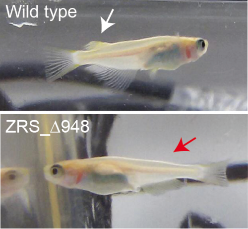
But, why were we all so excited? Well, the finding provided conclusive support for a shared regulatory logic controlling the development of paired (i.e pectoral) and unpaired (i.e. dorsal) appendages. This was indicating a common evolutionary origin for both types of appendages in stem gnathostomes. Since dorsal fins appear first in the fossil records, the discovery pointed to paired fins emergence by the exaptation of a developmental program first established in the median fins. The hypothesis had been formulated years before (5, 6), but this was a direct genetic demonstration entailing the functional conservation of a critical enhancer for more than 450 million years.


Finally, the finless fish

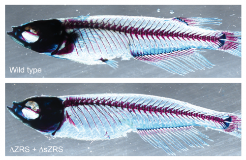
In the following months everybody did their best to complete the detailed analysis of the mutant phenotype and the ZRS expression in the different fins. In March 2017 we were ready to submit the paper, all believing the data deserved the best visibility. Happily, Tiago Faial, a senior editor in Nature Genetics, also saw the potential of the work and decided to send it for review. The review process was long but certainly improved substantially the quality of the paper. Although the reviewers found the work interesting, they requested us to delete also the shadow enhancer sZRS. They reasoned that if both ZRS and sZRS control shh expression in the pectoral fin bud, as our experiments suggested, their simultaneous deletion should result in a stronger phenotype. Again, Joaquín took the control and generated, in a record time, a larger deletion of 3.4 Kb encompassing both enhancers. There are many reasons for which such experiment would not yield the expected results. Among other things, additional unidentified enhancers may play a redundant role in paired fins. As a “defensive” experiment, we started another round of transgenics, but no additional fin enhancers were found. Then, to my absolute relief and delight, the results of the larger deletion came out. In the absence of ZRS and sZRS, shh failed to be expressed in the developing pectoral and pelvic fins, thus causing a very severe truncation of these extremities. Without paired and dorsal fins, the 3.4 Kb mutant larvae were able to swim quite decently using the remaining anal and tail fins. To our surprise, some of them managed to reach adulthood (See Figure 2) and even reproduced (See Video 1). This was the story behind this venture, or at least the way I witnessed it. The rest, either you can read it in the paper, or belongs to every participant’s personal experience.


I could not finish without mentioning the fantastic work that everybody did over the last years to push this story forward. Elisa de la Calle-Mustienes, Joyce Pieretti, and Silvia Naranjo performed a terrific job in the transgenesis and genomics front. Tetsuya Nakamura and Juan Pascual-Anaya helped a lot with the µCT and lamprey in situ experiments, respectively. And finally, Nacho Maeso, who was at the heart and soul of all scientific discussions. Without them, and the humble medaka fish, this work would have never been possible.


References.

1 Freitas et al 2014. J. Exp. Zool. B Mol. Dev. Evol. PMID:24677573

2 Shubin et al 2006. Nature. PMID:16598250

3 Sagai et al 2005 Development. PMID:15677727

4 Perri et al 2010. Current Biology. PMID: 20797865.

5 Freitas et al. 2006. Nature. PMID:16878142

6 Dahn et al. 2007. Nature. PMID:17187056


Thumbs up (10 votes)
Loading...

Tags: , , ,
Categories: Lab Life, Research

Laboratory Technician Position – Millman Lab

Posted by , on 23 April 2018

Closing Date: 15 March 2021

We are looking for someone to join our team in a NIH R01-funded laboratory technician position. The Millman lab is located at Washington University School of Medicine (St. Louis, MO) and is focused on developing a cellular therapy for diabetes using stem cell differentiation (https://endo.wustl.edu/millman-lab/). The primarily responsibilities of the position are assisting with mouse transplantations and performing assays to evaluate cells and tissues that we generate. If you are interested, please email your resume to jmillman [at] wustl [dot] edu or apply online at jobs.wustl.edu under job posting 39712.


Thumbs up (No Ratings Yet)
Loading...

Tags: ,
Categories: Careers, Jobs, Research

Postdoctoral position – Spence Laboratory, University of Michigan

Posted by , on 23 April 2018

Closing Date: 15 March 2021

Job Summary: The Spence laboratory is part of the Department of Internal Medicine, the Department of Cell and Developmental Biology and the Department of Biomedical Engineering at the University of Michigan Medical School. We are recruiting a highly motivated postdoctoral fellow to join our research team, and to conduct research focused on understanding human lung development. Our research seeks to understand species differences during development, cellular heterogeneity, cell-cell communication and progenitor/stem cell regulation in the lung. The position will include use of primary human tissue specimens and human pluripotent stem cells to address these topic areas.

The Spence lab is a highly energetic and collaborative group made up of undergraduate and graduate students, postdoctoral fellows and staff scientists. We conduct team-based science, while fostering and encouraging intellectual independence and creative approaches to addressing biological questions. As a laboratory, our whole team is committed to fostering a safe, peaceful and respectful environment for everyone to work towards common scientific goals. We actively support a diverse workforce.

We are interested in receiving applications from qualified applicants with training in a range of disciplines, including (but not limited to) developmental biology, cell biology, molecular biology, engineering, gemonics/epigenomics, systems biology and computational biology. The following skills are helpful, but not essential: pluripotent stem cell culture, organoid culture, gene editing (i.e. Cas9/Crispr), histology and immunofluorescence, confocal microscopy, FACS, analysis of large data sets (i.e. RNA-sequencing).

Required Qualifications: Qualified applicants will hold a Ph.D. in one of the aforementioned (or related) disciplines, will be self-motivated, and will have a demonstrated history of productivity in the form of peer-reviewed publications. Strong interpersonal communication skills are essential.

Background Screening: Michigan Medicine conducts background screening and pre-employment drug testing on job candidates upon acceptance of a contingent job offer and may use a third party administrator to conduct background screenings.  Background screenings are performed in compliance with the Fair Credit Report Act.

Mission Statement: Michigan Medicine improves the health of patients, populations and communities through excellence in education, patient care, community service, research and technology development, and through leadership activities in Michigan, nationally and internationally.  Our mission is guided by our Strategic Principles and has three critical components; patient care, education and research that together enhance our contribution to society.

Interested applicants should email Jason Spence: spencejr@umich.edu

Thumbs up (No Ratings Yet)
Loading...

Categories: Jobs

BSDB Medal & Award winners of 2018

Posted by , on 19 April 2018

This year was a special BSDB Spring Meeting! We celebrated the 70th anniversary of our society. If you weren’t there and would like to get a taste of the meeting, or you would like to relive the experience, please download the abstract book or listen to the especially composed history rap. As every year, the Spring meeting was the time of awards and medals! The BSDB would like to congratulate all awardees and prize winners, who are listed below. Some award presentations were filmed and the Company of Biologists team conducted a number of interviews with some of the prize winners. These will be linked out from here as they become available.


► Medal Awards

► Writing competition (PhD & postdoc – LINK)

  • winner (attendance at the 77th Annual SDB meeting in Portland, Oregon, USA): Daniyal Jafree (UCL) – “The paper in Developmental Biology that most inspired me
  • runner up: Laura Hankins (Dunn school, Oxford) – “Painting the embryo by numbers: how nature provided the tools for an inspirational experiment
  • runner up: Victoria Rook (Queen Mary, London) – “Is the future of developmental biology written in science fiction?

► PhD Poster Prizes

  • 1st Prize (attendance at the 77th Annual SDB meeting in Portland, Oregon, USA): Christian Louis Bonatto-Paesse (Oxford Brookes Univ.; McGregor group) – Poster 153 “A Sox gene is a key player in spider embryogenesis
  • 2nd Prize (£50 bank transfer): Natalie Kirkland (UCL, Paluch group) – Poster 4 “Investigating Mitotic Nuclear Dynamics of Pseudo-Stratified Epithelia in Drosophila melanogaster

► Postdoc Poster Prizes

  • 1st Prize (£200 bank transfer): Sarah Bowling (Imperial College London; Rodriguez group) – Poster 273 “P53 and mTOR signalling determine fitness selection through cell competition during early mouse embryonic development
  • 2nd Prize (£50 bank transfer): Michelle Percharde (UCSF) – Poster 135 “The LINE1 retrotransposon regulates early embryonic cell identity

► Other

  • BSDB Exhibition Passport Prize Winner (£200 Blackwell’s book tokens): Afnan Azizi (Univ. Cambridge)
  • The Dennis Summerbell Award will be announce at the next Autumn Meeting, but we would like to congratulate again the awardee of 2017: Helen Weaver (Univ. of Bristol)
Thumbs up (No Ratings Yet)
Loading...

Tags: , , , , ,
Categories: Events, Societies