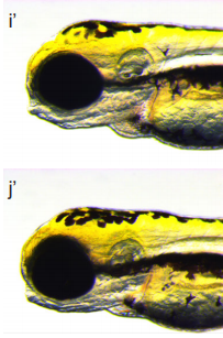
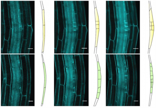
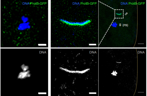
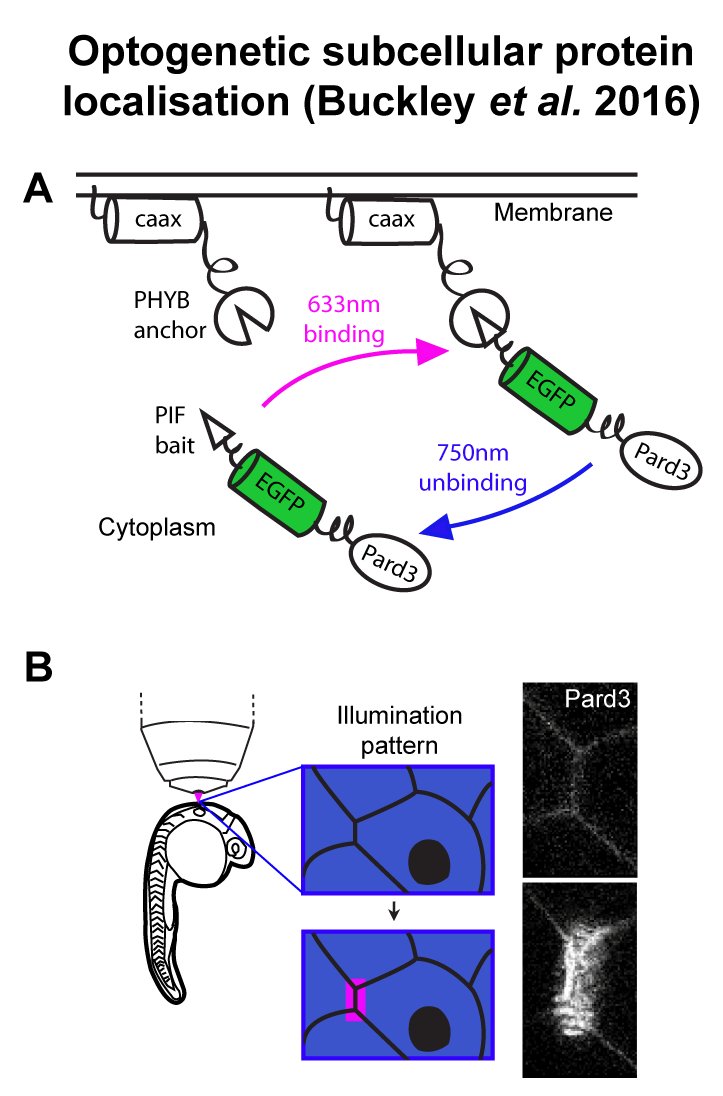
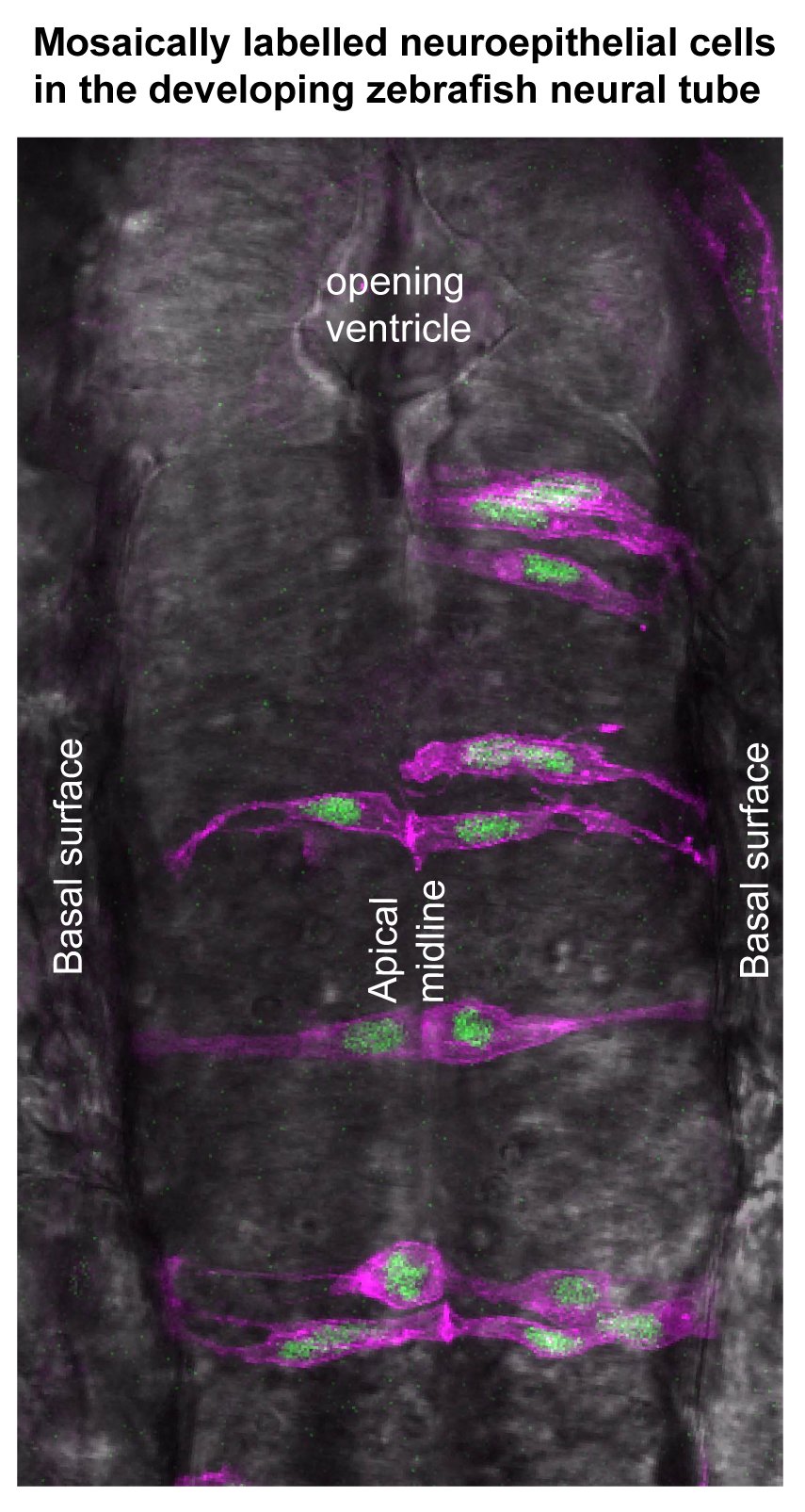
The Buckley lab at the department of Physiology, Development and Neuroscience (PDN), University of Cambridge is recruiting a postdoctoral research associate or research assistant. The lab uses cutting edge optogenetic and live confocal imaging approaches within the whole zebrafish neural tube to manipulate the polarity of single cells (Buckley et al., 2016, PMID: 26766447). In combination with CRISPR-mediated functional knock down experiments, we are directly testing the role of cell polarity in building epithelial integrity during organ development and in breaking it during developmental processes such as EMT and diseases such as carcinoma. The department of PDN is home to world-leading research in development, neuroscience, zebrafish live imaging and optogenetics. It hosts the Cambridge Advanced Imaging Centre (CAIC), which provides cutting edge microscopy systems, bespoke development of new imaging equipment and expert support.
We are seeking an enthusiastic and proactive candidate to join the team at the beginning of this exciting research. There are two main projects with which the successful candidate could be involved, depending on their interests and expertise. The first is to use optogenetics and tissue-specific CRISPR to determine how cell polarity and cell division are linked during epithelial establishment (we previously discovered a novel mechanism of cell polarisation that occurs independently to cell division: Buckley et al., PMCID: PMC3545300). We will do this within zebrafish embryos and, in partnership with our collaborators, in mammalian stem cell culture systems. The second project is to test the role of polarity dysregulation in tissue disruption. We will do this by optogenetically manipulating polarity-linked signalling pathways (such as the PI3K pathway) in the already established zebrafish neural tube epithelium. We will use 4D imaging to assess the cellular consequences of these manipulations and will model how signalling dynamics are propagated through the tissue in real time.
The successful candidate should have or be near completion of a PhD (or equivalent) in a relevant field and have a competitive history of research achievements. We are interested both in candidates with a background in developmental cell biology and those coming from a more biophysical background. Experience in molecular biology and genetics is essential and ideally the candidate should have experience in CRISPR technology. Candidates must also have a good understanding of data analysis and bioinformatics. Experience in advanced imaging and analysis would be a great advantage, as would specific knowledge of zebrafish genetics. Knowledge and interest in cell polarity and epithelial development, biochemical signalling pathways and optogenetic techniques would be desirable.
Although this is a full-time post, part-time working i.e. 80% of full-time over 4 days may be possible.
Fixed-term: The funds for this post are available for 3 years in the first instance.
Applications are invited from outstanding individuals to work under the supervision of Dr Rui Monteiro, Birmingham Fellow, on a BHF funded research project to study the role of TGFb signalling in angiogenic and haemogenic endothelial cell programming. The Monteiro Lab are interested in learning how extrinsic signalling impinges on lineage fate decisions in development and how progenitors and stem cells carry out those decisions, with a particular emphasis on the Transforming Growth Factor β (TGFβ) pathway. Previous work in the lab demonstrated that TGFb1 and TGFb3 play different roles in programming haemogenic endothelium to become of blood stem cells in vivo (Monteiro et al, 2016). The Research Fellow will study the gene regulatory network that carries out the ligand-specific functions for TGFb1 and TGFb3 ligands in haemogenic and angiogenic endothelium in vivo. They will make use of several approaches, including genome editing with CRISPR/Cas9, transgenesis, fluorescence activated cell sorting and transcriptional and epigenetic profiling using zebrafish as a model.
The successful applicant will have a first degree and a PhD in developmental biology, molecular genetics, biology or in a related discipline relevant to the project. They will also have a strong background in molecular biology and previous experience with model organism and/or analysis of transcriptomic and epigenetic data.
Informal enquiries should be directed to Dr. Rui Monteiro (R.Monteiro@bham.ac.uk)
Starting salary is normally on Grade 7 according to experience.
Closing date: 10 March 2018 Reference: 58652
To download the details of this position and submit an electronic application online please go to https://www.birmingham.ac.uk/staff/jobs/index.aspx. Please quote the appropriate Job Ref in all enquiries, alternatively information can be obtained from www.hr.bham.ac.uk
Project abstract – The ability of cells to self-organize into patterned tissues composed of multiple cell types is central to animal morphogenesis and relies on both biological and physical factors. We propose to investigate numerically and from a biophysical point of view the pattern formation observed in two different complex tissues respectively characterised by their stereotyped vs seemingly disorganised structure: the embryonic epithelium of Xenopus (with A. Pasini) and Drosophila brain tumors (with C. Maurange).
In Xenopus, we will study how multiciliated cells (MCCs) distribute in a regularly spaced pattern during intercalation into an epithelial layer, and explore how the pattern is established and maintained through a balance between mutual repulsion among MCCs and attraction between MCCs and epithelial layer cells. In Drosophila, we will study how clusters of brain cancer stem cells (CSCs) form and how they affect tumor progression. We will investigate how physical (tension, adhesion) and biochemical (growth and differentiation factors) cues contribute to segregate clusters of cells with different self-renewing potentials, regulate their size distribution and density, and thus determine tumor growth rate.
The computational tools envisaged for the project involve the numerical implementation of energy minimization algorithms such as the Cellular Potts Model (with R. Clément). We plan to model both systems with an energy function encompassing the different biological and physical interactions suspected to play a role in the processes. Such energy functions can comprise adhesion, tension, affinities or repulsions among cell types. Motility and cell proliferation can also be implemented at given rates, depending on cell types. Models with be implemented in light of the experimental results, and we expect that simulations will in turn guide the design of new biological experiments.
Expected profile – Candidates should have a robust background in physics and numerical simulations, and ideally be familiar with the Potts Model and its cellular version. As the project is strongly interdisciplinary and involves close collaboration with experimental biologists, previous experience in developmental biology or biophysics will be appreciated. A strong interest in biological questions, in particular in the principles of morphogenesis, is mandatory.
A postdoctoral position is available to study mechanisms of Sonic Hedgehog signal transduction in Stacey Ogden’s lab at St. Jude Children’s Research Hospital, Memphis, TN. The successful candidate will join a collaborative work group aimed at understanding how the Sonic Hedgehog pathway is regulated during development, and dissecting how its regulation is usurped in cancer. Areas of interest include biogenesis and secretion of the Hedgehog family ligands, contributions of lipid metabolism to pathway activity, regulation and signaling of the signal transducer Smoothened and investigation of the downstream effectors to which it signals. Research projects in the lab will entail use of biochemical and cell biological techniques and mouse model systems.
Applicants should have or expect a PhD degree at the time of application. The selected postdoctoral fellow will actively develop their own research project, perform laboratory experiments with minimal supervision, develop new procedures as needed and interact collaboratively with other members of the lab. The successful candidate will also actively participate in the publication and presentation of research results. Prior experience with signal transduction research, lipid metabolism or mouse model systems is preferred.
The San Francisco Declaration on Research Assessment (DORA) was conceived in 2012 at an ASCB meeting, and has since its launch in 2013 has garnered thousands of signatories from individuals and organisations. Its aim is to improve the way in which the quality of research output is evaluated, with a key recommendation being the elimination of journal-based metrics by funding agencies, institutions and publishers when judging research and researchers.
This year, DORA has had something of an upgrade: with the support of organisations including the Company of Biologists (the not-for-profit publisher that runs Development and funds the Node!), DORA now has a full time Community Manager Anna Hatch, a new steering committee, and a new website.
To find out more about these developments, we caught up with Stephen Curry, who chairs the DORA steering committee and is Professor of Structural Biology and Assistant Provost for Equality, Diversity and Inclusion at Imperial College London.
Hi Stephen! Can you tell us a bit about your science?
I’m a structural biologist and primarily use protein crystallography to work out the three-dimensional structures of interesting macromolecules. My main research efforts have been focused on virus and host-cell proteins involved in the replication of RNA viruses – principally foot-and-mouth disease virus and noroviruses. But I am in the process of winding down my research lab so that I can concentrate on other interests (discussed below) and my new role as Assistant Provost for Equality, Diversity and Inclusion at Imperial College.
You’re also passionate about science advocacy and communication – you are Vice Chair of the Science is Vital campaign group, contribute to The Guardian’s science blog Occam’s Corner, and have 16k followers on Twitter. Has this side of science – away from the lab bench – always been important to you?
It’s always been important but I’ve only really been properly active in this space since 2008 when I started my blog. I found that writing about science really made me think about what it means to be a scientist in 21st century Britain and that led me to learn a lot about scientific publishing and research funding, both of which are tied in rather convoluted ways to the business of research assessment. I have enjoyed getting involved in these debates and in campaigns to bring about positive change. Being involved in Science it Vital right from the very beginning has been a fantastic lesson in what can be done with modern communication tools if you just knuckle down and get organised.
You were one of the original signatories to DORA – why was DORA necessary in 2013?
It was already overdue in 2013. I wasn’t involved in the formulation of the declaration but was invited to sign prior to the launch and didn’t hesitate to do so. I had already become aware of the perverting effects of journal impact factors on science and scientists’ careers. And I knew that many other people shared my concerns. My 2012 blogpost, Sick of Impact Factors, remains on of the most ‘popular’ that I have ever written. It clearly struck a nerve.
And what do you think has been achieved in the years since then?
DORA has been really helpful in re-focusing the conversation on how the scientific community does research assessment. Without anyone designing the system, journal metrics have been co-opted for the evaluation of individuals to such a degree that publication in certain tiles (infamously Nature, Cell and Science in the biomedical sciences) are now seen as the key to success. DORA has helped to challenge that view – though we should certainly be mindful of parallel work on the Leiden Manifesto and The Metric Tide report (on which I was a co-author). So I think there is much greater awareness of the nature of the problem now and even some tentative steps to address it.
So what’s new – what does DORA’s new lease of life entail?
What’s new is that DORA now has a much higher level of material and financial support (from 9 organisations: ASCB, Company of Biologists, CRUK, eLife, EMBO, F1000, Hindawi, PLoS, and Wellcome). That has allowed us to hire a full-time community manager (Dr Anna Hatch) and refresh the steering group, which I now chair. We’ve also refreshed out web-site and have a new, easy-to-find URL (sfdora.org). This will allow us to raise the profile of DORA – we mean to get the word out much more proactively – but we are also determined to make a renewed effort to ignite the discussion around what constitutes robust and effective research assessment. We know that to change practice we need to figure out practical ways to help busy reviewers sift through job and grant applications and CVs without falling back on mis-use of the JIF.
Personally, what has it been like to be involved with DORA – is it challenging to get consensus from such a broad group of scientists and organisations?
There’s a lot of work to do because there is always resistance to change. DORA is not out to name and shame people or organisations that haven’t signed. But we want to challenge them to think about research assessment and do what we can to help them find a way forward. We aim to do a much more comprehensive job of discovering and disseminating good practice from around the world.
Why (and how) should young researchers get involved in DORA?
Because bad research assessment leads to bad research. DORA’s focus on improving research assessment fits very well with the ambitions that first attracts early career researchers to research: to understand and change the world. We will do that best if we are doing a proper job of recognising and rewarding the best research. That’s not just about publishing the best science (irrespective of journal name), but also meshes with parallel concerns about open science, data and code sharing and efforts to address deepening concerns about reproducibility, which are at least partly due to our over-reliance on metrics such as the JIF for judging individuals. However, we cannot simply expect young researchers to take the responsibility for change; it is up to the old guard (people like myself) and organisations like DORA to provide real support.
Primary supervisor: Professor Claudio Stern FMedSci FRS, Department of Cell and Developmental Biology, University College London
Project title: “Dynamics of cell behaviour during somite formation”
A studentship funded by the Anatomical Society is available in Claudio Stern’s lab. The project will study the mechanisms of somite formation, to elucidate the molecular and physical mechanisms that control somite size, shape and regional identity and the role of the “segmentation clock” and local cell-cell interactions in this process. It is a multi-disciplinary project and will involve advanced live imaging (including super-resolution microscopy in vivo), molecular biology, biophysics and some computational modelling. The project may include travel to the labs of collaborators in the USA and/or Singapore.
Conditions and requirements: This PhD studentship is open to British, Irish or European citizens who have spent at least three years at a British or Irish institute of higher education. It is funded by the Anatomical Society of Great Britain and Ireland and offers a stipend (tax free) of £16,553 per annum (revised annually), university fees (UK/EU rate) and a contribution to research expenses as well as funds to travel to meetings. Funding is for 3 years but it may be extended to a fourth year if necessary. Candidates should have a 2.1 (or equivalent) degree or better in a Biomedical, Physical or Computational science-related area and strong interest in developmental biology, ideally along with some laboratory experience working in a biomedicine-related research project. Experience with microscopy, programming (preferably PYTHON) and/or other computational/mathematical skills, are not essential but will be an advantage.
Starting date: 1 October 2018 or earlier by arrangement.
To apply (with a cover letter, CV a brief statement of your interests and the names and contact details of two academic referees), or for further information please contact Prof. Stern: c.stern@ucl.ac.uk
There is no formal closing date for applications but a student will be appointed as soon as a suitable, high quality candidate is identified.
For both young and established developmental biologists considering their next career move, choosing a model system with which to answer one’s research questions is a big decision. Of course, the most important thing to consider is whether or not a particular system is compatible with your research goals. But for a young scientist looking to make the move from one system to another, the ease of such a transition is an important concern. Having transitioned from sea urchin to zebrafish and then to chicken, I am often asked about my experience working with each of these three organisms, and what the practical, hands-on similarities and differences are among these creatures as model systems for development.
It began with the sea urchin
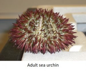
For my graduate research I studied skeleton formation in the sea urchin embryo. Most laboratories get adult sea urchins either by lab-organized diving trips if the lab is close enough to the ocean, or much more commonly by purchasing them from a supplier. When the sea urchins get to the lab, they need to be carefully acclimatized to the artificial sea water and the temperatures of lab aquaria, as any stress can make them spawn out their precious gametes prematurely. The maintenance of these aquaria and the monitoring of a new shipment of animals is an important and time-consuming job. Harvesting sea urchin sperm and eggs is as simple as injecting potassium chloride into the adults, after which they usually die (sad face!). In some species, however, adults can be shaken vigorously to make them spawn, and these animals can be used a few more times.
A major disadvantage of the sea urchin as a model system is the inability to establish stable transgenic lines or to easily create knockouts. Most analyses of development therefore depend on knockdown and overexpression experiments, which though easy to perform by microinjections, are not always clean. On the other hand, the ability to produce very large numbers of embryos from each round of fertilization gives the sea urchin a strong advantage for large-scale experiments. Since in situ hybridization and immunohistochemistry can be done on whole embryos, no time is spent sectioning embryos prior to analysis. Also, these embryos are very easy to analyze with several types of microscopy. Sea urchin embryonic development lasts only 2-4 days depending on the species, a definite benefit for moving a project along. An unexpected advantage of the sea urchin is the ability to replicate results in several closely-related species, which makes for a stronger case when putting together a story for publication. This also makes it an interesting model for evo-devo research especially when comparing multiple urchin species to other echinoderms.
Fish, fish and more fish
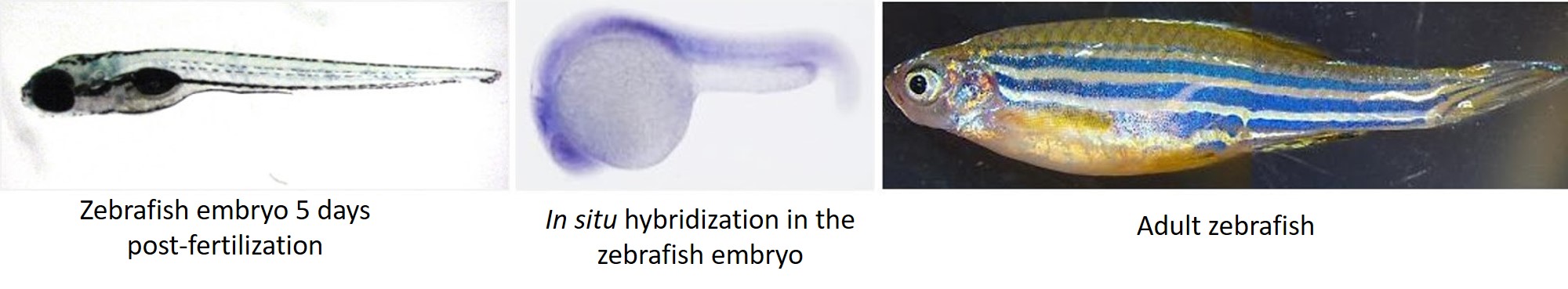
After my initial love affair with the sea urchin, I transitioned to zebrafish during my postdoc where I made zebrafish models for rare human diseases. Unlike the average sea urchin lab where adult animals are acquired from an external supplier, zebrafish labs often have several stable lines in-house, maintained by breeding adult fish and raising their offspring. While all this aquaculture can be time-consuming for a graduate student or postdoc, many institutions have fish core facilities and/or staff hired specifically for fish husbandry. Male and female zebrafish are usually kept separately, and to get them to breed, you only need to put them together in the same tank and they take over from there.
The zebrafish has many of the same advantages as the sea urchin, as it is easy to get large numbers of relatively transparent embryos. Adult animal and embryo care is similar to the sea urchin, as are methods of microinjection and analysis of results. The distinct advantage of the zebrafish (and indeed the main reason I found it attractive as a model system) is the immense power of transgenics in this system. Stable transgenic lines and knockouts are standard in the zebrafish field, and this opens up a world of possibilities for a researcher.
Another advantage of this system is the ability to culture embryos into adulthood. This lets you study a phenotype throughout the lifespan of the fish. Moreover, since the zebrafish is a closer relative to humans, antibodies raised against human proteins work better in the fish than they do in sea urchins. Aside from learning about zebrafish morphology and how to make the best use of the transgenic toolkit, my transition from sea urchin to zebrafish was seamless.
Chicken, anyone?
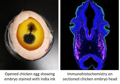
My current postdoctoral work is on neural crest and placode cells in the chicken embryo, which is distinct from the first two systems described. We get our embryos by incubating fertilized chicken eggs shipped in from a supplier, and as orders for eggs must be placed relatively far in advance, a researcher’s plan for experiments can fizzle out very quickly if egg quality is poor for a specific shipment. Also, a lot of time is spent on egg prep work, and you start out with fewer eggs, which means fewer embryos for your experiments.
Unlike aquatic embryos where you can just peek into a petri dish to tell what stage of development your embryos are, chicken eggs have to be opened and sometimes stained to check embryo development. Very early embryos especially do not take too kindly to this abuse, and may stop growing. My favorite thing about chick work is that you can stop and restart development multiple times simply by changing incubation temperature. This means that you can set a timer on an incubator and have your embryos ready at the stage at which you want them, when you want them. This cuts down on those infamous late nights in the lab.
The toolkit available to a chicken embryologist is relatively large and effective, and of the three systems described, antibodies generally work best in the chick. It is possible (but not at all common) to create stable transgenic lines of chicken, and other more common gene manipulation techniques such as morpholinos also work well. Since chicken embryos are relatively large, they often must be sectioned to observe the results of experiments.
The one thing I couldn’t escape with all three systems was the fact that embryo quality changed with the weather and the seasons.
The verdict?
Through all these transitions, I have found that studying development is a state of mind that transcends any specific model system, and the expertise you gain from one system is very easily transferable to another. However, if forced to pick a favorite, I would confess that my heart still belongs to the sea urchin.
The deadline to apply for the 2018 Mouse Development, Stem Cells & Cancer course at Cold Spring Harbor Laboratory (CSHL) is March 15th. If you don’t know much about the course or are on the fence about applying, I want to give you some background about my experiences from 2017, in hopes that I can convince you to apply, too.
The mouse course was founded in 1983 by superstar mouse wranglers Frank Costantini, Brigid Hogan, and Elizabeth Lacy. They recognized the power of emerging techniques to generate transgenic mice and knew they had to create an opportunity to pass these skills on to scientists of all levels and from around the world. Every year, the mouse course brings together 14 students at various stages of their careers and from diverse backgrounds to blast through an intense curriculum packed with hands-on lab work. The course is hosted by the historic Cold Spring Harbor Laboratory, established in 1890: the home of many major scientific discoveries that have shaped modern biology.
In June 2017, I was fortunate to take the mouse course. During my 3 weeks at the CSHL campus, I learned a host of experimental techniques and made tons of new professional contacts and friends. Exposure to these new tools and conversations with the many faculty speakers brought fresh insight into problems I face in my current research, giving me ideas for future projects and collaborations that will benefit me for the rest of my career. I emerged from the mouse course a better scientist equipped, not just with new skills, but the belief I can learn whatever I set out to learn.
Applying for the course
Who should apply for the mouse course? You! Graduate students at all levels, postdoctoral fellows, and new PIs. We had all 3 categories in our class, and we came from different backgrounds within the life sciences: genetics, neuroscience, developmental biology, cancer biology, reproductive biology, and medicine, among others. I am an early graduate student, as were several other participants. The 14 of us had different levels of experience with lab and mouse work, but everyone had the chance to learn and try each new skill, and even the more seasoned students picked up new tricks.
[An aside: If you’re worried you might not have enough experience to apply, consider my case. I had virtually no experience with most techniques taught during the course, and (have to admit), I have a fear of handling mice. As long as you’re willing to grit your teeth and really throw yourself into the course, any level of background experience (or mouse-phobia) is ok.]
For each applicant, the course has different albeit overlapping benefits. For example, junior graduate students can establish a solid experimental foundation for their graduate research. Senior graduate students and new postdocs moving into fresh projects in mouse labs can use the course to quickly gain experience with a new model system. Finally, senior postdocs and new PIs can learn more about the experimental techniques and equipment they’ll need for their new labs, not to mention learning how to design mouse protocols and experiments essential for development of their research programs. In sum: if you want to get ahead in mouse research, you should apply, regardless of your current position along whatever scientific trajectory you are following.
With an intensive, 3-week long course like this, you might find reasons to talk yourself out of applying. Maybe you worry about being away from your regular work schedule for 3 weeks. Sure, 3 weeks away can be disruptive, but in my opinion it’s worth it, and you can plan around it. The mouse course only happens once a year, and your lab will still be there when you come back! Plus, you’ll be returning with way more expertise and new ideas than when you left. If you’re worried about the cost of the course, remember that many institutions offer travel awards and funds to cover the costs. CSHL also provides generous financial aid to students who need it, so there’s no need to worry about funding, and financial concerns should not stop you from applying. Finally, if you’re pretty convinced the mouse course is a good idea, but you don’t know whether this is the right year, or maybe next year would be more convenient, here’s the truth: it will never be perfectly convenient (because science is busy!). That said, applying to the mouse course this year can provide you with the expertise and networking opportunities that will strongly affect how you tackle and succeed next year. So don’t wait.
Taking the course
The mouse course is jam-packed with experiments, some lasting several days, and all requiring specific lab supplies and equipment. The course instructors and teaching assistants lovingly and painstakingly organize all the protocols into a tight schedule that flows smoothly and follows evolving themes over the course of the 3 weeks. At the beginning of the course, the experimental focus is on the tools required to create a transgenic mouse; specifically, work with very early embryos. This section includes techniques like electroporation and microinjection of 1- and 2-cell embryos, as well as CRISPR workshops. During my course, we moved along the developmental timeline and towards tools required to manipulate and characterize older embryos, including roller bottle culture and electroporation of mid-gestation embryos, tissue sectioning and staining, and fluorescence-activated cell sorting (FACS). Aside from the lab work, there were guest lectures once or twice a day. Each speaker would cover background topics related to the day’s lab work, and then talk about new research being carried out by their group(s). Usually, the guest speaker would also join us in the lab, and sometimes even show off their skills by demonstrating experiments and giving students hands-on help.
The course also included great ways to connect with other students and faculty. Students presented their work in a mini-seminar early on in the course, which allowed us to get to know each other and understand our various backgrounds and scientific inclinations. We enjoyed weekly social events organized either by the instructors or the students, as well, providing a perfect place to unwind (and sing karaoke, if that’s your thing). One of my favorite parts of the course was the opportunity to meet the guest lecturers. Students would sign up to take speakers out for their “first drink” at the bar; basically, this was a chance to sit down and get to know each other in an informal setting. These get-togethers at the campus pub were a fantastic way to relax after a long day in the lab, not to mention giving us a chance to chat about anything from science and careers to English bulldogs and pink fairy armadillos. (Don’t ask.)
After the course (and some final reasons why you should apply if you aren’t already convinced)
After my 3 weeks at CSHL, I was mentally and physically exhausted, but excited about my work in a way I hadn’t felt before. I left the course equipped with new skills and heaps of reading materials and protocols, all of which I was enthused to share with my labmates back home. In the months since the course, I’ve had the chance to apply protocols taught by the instructors and to follow up on ideas that came up in conversations with guest lecturers. I’ve even spent some idle time dreaming about and planning out a potential post-doc project (which is still several years away for me), based on discussions that took place during the course. The many benefits I reaped overall extended beyond just my own feelings about research; the course was also a place I could start to build a network of scientific contacts and friends. At conferences since my time at CSHL, I have encountered so many familiar faces and reconnected with fellow students, teaching assistants, and guest lecturers. My experiences and friendships from the mouse course have helped me become a part of and feel at home within a larger scientific community.
So: if you want to add an injection of energy and excitement to your scientific research and career that will last for years (and why wouldn’t you?!), then apply. You won’t regret it.
Our latest monthly trawl for developmental biology (and other cool) preprints. Let us know if we missed anything.
Here at the Company of Biologists we are very excited about our soon to launch biology preprints highlighting service, preLights. Before the site goes live, you can sign up for email updates here and follow preLights on Twitter here.
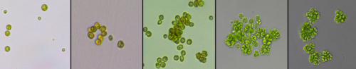
This month was notable for its plant content (see the many beautiful images below), plus a bumper ‘Research practice’ section with discussion of gender, ethics, education, and the future of US science.
The preprints were hosted on bioRxiv, PeerJ, andarXiv. Use these links to get to the section you want:
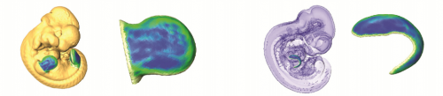
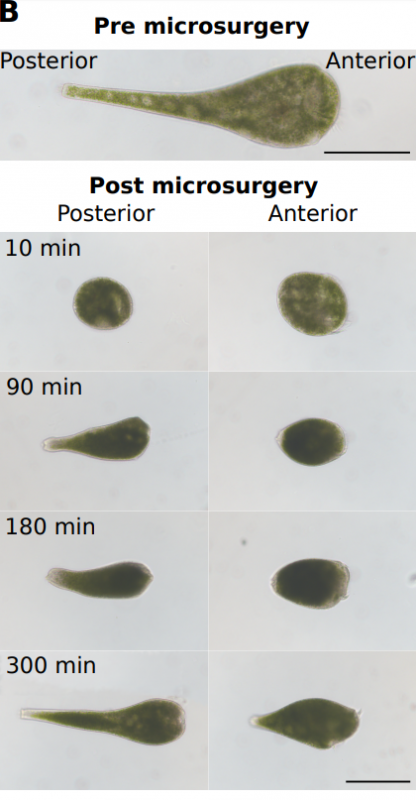
Pcdh18a-positive tip cells instruct notochord formation in zebrafish. Bernadett Bosze, Benjamin Mattes, Claude Sinner, Kathrin Stricker, Victor Gourain, Thomas Thumberger, Sham Tlili, Sabrina Weber, Joachim Wittbrodt, Timothy E Saunders, Uwe Straehle, Alexander Schug, Steffen Scholpp
GSK3 Controls Migration of the Neural Crest Lineage. Sandra G Gonzalez Malagon, Anna Lopez Munoz, Daniel Doro, Triona Bolger, Evan Poon, Elizabeth Tucker, Hadeel Adel Al-Lami, Matthias Krause, Christopher Phiel, Louis Chesler, Karen J Liu
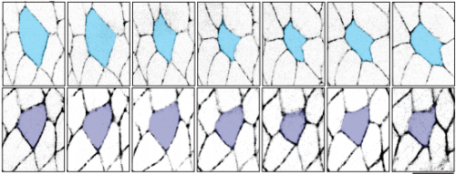
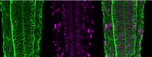
Mapping and dynamics of regulatory DNA during seed development. Alessandra M Sullivan, Andrej A Arsovski, Agnieszka Thompson, Richard Sandstrom, Robert E Thurman, Shane Neph, Audra K Johnson, Shawn T Sullivan, Peter J Sabo, Fidencio V Neri III, Molly Weaver, Morgan Diegel, Jennifer L Nemhauser, John A Stamatoyannopoulos, Kerry L Bubb, Christine Queitsch
The contribution of non-canonical splicing mutations to severe dominant developmental disorders. Jenny Lord, Giuseppe Gallone, Patrick J. Short, Jeremy F. McRae, Holly Ironfield, Elizabeth H. Wynn, Sebastian S. Gerety, Liu He, Bronwyn Kerr, Diana S. Johnson, Emma McCann, Esther Kinning, Frances Flinter, I. Karen Temple, Jill Clayton-Smith, Meriel McEntagart, Sally Ann Lynch, Shelagh Joss, Sofia Douzgou, Tabib Dabir, Virginia Clowes, Vivienne P. M. McConnell, Wayne Lam, Caroline F. Wright, David R. FitzPatrick, Helen V. Firth, Jeffrey C. Barrett, Matthew E. Hurles, on behalf of the DDD study
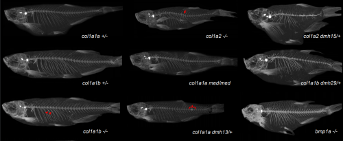
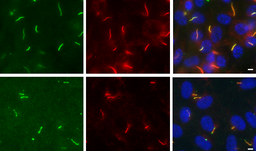
Zebrafish type I collagen mutants faithfully recapitulate human type I collagenopathies. Charlotte Gistelinck, Ronald Y Kwon, Fransiska Malfait, Sofie Symoens, Matthew P Harris, Katrin Henke, Shannon Fisher, Patrick Sips, Brecht Guillemyn, Jan Willem Beck, Petra Vermassen, Hanna De Saffel, MaryAnn Weis, Anne De Paepe, David R Eyre, Andy Willaert, Paul Coucke
A high-quality sequence of Rosa chinensis to elucidate genome structure and ornamental traits. Laurence Hibrand, Tom Ruttink, Latifa Hamama, Ilya Kirov, Deepika Lakhwani, Ning-Ning Zhou, Peter Bourke, Nicolas Daccord, Leen Leus, Dietmar Schulz, Henri Van deGeest, Thamara Hesselink, Katrijn Van Laere, Sandrine Balzergue, Tatiana Thouroude, Annie Chastellier, Julien Jeauffre, Linda Voisine, Sylvain Gaillard, Theo Borm, Paul Arens, Roeland Voorrips, Chris Maliepaard, Enzo Neu, Marcus Linde, Marie-Christine Le Paslier, Aurelie Berard, Remi Bounon, Jeremy Clotault, Nathalie Choisne, Hadi Quesneville, Koji Kawamura, Sebastien Aubourg, Soulaiman Sakr, Rene Smulder, Elio Schijlen, Etienne Bucher, Thomas Debener, Jan De Riek, Fabrice Foucher
The genome of the water strider Gerris buenoi reveals expansions of gene repertoires associated with adaptations to life on the water.David Armisen, Rajendhran Rajakumar, Markus Friedrich, Joshua B Benoit, Hugh M Robertson, Kristen A Panfilio, Seung-Joon Ahn, Monica F Poelchau, Hsu Chao, Huyen Dinh, HarshaVardhan Doddapaneni, Shannon Dugan-Perez, Richard A Gibbs, Daniel ST Hughes, Yi Han, Sandra L Lee, Shwetha C Murali, Donna M muzny, Jiaxin Qu, Kim C Worley, Monica Munoz-Torres, Ehab Abouheif, Francois Bonneton, Travis Chen, Christopher Childers, Andrew G Cridge, Antonin JJ Crumiere, Amelie Decaras, Elise M Didion, Elizabeth Duncan, Elena N Elpidina, Marie-Julie Fave, Cedric Finet, Chris GC Jacobs, Alys Jarvela, Emily J Jennings, Jeffery W Jones, Maryna P Lesoway, Mackenzie Lovegrove, Alexander Martynov, Brenda Oppert, Angelica Lilico-Ouachour, Arjuna Rajakumar, Peter N Refki, Andrew J Rosendale, Maria Emilia Santos, William Toubiana, Maurijn van der Zee, Iris M Vargas Jentzsch, Aidamalia Vargas Lowman, Severine Viala, Stephen Richards, Abderrahman Khila
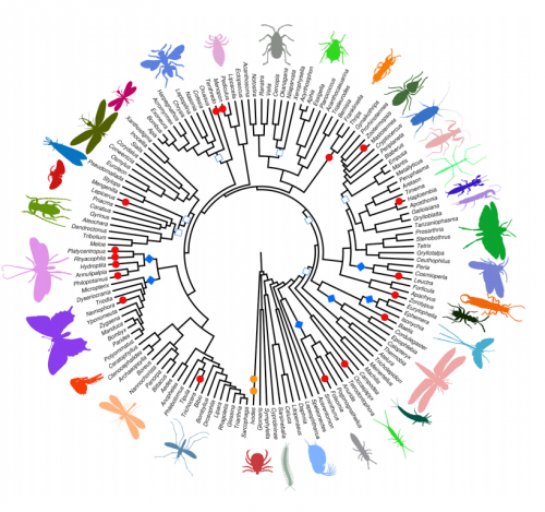
Embracing heterogeneity: Building the Tree of Life and the future of phylogenomics. Gustavo A Bravo, Alexandre Antonelli, Christine D Bacon, Krzysztof Bartoszek, Mozes Blom, Stella Huynh, Graham Jones, L. Lacey Knowles, Sangeet Lamichhaney, Thomas Marcussen, Hélène Morlon, Luay Nakhleh, Bengt Oxelman, Bernard Pfeil, Alexander Schliep, Niklas Wahlberg, Fernanda Werneck, John Wiedenhoeft, Sandi Willows-Munro, Scott V Edwards
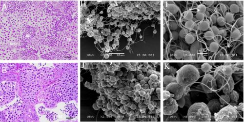
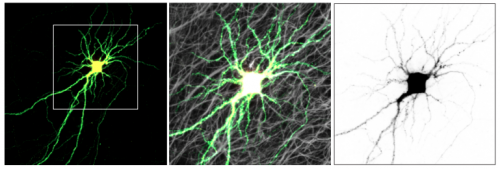
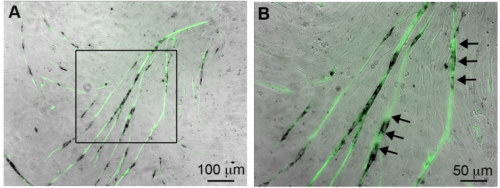
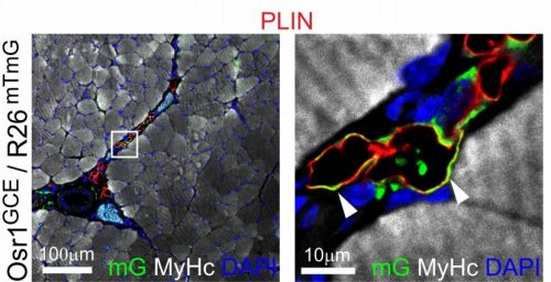
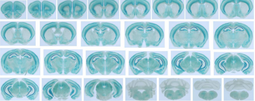
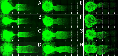
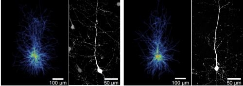
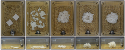
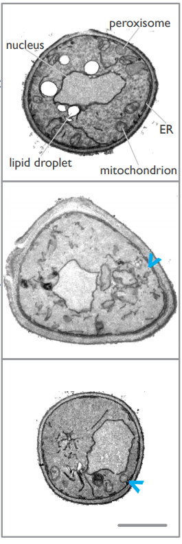
Observing the Cell in Its Native State: Imaging Subcellular Dynamics in Multicellular Organisms.Tsung-li Liu, Srigokul Upadhyayula, Daniel E Milkie, Ved Singh, Kai Wang, Ian A Swinburne, Kishore R Mosaliganti, Zach M Collins, Tom W Hiscock, Jamien Shea, Abraham Q Kohrman, Taylor N Medwig, Daphne Dambournet, Ryan Forster, Brian Cunniff, Yuan Ruan, Hanako Yashiro, Steffen Scholpp, Elliot M Meyerowitz, Dirk Hockemeyer, David G Drubin, Benjamin L Martin, David Q Matus, Minoru Koyama, Sean G Megason, Tom Kirchhausen, Eric Betzig
Digital museum of retinal ganglion cells with dense anatomy and physiology. J. Alexander Bae, Shang Mu, Jinseop S. Kim, Nicholas L. Turner, Ignacio Tartavull, Nico Kemnitz, Chris S. Jordan, Alex D. Norton, William M. Silversmith, Rachel Prentki, Marissa Sorek, Celia David, Devon L. Jones, Doug Bland, Amy L. R. Sterling, Jungman Park, Kevin L. Briggman, H. Sebastian Seung, the EyeWirers
Resolving the Full Spectrum of Human Genome Variation using Linked-Reads. Patrick Marks, Sarah Garcia, Alvaro Martinez Barrio, Kamila Belhocine, Jorge Bernate, Rajiv Bharadwaj, Keith Bjornson, Claudia Catalanotti, Josh Delaney, Adrian Fehr, Brendan Galvin, Haynes Heaton, Jill Herschleb, Christopher Hindson, Esty Holt, Cassandra B. Jabara, Susanna Jett, Nikka Keivanfar, Sofia Kyriazopoulou-Panagiotopoulou, Monkol Lek, Bill Lin, Adam Lowe, Shazia Mahamdallie, Shamoni Maheshwari, Tony Makarewicz, Jamie Marshall, Francesca Meschi, Chris O’keefe, Heather Ordonez, Pranav Patel, Andrew Price, Ariel Royall, Elise Ruark, Sheila Seal, Michael Schnall-Levin, Preyas Shah, Stephen Williams, Indira Wu, Andrew Wei Xu, Nazneen Rahman, Daniel MacArthur, Deanna M. Church
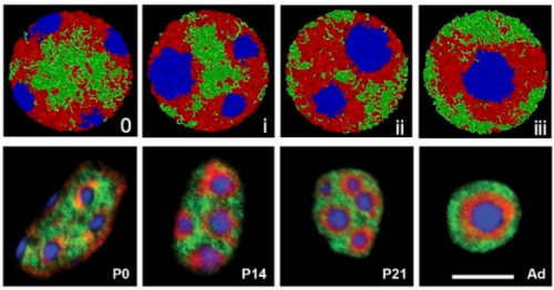
Equivalent high-resolution identification of neuronal cell types with single-nucleus and single-cell RNA-sequencing. Trygve E Bakken, Rebecca D Hodge, Jeremy M Miller, Zizhen Yao, Thuc N Nguyen, Brian Aevermann, Eliza Barkan, Darren Bertagnolli, Tamara Casper, Nick Dee, Emma Garren, Jeff Goldy, Lucas T Gray, Matthew Kroll, Roger S Lasken, Kanan Lathia, Sheana Parry, Christine Rimorin, Richard H Scheuermann, Nicholas J Schork, Soraya I Shehata, Michael Tieu, John W Phillips, Amy Bernard, Kimberly A Smith, Hongkui Zeng, Ed S Lein, Bosiljka Tasic
Mapping nonapoptotic caspase activity with a transgenic reporter in mice. Peter Nicholls, Thomas Pack, Nikhil Urs, Sunil Kumar, Gabor Turu, Evan Calabrese, Wendy Roberts, Ping Fan, Valeriy Ostapchenko, Monica Guzman, Flavio Beraldo, Vania Prado, Marco Prado, Ivan Spasojevic, Joshua Snyder, Kafui Dzirasa, G. Allan Johnson, Marc Caron
Harmonizing semantic annotations for computational models in biology. Maxwell L Neal, Matthias König, David Nickerson, Goksel Mısırlı, Reza Kalbasi, Andreas Dräger, Koray Atalag, Vijayalakshmi Chelliah, Michael Cooling, Daniel L Cook, Sharon Crook, Miguel de Alba, Samuel H Friedman, Alan Garny, John H Gennari, Padraig Gleeson, Martin Golebiewski, Michael Hucka, Nick Juty, Nicolas Le Novère, Chris Myers, Brett G Olivier, Herbert M Sauro, Martin Scharm, Jacky L Snoep, Vasundra Touré, Anil Wipat, Olaf Wolkenhauer, Dagmar Waltemath
Opportunities And Obstacles For Deep Learning In Biology And Medicine. Travers Ching, Daniel S. Himmelstein, Brett K. Beaulieu-Jones, Alexandr A. Kalinin, Brian T. Do, Gregory P. Way, Enrico Ferrero, Paul-Michael Agapow, Michael Zietz, Michael M Hoffman, Wei Xie, Gail L. Rosen, Benjamin J. Lengerich, Johnny Israeli, Jack Lanchantin, Stephen Woloszynek, Anne E. Carpenter, Avanti Shrikumar, Jinbo Xu, Evan M. Cofer, Christopher A Lavender, Srinivas C Turaga, Amr M Alexandari, Zhiyong Lu, David J. Harris, Dave DeCaprio, Yanjun Qi, Anshul Kundaje, Yifan Peng, Laura K. Wiley, Marwin H. S. Segler, Simina M Boca, S. Joshua Swamidass, Austin Huang, Anthony Gitter, Casey S. Greene
Recently, Nature published my correspondence “Dispense with redundant P values”. It highlights my concern that p-values are often calculated because “everybody does it”. This reminded me of the mechanical repetition that parrots are well-known for (footnote 1). Parroting of p-value reporting should stop and I suggest to only present a p-value in a figure if it is necessary for interpretation.
During the editing process of my contribution a specific example of a redundant p-value was removed. The reasoning was that it seemed unfair to single out only one paper. I agreed and I would like to stress that parroting of p-value reporting is not restricted to a specific paper, a specific issue of Nature or to some specific journal. It’s just that I found it very ironic that in the same issue of Nature that proposes “Five ways to fix statistics” (Leek et al., 2017) there are several clear examples of figures (in different papers) with meaningless p-values.
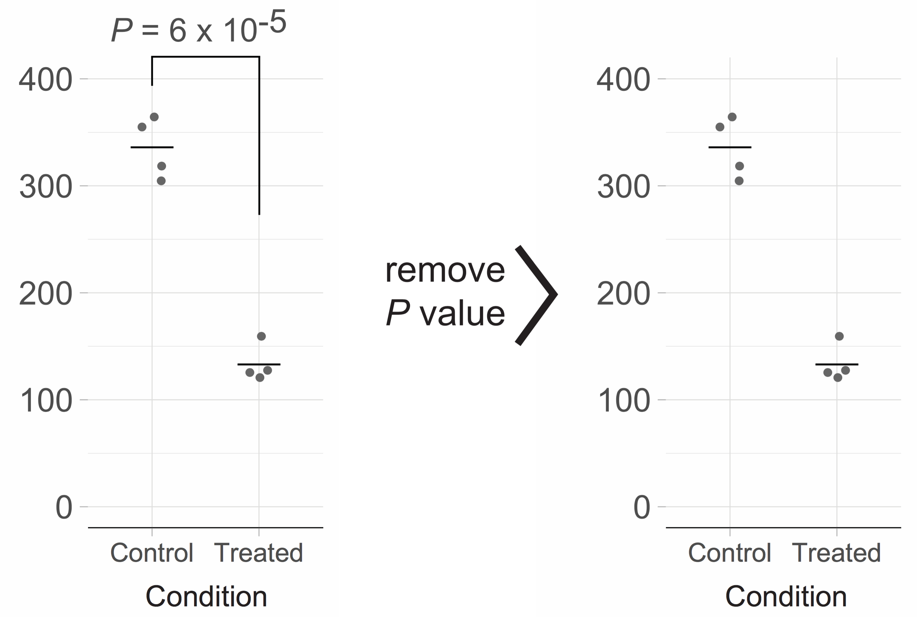
Since I am convinced that an example will clarify my point, I have extracted the data (see footnote 2) from one of the papers in the aforementioned issue (without disclosing the nature of the paper). I performed a t-test (two-tailed, unequal variances) as described in the paper and reproduced the p-value (footnote 3). The resulting figure is shown below on the left and closely mimics the figure of the original paper.
Clearly, there is a large difference between the ’Control’ and ‘Treated’ condition, reflecting a large effect of the treatment. To reach that conclusion, there is no need for a p-value. Moreover, the p-value does not convey any relevant information for interpretation of the figure. As such, the p-value qualifies as chartjunk (E.R. Tufte, 1983) and should be omitted. This will generate a cleaner figure (shown above on the right) that emphasizes the data.
The problems with p-values are larger than their meaningless use in figures whenever the effects are large. The way that p-values are defined has some confusing backward logic (footnote 4). Consequently, p-values are often misinterpreted (Greenland et al., 2016, Lakens, 2017) or misused as a ‘measure of credibility’ (Goodman, 2016). The misconception that p-values represent the strength of evidence is reinforced by catogerizing p-values, e.g. by using increasing number of asterisks (e.g. * P < 0.05; ** P < 0.01; *** P < 0.001). P-values cannot be used as a rating system (Wasserstein and Lazar, 2016) and categories should be avoided at all times.
To avoid the unnecessary, and at times misleading, use of p-values, the mechanical repetition of current practices should stop. Whether p-values are important for the interpretation of the figure should be a central question. Before that question can be answered, the correct definition of a p-value needs to be thoroughly understood. In addition, the correct interpretation and common misconceptions (Greenland et al., 2016) of p-values should be considered. I hope that careful reflection on the meaning of p-values will decrease their use and improve figures.
Acknowledgments: I am indebted to Marten Postma for the many discussions about statistical concepts and applied statistics, that have increased my understanding of the topic.
Footnotes
Footnote 1: The original title of my correspondence was “Prevent p-value parroting”. This title was changed to “Dispense with redundant P values” by Nature after I returned the proofs and without consulting me.
Footnote 2: The raw data that I extracted is listed below in csv format:
Footnote 3: This is an example of yet another questionable practice, i.e. calculating a p-value for a dataset with only a couple of datapoints per condition. Ironically, several examples can be found in the aforementioned issue while this matter has also been addressed previously by David Vaux (2012) in Nature (and by many others as well).
Footnote 4: The p-value is the probability of the observed data (or more extreme values), assuming that the null-hypothesis (there is zero difference between the two conditions) is true.
 (No Ratings Yet)
 recommendation being the elimination of journal-based metrics by funding agencies, institutions and publishers when judging research and researchers.
 (2 votes)
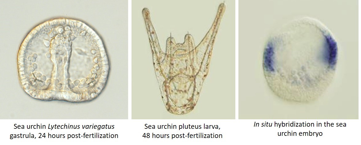
 (13 votes)