Do you play bass? Guitar? Keys? Sax? Brass? We’re putting together an SDB all-star jazz combo to play at the SDB meeting in Portland, and we’re looking for a couple more players! Feel free to reach out to me at david.matus@stonybrook.edu if you’re interested!
Adult neurogenesis is distinct from embryonic forms of neurogenesis in that neuronal development underlies extrinsic regulation, from physiological to pathological. This regulation targets different stages of neuronal development, between cell cycle entry and proliferation of radial glia-like stem cells to the lasting integration of the newly generated neurons. There are obvious feedback loops in that regulation of neurogenesis might alter behavior, which in turn acts back on adult neurogenesis. But how does this all work? How does regulation occur across scales? What constitutes ‘causes’ and ‘mechanisms’ in such complex system? This conference uses these key questions as entry point into discussions about how activity and development are linked in adult neurogenesis.
Free registration up for grabs!
Abcam and the Node are looking for an official meeting reporter to attend this meeting. The Reporter will be responsible for providing regular updates of interesting talks/discussions for twitter/tweets (by Abcam), plus a meeting report of their experience and the sights and sounds of the meeting (for publishing on The Node and Abcam website). Check out the article written by the last conference’smeeting reporter winner, Nambirajan Govindarajan.
To apply to be the meeting reporter, please send a short paragraph (written for a scientific audience, max. 200 words) toevents@abcam.com, telling us what makes adult neurogenesis fascinating to you! Application deadline: February 2, 2018. The winner will receive free registration to the meeting (travel and accommodation not included).
Meeting information
Organizer: Gerd Kempermann (CRTD – Technische Universität Dresden)
Confirmed speakers: Nora Abrous, Laure Bally-Cuif, Benedikt Berninger, Michael Brand, Federico Calegari, Fred H. Gage, Sebastian Jessberger, H. Georg Kuhn, Alejandro Schinder, Jason Snyder, Sandrine Thuret, and Henriette van Praag
Call for abstracts: Abstracts are invited for short talks or poster presentations and can be submitted during registration (see meeting website for more details)
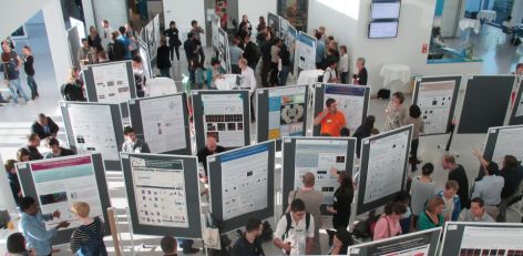
Poster session at the 2015 meeting
Terms and conditions
Effective November 2017
The contest is open to persons aged 21 years or over, except employees of, and consultants to Abcam, their families, government employees and any third party
associated with administration of the contest.
The contest is free to enter and no purchase is necessary. VOID WHERE PROHIBITED BY LAW.
All entries must be submitted by February 2, 2018. Entries received after this time will not be accepted.
The winner will be selected by a judging panel and announced after the entry deadline of February 2, 2018. The winner will receive one free registration to the Adult Neurogenesis 2018 conference.
The prize is non-exchangeable, non-transferable and no cash alternative is offered.
The decision of the judges regarding any aspect of the contest is final and binding and no correspondence will be entered into about it.
By entering this competition, you are agreeing to be bound by these terms and conditions, and you are representing and warranting to Abcam that you meet
the eligibility requirements of the contest. You are also agreeing to be contacted by Abcam in relation to the competition. We will not share your details with
anyone else except the winner’s announcement.
By entering this competition, you agree to receive further information regarding Abcam products and services. You can opt out of receiving communications
from us at any time by unsubscribing from our newsletter.
Insofar as is permitted by law, Abcam reserves the right to hold void, cancel, suspend, or amend the contest where it becomes necessary to do so.
Insofar as is permitted by law, Abcam, its agents or distributors will not in any circumstances be responsible or liable to compensate the winner or accept any
liability for any loss, damage, personal injury or death occurring as a result of taking up the prize except where it is caused by the negligence of Abcam, its agents or distributors or that of their employees. Your statutory rights are not affected.
Personal data supplied during this contest maybe passed onto third party suppliers only insofar as required for fulfilment/delivery/arrangement of the prize.
This offer is void if it conflicts with the policy of the recipient.
The contest will be governed by law of England and Wales and entrants to the contest agree to submit to binding arbitration to resolve any disputes relating to the contest.
Abcam plc located at 330 Cambridge Science Park, Cambridge CB4 0FL is the sponsor of this contest.
With new findings, scientists may be poised to break a long impasse in research on Huntington’s disease, a fatal hereditary disorder for which there is currently no treatment.
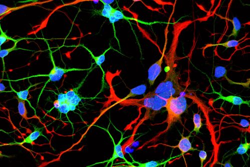
Huntington’s neurons show multiple nuclei (blue) within the same cell, and other signs of trouble, long before symptoms emerge.
One in 10,000 Americans suffer from the disease, and most begin to show symptoms in middle age as they develop jerky movements—and as these patients increasingly lose brain neurons, they slide into dementia. But the new research suggests that these symptoms may be a late manifestation of a disease that originates much earlier, in the first steps of embryonic development.
A team at Rockefeller led by Ali Brivanlou, the Robert and Harriet Heilbrunn Professor, developed a system to model Huntington’s in human embryonic stem cells for the first time. In a report published in Development, they describe early abnormalities in the way Huntington’s neurons look, and how these cells form larger structures that had not previously been associated with the disease.
“Our research supports the idea that the first domino is pushed soon after fertilization,” Brivanlou says, “and that has consequences down the line. The final domino falls decades after birth, when the symptoms are observable.”
The findings have implications for how to best approach treating the disorder, and could ultimately lead to effective therapies.
A new tool
Huntington’s is one of the few diseases with a straightforward genetic culprit: One hundred percent of people with a mutated form of the Huntingtin (HTT) gene develop the disease. The mutation takes the form of extra DNA, and causes the gene to produce a longer-than-normal protein. The DNA itself appears in the form of a repeating sequence, and the more repeats there are, the earlier the disease sets in.
Research on Huntington’s has thus far relied heavily on animal models of the disease, and has left many key questions unanswered. For example, scientists have not been able to resolve what function the HTT gene serves normally, or how its mutation creates problems in the brain.
“We started seeing things that were completely unexpected”
Suspecting that the disease works differently in humans, whose brains are much bigger and more complex than those of lab animals, Brivanlou, along with research associates Albert Ruzo and Gist Croft, developed a cell-based human system for their research. They used the gene editing technology CRISPR to engineer a series of human embryonic stem cell lines, which were identical apart from the number of DNA repeats that occurred at the ends of their HTT genes.
“We started seeing things that were completely unexpected,” says Brivanlou. “In cell lines with mutated HTT, we saw giant cells. It looked like a jungle of disorganization.”
When cells divide, they typically each retain one nuclei. However, some of these enlarged, mutated cells flaunted up to 12 nuclei—suggesting that neurogenesis, or the generation of new neurons, was affected. The disruption was directly proportional to how many repeats were present in the mutation: The more repeats there were, the more multinucleated neurons appeared.
“Our work adds to the evidence that there is an unrecognized developmental aspect to the pathology,” Brivanlou says. “Huntington’s may not be just a neurodegenerative disease, but also a neurodevelopmental disease.”
Toxic or essential?
Treatments for Huntington’s have typically focused on blocking the activity of the mutant HTT protein, the assumption being that the altered form of the protein was more active than normal, and therefore toxic to neurons. However, Brivanlou’s work shows that the brain disruption may actually be due to a lack of HTT protein activity.
To test its function, the researchers created cell lines that completely lacked the HTT protein. These cells turned out to be very similar to those with Huntington’s pathology, corroborating the idea that a lack of the protein—not an excess of it—is driving the disease.
“We should rethink our approach to treating Huntington’s”
The findings are significant, Brivanlou notes, since they indicate that existing treatments that were designed to block HTT activity may actually do more harm than good.
“We should rethink our approach to treating Huntington’s,” he says. “Both the role of the HTT protein and the timing of treatment need to be reconsidered; by the time a patient is displaying symptoms, it may be too late to medicate. We need to go back to the earliest events that trigger the chain reaction that ultimately results in disease so we can focus new therapies on the cause, not the consequences.”
The researchers hope their new cell lines will be a useful resource for studying the cellular and molecular intricacies of Huntington’s further, and suggest they may provide a model for examining other diseases of the brain that are specific to humans.
# # #
Contact:
Katherine Fenz, Media Relations Manager at The Rockefeller University
Tel: +1 212 327 7913; Email: kfenz@rockefeller.edu
About The Rockefeller University
The Rockefeller University is the world’s leading biomedical research university and is dedicated to conducting innovative, high-quality research to improve the understanding of life for the benefit of humanity. Our 82 laboratories conduct research in neuroscience, immunology, biochemistry, genomics, and many other areas, and a community of 1,800 faculty, students, postdocs, technicians, clinicians, and administrative personnel work on our 14-acre Manhattan campus. Our unique approach to science has led to some of the world’s most revolutionary and transformative contributions to biology and medicine. During Rockefeller’s 115-year history, 25 of our scientists have won Nobel Prizes, 22 have won Albert Lasker Medical Research Awards, and 20 have garnered the National Medal of Science, the highest science award given by the United States.
DanStem seeks two laboratory technicians or biomedical laboratory scientists for tasks related to a research project in stem cells and type 1-diabetes.
About DanStem
The Novo Nordisk Foundation Center for Stem Cell Biology – DanStem was established as a result of a series of international recruitments coupled with internationally recognized research groups focused on insulin producing beta cells and cancer research already located at the University of Copenhagen. DanStem addresses basic research questions in stem cell and developmental biology and has activities focused on the translation of promising basic research results into new strategies and targets for the development of new therapies for cancer and chronic diseases such as diabetes and liver failure. Learn more about DanStem at http://danstem.ku.dk/
Job description
You will be responsible for growing and expanding human stem cells and in close collaboration with the group’s researchers; you will help develop new methods and protocols for differentiation of hPSC. You will also perform basic characterization of hPSC using karyotyping, immunohistochemistry, qPCR and FACS, to ensure quality and reproducibility. In addition, you will be responsible for maintaining equipment in the hPSC laboratory.
The goal of the research project is to develop therapeutic active islet-like aggregate for future cell therapy in clinical phase 1 trials.
Your qualifications
We expect you to hold a degree as either laboratory technician or biomedical laboratory scientist and that you have several years of experience working as a technician in a research lab. Furthermore, we expect the following qualifications:
Minimum 5 years of experience in cellular work, preferably with stem cells
Experience with transfection, immunohistochemistry, qPCR and FACS
That you work systematically and are flexible
Good cooperation and communications skills as the project is carried out within a team of scientists and technicians
Good English skills in both speech and writing, as our working language is primarily English
For further information, please contact Professor Henrik Semb by e-mail semb@sund.ku.dk
Terms of salary and employment
The employments are planned to start as soon as possible upon agreement with the chosen candidates. The place of work is at DanStem, University of Copenhagen, Blegdamsvej 3B, Copenhagen. The average working hours are 37 hours per week. The positions are time limited to the end of 2020 with a possibility of further employment.
Terms of salary and employment are in accordance with the collective agreement between the Danish Ministry of Finance and the Organizations of Public Employees – the Governmental area (OAO-S) and the HK organization agreement with placement in salary group 5, or The Danish Association of Biomedical Laboratory Scientists and the Organization Agreement for Biomedical laboratory scientists.
Furthermore a salary supplement can be negotiated based on individual qualifications.
Application
Send your application electronically by clicking “Apply online” below or via this advertisement on http://employment.ku.dk/staff/. The application must include the following documents/attachments:
Motivated letter of application (max 1 page)
Curriculum vitae incl. education, experience, previous employments, language skills and other relevant skills
Certified copy of diplomas/degree certificate(s)
Letter of recommendation and/or contact details of referees
Application deadline: 20 February 2018
The University of Copenhagen wishes to reflect the diversity of society and welcomes applications from all qualified candidates regardless of personal background.
preLights is a new preprint highlights service supported by The Company of Biologists, the not-for-profit publisher which hosts the Node via one of its five journals, Development.
The site is currently being built and will be launched soon, but before then you can keep up-to-date by signing up for news here:
The Company of Biologists Gurdon Summer Studentship scheme was initiated by the BSDB in 2014 to provide highly motivated undergraduate students with an opportunity to engage in practical research during their summer vacation. Each year, ten successful applicants spend eight weeks in the research laboratories of their choices. For the last four years, the quality of projects and feedback we receive has been outstanding – as is clearly illustrated by reports from previous awardees.
The new round of applications is now open and will close Friday, 30 March 2018. Please, look at our dedicated web page for eligibility and application procedures.
Our recent paper in “Nature”[1] deconstructs molecular arguments that have been used to homologize bilaterian nerve cords. Our work illustrates well the strength of the comparative approach and the broad sampling across the animal tree of life that we use in my research group at the Sars Centre for Marine Molecular Biology.
Evo-Devo branched off as a discipline from Developmental Biology. Therefore Evo-Devo inherited concepts and narratives from Developmental Biology that dominated over those of its cousin-discipline Evolutionary Biology. This was in part because Evo-Devo began with the comparison of data from a handful of well-studied genetic models, such as Drosophila, C. elegans, and vertebrates, which were used as a quite limited representation of the animal diversity. At this time, Evo-Devo was more “Devo” than “Evo”.
I entered Evo-Devo as a trained comparative zoologist who was –and still is– fascinated by animal diversity, evolution, and the many developmental pathways animals can display. I was attracted by the new discoveries of developmental biology, but being well familiar with the comparative method, I was also skeptical about their potential evolutionary implications, given the low taxon sampling (that is, the sparse number of species investigated across a tree).
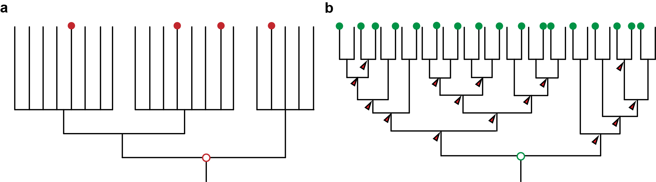
At about the same time, molecular phylogenetic methods were shaking and uprooting the Animal Tree of Life. New molecular trees provided evidence for the monophyletic Ecdysozoa [2] (e.g. nematodes and arthropods), Lophotrochozoa [3] (e.g. annelids, molluscs) and Deuterostomia (e.g. urchins and frogs). Traditional relationships, such as the close relationship of annelids and arthropods, were questioned. However, the order of the internal branches remained unresolved. Such polytomies, together with the low taxon sampling by that time, obscured evolutionary conclusions about ancestral characters in these major groups, and thus hampered the detection of evolutionary change in the animal tree of life (Figure 1a). Instead, this situation sparked anthropogenic speculations.
Evo-Devo was thus influenced by two problems: the low number of studied species, and the lack of a phylogenetic framework that allows hypotheses testing.
This situation rapidly changed in areas of the Animal Tree of Life where taxonomical sampling was denser, and where the phylogenetic relationships were better resolved – such as in the vertebrates, and partly in the arthropods. This favored hypothesis testing, and improved our understanding of morphological evolution, as seen for instance with the evolution of limbs from fins [4]. But scientists interested in squishy invertebrates had much harder times.
Advances in sequencing technologies, imaging, and molecular methods have radically changed Evo-Devo, opening up new opportunities, and becoming a continuous source of innovation in evolutionary biology. Next generation sequencing has hugely impacted phylogenomics, allowing much better resolved animal relationships, thus providing the necessary phylogenetic frame to test evolutionary hypotheses [5]. These same sequencing technologies also make it now much easier to take animals from the field and implement molecular techniques faster for their study, which enables a much denser taxonomical sampling of animal diversity. And as sampling more data points provides a better measurement in physics, sampling more species provide a better understanding of evolution (Figure 1b). A denser sampling within a resolved phylogeny allows a more solid reconstruction of ancestral characters and offers a much better picture about character changes during evolution [6]. This approach is common practice in evolutionary biology [7] – but has not yet been extensively used in Evo-Devo.
Figure 1: a) An unresolved phylogeny with only few taxa sampled (red dots) can only provide vague and untestable hypotheses. b) A well resolved phylogeny with large sampling allows the reconstruction at many more nodes and thus also insights about the evolution along the branches that connect the nodes. It also provides a framework for testing hypotheses.
The approach allows to test previous hypotheses that have been developed when low taxon sampling and/or unresolved phylogenies prevailed. For instance, our study of nine species published in “Nature” we show that the expression of the transcription factors traditionally used to call for a single condensed nerve cord in the last common ancestor of bilateral animals [8] evolved not only independently from each other, but also independently from the nervous system architecture and neuronal cell types. This implies that statements such as “this organ is so complex, it is unlikely to have evolved twice” are human presumptions, and that shared similarities might also be the result of independent adaptation processes (that is homoplasy). The comparative method strongly reduces human bias and also helps to avoid wrong conclusions [9].
The advantages of our approach not only extend to the study of organ system evolution, but simply affects all other characters that undergo evolutionary changes: tissues, cell types, developmental modes, genome architectures, gene expression [9]. Increasing the number of research organisms and the interpretation of the data on the basis of a resolved phylogeny provides a powerful toolkit for understanding evolution. It opens up the horizon for addressing new questions on when and how things changed in evolution, which is indeed where our curiosity lays.
Please, have a look at the newest issue of the BSDB newsletter, which can be downloaded here. It covers two eventful years of our society’s history and is by far the longest ever published! This seems only appropriate considering that 2018 marks the 70th anniversary of the BSDB’s foundation. In recognition of its history, the BSDB has decided to digitise all its newsletters which have recently been re-discovered in an almost forgotten archive. They will be gradually uploaded on our archive page (bsdb.org/about-us/bsdb-newsletters) where issue no. 1 from 1979 has already been linked out to get the ball rolling. We have now first lists of the archive’s contents which seem to date back to the early 60s. If you have an interest in these, please contact comms@bsdb.org. Also, if you have further thoughts on how to catalogue and store the archive safely for the future, please let us know.
Editorial: My final newsletter – and some thoughts about communication
This is my final newsletter as communications officer of the BSDB. I must admit that I enormously enjoyed the task, and can only hope that the changes to our website and the ways in which the society has been represented during my time in office are seen positively by our members. It is my pleasure to announce that Ben Steventon (p.6) has agreed to take over as BSDB communication officer from autumn 2018. I am confident that he will do a brilliant job.
Unlike previous editions, this newsletter covers two years of our society’s life. But honestly, did you even notice the delay? Extrapolating from the download metrics of former newsletters, the long gap is very likely to have gone widely unnoticed, and I can see two reasons for this. Firstly, on the positive side, all our society news is now being published more promptly on our website or on The Node. This changes the nature of the newsletter from being a source of primary information, and turns it into a legacy item or even a historical document for future BSDB generations. This is reason enough for the BSDB to continue with its newsletter. Secondly, a reason of more concern is that the low viewer numbers likely reflect a tendency of communication fatigue in our community. Let me briefly extend on these thoughts before outlining the content of this newsletter.
The BSDB informs promptly online
If I have done my job well for the last two years, the contents of this newsletter should no longer be news to you, but rather be a reminder of our active society life during this period. I have made every effort that news was brought to you promptly via our website or through The Node. Of these, The Node has become an increasingly important medium.
Maintained by The Company of Biologists as a communication platform for the community of biomedical scientists, The Node has taken on the form of a modern electronic newsletter that reaches out internationally. As explained in a recent publication, The Node’s community manager Aidan Maartens either authors or commissions meeting reports, book reviews, obituaries or interview transcripts, and collates community-relevant information about meetings and workshops. Importantly, we as individuals or societies can use The Node as a communication platform to advertise jobs and events, write about science-related topics, explain our latest publications, or share community-relevant experiences. Once registered, we can publish freely and ‘uncensored’ as long as we keep within The Node’s reasonable rules. In this way, we can capitalise on a very well established communication platform, which is further enhanced by The Node’s Twitter and Facebook accounts, each with thousands of followers. Much of the science-related news that would previously have been disseminated via the BSDB newsletter or website, is being covered by The Node, taking an enormous work load off my shoulders – and the same can likely be said for Developmental/Cell Biology societies worldwide. There is true synergy and, naturally, more community members visit The Node’s website than the BSDB’s. Consequently, I closely collaborate with Aidan and follow a strategy in which many of our news posts are published on The Node – of course always making sure that they are likewise accessible through our own site.
This said, the BSDB website still has an important purpose. User metrics indicate that our site has its prime function in BSDB-specific information, such as our conference and travel grants, awards and meetings and committee information. I take great care that these pages are updated as soon as new information is available. Please, let me or Ben know if there is anything that’s wrong or missing – we’ll act swiftly.
Is our communication failing?
Regardless of whether newsletters occur in static journal format or as dynamic websites, they often are important motors for scientific communities and their science, as is well explained in an article by C. M. Kelty from 2012. But does this strategy still work effectively? I sometimes feel that we were better informed in pre-internet times when information was less abundant but focussed on the essentials, and when dissemination was easier because the readership was hungry for information. Today, we tend to see quantity over quality: more than a hundred emails rain into our accounts every day, and social media timelines have become so busy that there is hardly time to view even a fraction of the messages. And, even if information is being read, I see little evidence that this leads to impactful social media debates. Furthermore, the sharing of information on social media is short-lived and often ineffective as can be deduced from viewer metrics of shared links. Even the use of social media as a mere source of professional information seems to fail: a survey by The Node showed that most members of our community have no Twitter account. Even more alarmingly, subscription rates to websites are low (~900 for The Node and ~150 for the BSDB), when considering that the BSDB alone has a ~1400 strong membership. In a nutshell, besides not reading the newsletter, many of us are not tapping into the existing online community news channels.
In consequence, important information can no longer be disseminated effectively and reliably, and our community no longer has the means to develop a common voice. This clearly weakens us in times where the need for communicating the importance of fundamental science is perhaps greater than ever. In recognition of this challenge, many contributions in this issue are dedicated to the topic of science communication: our chair Ottoline Leyser speaks about the importance of communication (p.4); a dedicated article explains the BSDB’s advocacy campaign in collaboration with The Node (p.23); an overview of a recent special issue on science communication in the field of biomedical science is being provided (p.27); our student and postdoc representatives announce a writing competition aiming at advocacy (p.31); the example of an advocacy article for Open Access Government is given (p.78).
Further contents of this issue
As usual, the start of this newsletter is made by the chair’s and officers’ reports. In the chair’s address (p.4), Ottoline Leyser reflects on the BSDB as an inclusive, co-operative, and outward looking society, and the need to uphold these traits in times of Brexit and worldwide political tendencies of isolationism; Ottoline ends her address with some thoughts on the importance of communication. The secretary’s report by Kim Dale (p.5) highlights the positive developments of the BSDB in terms of its steadily growing membership. She notes that, in 2018, the committee will see a turn-over of 5 members (see also p.7) – so await a call for nominations before the next Spring Meeting. In the same vein, the three new members that have joined the committee in 2017 are being introduced on page 6. The meetings officer Joshua Brickman (p.9) looks back at the BSDB’s excellent meeting record of the last two years (see also meeting reports on pages 11 and 14), and gives an outlook on the exciting meetings planned for the next three years (see also p.10), including the BSDB’s 70th anniversary meeting taking place 15-18 April 2018 in Warwick (see this issue’s cover image). The treasurer’s report by Chris Thompson (p.19) is an impressive account of the high number of members that were supported to attend meetings or workshops in 2016 and 2017, and sends out the reconfirming message that our financial status remains solid, providing the BSDB with continued capacity to support its members and their science-related activities. Our student & postdoc representatives, Alexandra Ashcroft and Michelle Ware (p.30), report about their outstanding efforts to deliver on the requests of junior members expressed during the survey from 2015. In response, Alex and Michelle introduced a very successful career workshop (see the respective reports on p.32) and a new career website (detailed on p.35). Finally, the newsletter concludes with reports about our main awardees of the last two years, in particular the winners of the Waddington (p.38), Cheryll Tickle (p.41) and Beddington medals (p.45), as well as the two Dennis Summerbell awardees (p.47), and 14 project reports by students who were supported by the Gurdon/The Company of Biologists Summer Studentship scheme. See a complete overview of all awardees, including poster prize winners (p.36) – and remember that most award lectures were documented and are available on our YouTube channel.
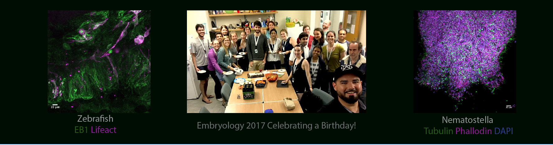
The deadline to apply for the 2018 Embryology course at the Marine Biological Laboratories is just one week away (February 1). That means it is time to get your application submitted!
If you have read the previously posted articles, you learned about this course helping Ania over come fears, its life changing capabilities from Tessa and the benefits of a collaborative environment from Steve.
Don’t pass up the opportunity to be in this environment and place of personal and scientific growth.
I am currently finishing up my PhD at UNC in Molecular, Cell and Developmental Biology. However, before taking the Embryology course I considered myself purely a Biochemist, Cell and Structural Biologist. My research focuses on understanding the structural and functional role of the cytoskeleton at the atomic to cellular level. Due to this skill set many of my colleagues (myself included) thought I would be a Physiology student (another wonderful summer course at MBL – oh and the two courses are kind of rivals). However, as I proceeded through my graduate work I knew that I wanted to understand the role of the cytoskeleton at the tissue and whole animal level. I didn’t know a lot about the work being done in this area and the scientists doing this work. I knew that the Embryology course would offer me the best way to gain technical knowledge, connections to developmental biologists and to learn about the work currently being done in this field. The course provided me with all of this and so much more. The connections I made at the course helped me to land a wonderful postdoc in Developmental Biology!
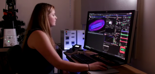
The connections and personal growth the course fosters and the skills acquired are far beyond any other experience I have had thus far. Honestly, I was worried that I wouldn’t fit in – or I would be so lost and so far behind all the time – I couldn’t have been more wrong. I felt right at home, every single person was fabulous, willing to help out and answer any and all of my questions no matter how basic or crazy. One of my favorite things about the course was the access to so many amazing microscopes. I had such a blast imaging on a dozen or so different systems!
No matter your background, training or knowledge base you will learn so much from this course and you will think about science in new and different ways. Do not hesitate to apply!
For more information on the course check out my blog posts from last summer and #embryo2017 on twitter and instagram.
POSTDOCTORAL POSITION is immediately available to study different aspects of lymphatic vasculature development using a variety of available mouse models. Highly motivated individuals who recently obtained a PhD. or MD degree and have a strong background in mammalian vascular, molecular and developmental biology are encouraged to apply. Interested individuals should send their curriculum vitae, a brief description of their research interests, and the names of three references to:
Guillermo Oliver, Ph.D
Thomas D Spies Professor of Lymphatic Metabolism
Director Center for Vascular and Developmental Biology
Northwestern University Feinberg School of Medicine
Northwestern University is an Equal Opportunity, Affirmative Action Employer of all protected classes, including veterans and individuals with disabilities. Women, underrepresented racial and ethnic minorities, individuals with disabilities, and veterans are encouraged to apply. Hiring is contingent upon eligibility to work in the United States.
 (No Ratings Yet)
 (8 votes)
 This is my final newsletter as communications officer of the BSDB. I must admit that I enormously enjoyed the task, and can only hope that the changes to our website and the ways in which the society has been represented during my time in office are seen positively by our members. It is my pleasure to announce that Ben Steventon (p.6) has agreed to take over as BSDB communication officer from autumn 2018. I am confident that he will do a brilliant job.