Laboratory of Protein Metabolism in Development and Aging
International Institute of Molecular and Cell Biology in Warsaw
is seeking PhD student
Location: Warsaw, a vibrant city with an international academic research environment. International Institute of Molecular and Cell Biology (www.iimcb.gov.pl) – one of the most dynamic and top ranked Polish research institutes.
Job description: Laboratory of Protein Metabolism in Development and Aging, which will be opening on August 2017, is seeking highly motivated PhD candidates to join our young team investigating the protein homeostasis in development and aging. We use both genetic, molecular and biochemical approaches, primarily in the C. elegans, to study proteolytic networks. PhD fellowship is funded in frame of National Science Centre OPUS grant.
Summary: Organismal development or environmental stimuli challenge the homeostatic protein balance (proteostasis) of individual cells, tissues or the entire organism. The ubiquitin proteasome system (UPS) is a key determinant of proteostasis as it regulates the turnover of damaged proteins supporting cellular protein homeostasis and thereby maintains the proteome during stress and aging. Our long-term objective is to understand the mechanistic and developmental aspects of protein degradation pathways defined by combinations of particular ubiquitin ligases (E3). The identification of stress and aging-induced signals that coordinate the interplay between specific E3s will offer intriguingly new mechanistic insights how proteolytic networks are fine-tuned to maintain the cellular proteome and support development and longevity.
Qualifications:
• master degree in any field related to biological sciences obtained within the last two years;
• general laboratory experience;
• expertise in either of the following areas will be preferred: cell biology, molecular biology, genetics, biochemistry, fluorescence microscopy
• analytical and creative thinking;
• ability to communicate knowledge in English (written and spoken);
• motivation and passion for experimental work;
• excellent interpersonal skills.
How to apply:
Please send your application to the e-mail address: wpokrzywa@iimcb.gov.pl, until 20th September 2017. The application should include cover letter, CV including candidate’s research achievements, university scores, and marks for candidate’s master degree. Thanking all applicants for their interest, we will contact only selected candidates for an interview.
Please include in your application the following statement: “In accordance with the personal data protection act from the 29th of August 1997, I hereby agree to process and to store my personal data by the Institution for recruitment purposes”.
The recruitment procedure fulfills the National Science Centre’s regulations on granting the scholarships to young scientists.
Selected publications:
Riga T*, Pokrzywa W*, Kevei E, Akyuz M, Vishnu Balaji, Svenja Adrian, Hoehfeld J, Hoppe T. (2017). The ubiquitin ligase CHIP integrates proteostasis and aging by regulation of insulin receptor turnover. Cell. 169: 470-482.
Ackermann L, Schell M, Pokrzywa W, Gartner A, Schumacher B, Hoppe T. (2016). E4 ubiquitin ligase specific degradation hubs coordinate DNA double strand break repair and apoptosis. Nat Struct Mol Biol. 23: 995-1002
Kaushik S, and Cuervo AM (2015). Proteostasis and aging. Nat Med. 21, 1406-15
Frumkin A, Dror S, Pokrzywa W, Bar-Lavan Y, Karady I, Hoppe T, Ben-Zvi A. (2014). Challenging muscle homeostasis uncovers novel chaperone interactions in Caenorhabditis elegans. Front Mol Biosci., doi: 10.3389
van Oosten-Hawle P, and Morimoto RI (2014). Organismal proteostasis: role of cell-nonautonomous regulation and transcellular chaperone signaling. Genes & Dev. 28: 1533-43
Segref A, Kevei E, Pokrzywa W, Mansfeld J, Schmeisser K, Livnat-Levanon N, Ensenauer R, Glickman M.H, Ristow M, Hoppe T. (2014). Pathogenesis of human mitochondrial diseases is modulated by reduced activity of the ubiquitin/proteasome-system. Cell Metab. 4:642-652
Pokrzywa W. and Hoppe T. (2013). Chaperoning myosin assembly in muscle formation and aging. Worm. 2:e25644
Gazda L*, Pokrzywa W*, Hellerschmied D, Loewe T, Forné I, Mueller-Planitz F, Hoppe T, Clausen T. (2013). The myosin chaperone UNC-45 is organized in tandem modules to support myofilaments formation in C. elegans. Cell. 1, 183-195.
Kuhlbrodt K, Janiesch PC, Kevei E, Segref A, Barikbin R, and Hoppe T (2011). The Machado-Joseph disease deubiquitylase ATX-3 couples longevity and proteostasis. Nat Cell Biol. 13, 273-81
With a new grant of almost 15 million EUR from the Novo Nordisk Foundation, DanStem scientists will be focused on a new programme for translational hematology.
The Programme could have a major impact on treatment of haematological malignancies. The primary aim is to identify novel treatments for patients with blood cancers AML (Acute Myeloid Leukaemia) and MDS (Myelodysplastic Syndrome) for which treatments and success rates have changed little in the last decades.
Professor Kristian Helin will head the Programme, which will include collaboration with researchers from Rigshospitalet, Denmark’s largest hospital. Major activities include:
Identifying and characterizing cancer stem cells from patients with blood cancer;
Identifying the best available treatment for individual patients by screening patient-derived cancer stem cells for sensitivity toward a panel of approved drugs (personalized medicine); and
Collaborating with companies to develop new drugs for treating blood cancer.
The Division of Developmental Biology at the FAU Erlangen-Nürnberg, invites applications for a
PhD student position (Salary Scale E13 TV-L/65%) on
Molecular mechanisms of muscle lineage reprogramming
in the group of Dr. Christoph Schaub. The position will start at the earliest possible date and will be limited to three years with a possible extension.
The Schaub group is interested in the molecular mechanisms that regulate syncytial muscle cell lineage commitment, maintenance and plasticity using the Drosophila embryonic and adult musculature as a model. The PhD project will focus on the molecular mechanisms that guide a naturally occurring direct lineage reprogramming process during the metamorphosis of the Drosophila musculature. In particular, the project will define the molecular processes that initiate and execute the dedifferentiation of syncytial embryonic muscles into mononucleate myoblasts which in turn are reprogrammed into the progenitors of adult heart associated muscles (Schaub et al. 2015, Curr Biol (25), 488-494). The PhD student will use a broad spectrum of state of the art techniques ranging from genome editing to modern live imaging approaches to analyse these questions.
We are looking for a highly motivated candidate with experience in Drosophila genetics and/or cell and molecular biology. Experience in microscopic tissue dissections is an advantage but not a requirement. We offer an exciting project utilizing our combined expertise in muscle cell biology and Drosophila genetics in a well-equipped lab. The project is embedded in an interdisciplinary scientific landscape in association with the Muscle Research Center Erlangen (MURCE, http://www.murce.fau.de/) and the Optical Imaging Center Erlangen (OICE, http://www.oice.uni-erlangen.de/) and will have access to high end imaging microscopes (Confocal, Spinning disk and Light-sheet microscopy).
If you are interested in the position please send a cover letter stating your motivation, your curriculum vitae, copies of Bachelor/Masters certificate (or equivalent) and two letters of recommendation or contact information for two scientific references in a single PDF to christoph.schaub@fau.de.
The Marsden Lab in the Department of Biological Sciences at North Carolina State University is seeking a full-time Research Assistant/Lab Manager. The successful applicant will facilitate the lab’s day-to-day operations, participate in training undergraduate and graduate students, and engage in research projects focused on understanding the genetic and neural circuit basis of behavior.
This is a 12-month, full-time, exempt position with a comprehensive benefits package. The position will present opportunities for co-authorship of publications, and training in essential techniques will be provided.
Who you are:
You are a biologist, preferably a neuroscientist, with previous experience working with zebrafish.
You have excellent organizational skills and pay keen attention to detail.
You think strategically and communicate clearly.
You enjoy working in a team, teaching, and mentoring others.
You are curious and love to learn.
Your essential duties:
Care for and maintain a zebrafish colony, including daily feeding and water quality monitoring
Perform routine zebrafish husbandry to generate larvae for experiments
Order and manage inventory of laboratory supplies
Conduct research, working with the principal investigator to plan, design, and perform experiments in 3 areas:
Analyze the behavior of zebrafish in response to visual, auditory and olfactory stimuli using high-speed imaging and automated tracking software; develop new behavioral assays to measure learning, anxiety, and social behavior
Use molecular biology techniques to assay gene expression, design and create transgenic constructs, and microinjections to establish transgenic zebrafish lines for visualization of neural circuit connectivity and activity
Use confocal microscopy to image neural development, connectivity and activity, as well as gene expression patterns using in situ hybridization and immunohistochemistry
Interact with and participate in training students in the laboratory
Qualifications:
Master’s degree or Bachelor’s degree plus 3 or more years of relevant experience in a research laboratory
Experience feeding and maintaining zebrafish
Basic laboratory skills as well as familiarity with genetics, molecular biology techniques including basic cloning and PCR, and microscopy
Application instructions:
Go to https://jobs.ncsu.edu/postings/86205 and follow the instructions. You will need to submit a Cover Letter, CV, and list of References. For further questions, contact the principal investigator at kcmarsde@ncsu.edu.
“Forget the textbook picture” is what I proclaim when I teach master students in a course on Cell biology and Advanced Microscopy. Although the textbook is a fantastic resource for teaching, it largely fails to convey the complexity of cells, including their size, dynamics and structure. To fully appreciate the intricacies of cells, one needs to get in touch with the material. This involves going to the lab, prepare samples, observe the cells through a microscope and acquire images.
One of the most fun experiments that I supervise during a cell biology course deals with the size and shape of organelles. In this experiment, the organelles of cells are labeled with fluorescent markers that are genetically encoded on a plasmid. The students have to examine five different samples and infer which organelles are highlighted in each of those samples. The eagerness of getting it right motivates the students to carefully observe the cells and compare between different patterns of localization. Once every group of students has reported their findings on a white board, the results are discussed. The public reporting and discussion brings an element of competition to this experiment and it adds to the motivation to correctly identify the organelles.
Two examples of markers that highlight organelles in a human cell. Samples and images made by students
To perform this experiment, we use plasmids that encode different ‘cellular markers’, each capable of highlighting an organelle in a mammalian cell*. The plasmids are isolated from bacteria by the students in a blind fashion, i.e. the students do not know what organelle is labeled by the markers they isolate. The students transfect the cells with the unknown markers (we use HeLa cells for ease of culture and robust transfections). The next day, the students are given the task to prepare samples, observe cells with the different markers and identify them**. Many iterations of this experiment are imaginable, depending on the time and equipment*** that is available. For instance, endogenously tagged human stem cells (e.g. from the Allen cell collection) can be used to make the assignment more relevant from a biomedical perspective.
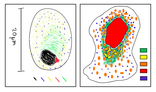
Two examples of cartoons of cells that display the distribution of organelles in cells. These drawings are made by students based on their observations of five fluorescent markers (the explanation of the colors is hidden on purpose).
In addition to identifying the organelles that are labeled, I ask the students to compare their results to a textbook image and generate an improved picture based on their observations. Altogether, this assignment is fun and I’m convinced that observing fluorescently labeled organelles (or other structures) in cells results in a better understanding of cellular function. I’d love to hear about your experiences in teaching cell biology and the experiments that students perform to learn about the intricacies of cells.
**I decided not to disclose which markers we use for obvious reasons. If you would like to know which markers we use, or if you would like more background on the experiment feel free to contact me by email or a tweet.
***The possibilities to implement a practical course in cell biology will depend primarily on the equipment that is available. At our university, we are lucky enough to have 14 basic fluorescence microscopes available for teaching 40 students at a time (160 in total). These microscopes are equipped with basic fluorescence filter sets (DAPI/FITC/TRITC) and simple CCD cameras.
it is our great pleasure to announce the 2nd International FishMed Conference on Zebrafish Research(FishMed2018), which will be held on March 25-27, 2018 at the International Institute of Molecular and Cell Biology in Warsaw (IIMCB), Poland. We organize this event to share with you the most recent knowledge and developments in zebrafish research and give you the opportunity to meet and discuss your work. The program will include keynote lectures, plenary talks, short oral presentations, poster sessions, and extensive time for discussions.
The special focus of the conference are early stage researchers, for whom dedicated competitive sessions called Young FishMed have been designed. Thus, fourteen early stage researchers who submit the best abstracts will be invited to give short presentations: Young FishMed lectures. Moreover, three best posters will be awarded. Registration starts on October 1, 2017, so please mark your calendars.
For more information including preliminary program, please visit the conference website: http://fishmed2018.pl/.
Confirmed Speakers
Keynote Speaker
Randall Peterson, University of Utah, USA
Young FishMed Keynote Speaker
Benoit Vanhollebeke, Université libre de Bruxelles, Belgium
Speakers
Catherina G. Becker, University of Edinburgh, UK
Filippo del Bene, Institut Curie – Centre de Recherche, France
Peter Currie, Monash University, Australia
Carl-Philipp Heisenberg, Institute of Science and Technology, Austria
Corinne Houart, Kings College London, UK
Adam Hurlstone, University of Manchester, UK
Anna Huttenlocher, University of Madison, USA
Hernan Lopez-Schier, Helmholtz Zentrum München, Germany
Ferdinand le Noble, Max Delbrück Center for Molecular Medicine, Germany
Paul Martin, University of Bristol, UK
Annemarie Meijer, Leiden University, The Netherlands
Marina Mione, University of Trento, Italy
Claire Russell, University of London, UK
Karuna Sampath, University of Warwick, UK
Stefan Schulte-Merker, University of Münster, Germany
Kristin Tessmar-Raible, University of Vienna, Austria
Tanya Whitfield, University of Sheffield, UK
Cecilia L. Winata, International Institute of Molecular and Cell Biology in Warsaw, Poland
Mehmet Fatih Yanik, ETH Zurich, Switzerland
Scientific Committee
Lilianna Solnica-Krezel, Washington University School of Medicine, USA
Steve Wilson, University College London, UK
Ewa Snaar-Jagalska, Leiden University, The Netherlands
Uwe Strähle, Karlsruhe Institute of Technology, Germany
Vladimir Korzh, International Institute of Molecular and Cell Biology in Warsaw, Poland
Jacek Kuźnicki, International Institute of Molecular and Cell Biology in Warsaw, Poland
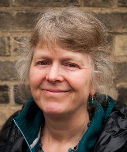
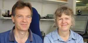
Jennifer Nichols is a Principal Investigator at the Cambridge Stem Cell Institute and Department of Physiology, Development and Neuroscience at the University of Cambridge, UK. Her lab works on lineage segregation and the establishment of pluripotency in the mammalian embryo. In 2017 she was awarded the British Society for Developmental Biology’s Cheryll Tickle Medal, given to mid-career female scientists with outstanding achievements in developmental biology. We met Jenny in her Cambridge office to talk about pluripotency in vitroand in vivo, the importance of collaboration in her career path, and what playing a musical instrument has in common with research.
Jenny Nichols
This year you were awarded the Cheryll Tickle Medal. What does the award mean to you?
Well it really meant a huge amount to me – the nominations come voluntarily from our peers, and then it’s voted on by people on the committee, so I was just really touched by that. And I hadn’t really thought of myself at all as someone who would receive medals.
Let’s start at the beginning: what got you into science in the first place?
My father was a naturalist interested in marine biology, and so when we went to the beach he’d be fishing around in rock pools and I’d tag along with him. So I was kind of brought up with biology that way – I always loved animals and finding things under rocks. I then went through education knowing that I wanted to do some kind of research: I quite liked physics and maths in the early days, but I could just see myself doing biology because I’d have the chance to work with animals.
In the final year of my degree, I knew I wanted to do developmental biology, and I wrote to various developmental biologists, one of whom was Richard Gardner. He wrote back to say that although he didn’t have anything open at the moment, he was always glad to meet people who were interested in developmental biology. So I hitchhiked up to visit him, and talked to him about the kind of work I was doing at the time, and it was great – I really liked him and liked Oxford. And then a couple of months later he wrote to tell me that his postdoc, Ginny Papaioannou, was going to be leaving. This was in the good old days when there were positions and resources available in universities and one didn’t have to depend on grants, so when Ginny left there was space in the lab. Richard asked whether I was interested in becoming his research assistant, and I arrived in his lab in 1981.
Figure 1 from Jenny’s first paper
In terms of how the paper came about, in the 1950s and 1960s the work of Tarkowski and colleagues had suggested that the development of the trophectoderm was very much dependent upon position within the early embryo. By the time I joined Richard’s lab things had moved on a little bit – for instance, it became possible to isolate the ICM and culture it, and that’s what Richard and other people such as Alan Handyside had been doing. This work led to the notion that, in addition to position, timing was crucial too: isolated ICMs from very early blastocysts could regenerate the trophectoderm, whereas those from later embryos gave rise only to epiblast and primitive endoderm. When you cultured ICMs from an intermediate stage, you could get both trophectoderm and primitive endoderm, at least by morphology, and that first paper focussed on the question of whether the outer layer of the ICM was a mosaic of the two cell types. We never really got to the bottom of what it all meant at the time, but the most important aspect of that work for my career was that I became an expert in dissecting embryos and culturing ICMs. And because I’d inherited the lab space from Ginny, there was a micromanipulation rig and a dissecting microscope for me: I had all this kit, and the mice were provided by the institution, so I had the opportunity while I was there to develop hand skills.
In Gardner’s lab you worked alongside Rosa Beddington: I understand she was a great influence on you?
Rosa was just finishing her PhD as I joined. She was incredibly clever and very good at manipulating embryos, and wouldn’t take no for an answer – if she wanted to do something, she’d find a way, and that made you think that if Rosa could do it, perhaps you could do it too.
For me, I think the most important thing she did was to invite me to help on the Cold Spring Harbor course ‘Molecular Embryology of the Mouse’. I started there as a teaching assistant just before I left Oxford in 1990. At the time I didn’t really have a great deal of confidence, being overshadowed by all the brilliant people in Oxford, and being a teaching assistant gave me something that I could do usefully. I also got to meet all of these amazing embryologists, and of course to mix with the participants. That gave me a big confidence boost and got me in contact with people in the field.
You moved from Oxford to Edinburgh with Austin Smith, getting your PhD in 1995 and then working as a postdoc. How did you come to meet Austin and, in the early days of his lab, what were the key questions you were aiming to answer?
When I was with Richard, Austin had come to work with John Heath across the road – he’d been interested in embryonic stem cells (ESCs), and his PhD had been devoted to finding what it was in the feeder layer on which mouse ESCs were grown that allowed them to self-renew and propagate. Being a biochemist, Austin wanted to understand what signalling pathways were involved, and then figure out how to make the process more efficient and relate it better to the embryo. John Heath introduced me to Austin and suggested that we might want to work together, and it took off from there. Being in Richard’s lab, everyone was a brilliant embryologist, but working with Austin – who was a biochemist and didn’t do embryo work – provided me with a niche. Then, when Austin got the job in the Centre for Genome Research in Edinburgh – another core-funded venture – he asked if I would go with him.
During his postdoc Austin had done HPLC to analyse what components in the medium secreted from the feeder layer promoted ESC renewal and found the active agent, which he called differentiation inhibitory activity (DIA) and turned out to be leukaemia inhibitory factor (LIF). So then the question was: if LIF is able to maintain ESCs in a self-renewing state in vitro, does it have any relevance in the embryo? When I started working with him, we wanted to look at the cells in culture, but also to understand how precursor ESCs in mouse embryos can self-renew. During my PhD I did in situ hybridisation on components of the LIF pathway in the embryo. In situ hybridisations had been going for a few years but were radioactive and quite laborious, and I spent a lot of time cutting sections of embryos from wax in preparation.
After many years in Austin’s lab you became an independent Principal Investigator (PI) in 2006 at the Cambridge Stem Cell Institute. How did you find the transition?
When Austin was planning to move down to Cambridge, I had a bit of uncertainty over whether to go with him, but decided in the end that it was the right move – we got on very well and made a very good partnership, and of course we still had so much left to discover about the system. It was an unusual transition to being a PI – all of a sudden, I was! But I hadn’t really thought about it; as far as I was concerned I worked with Austin. We did, by then, have joint grants, but my purpose at that time was to set up the transgenics facility. I didn’t have a fellowship but I did have a permanent job, and independence was a gradual transition from then, so I didn’t really notice any difference.
It seems that your research has been collaborative from the start. Does this reflect how you see science?
It’s how I see science going. In the old days people could get single-author papers, but I never really felt up to that, and so was always glad to collaborate. And I think that now it’s good advice that you collaborate – you need to have so many aspects to what you are doing and you can’t possibly be an expert in every one. The expectations for what is supposed to go into a paper are also very different from when I started. Plus, I enjoy collaborating and working with other people.
It’s good advice that you collaborate – you need to have so many aspects to what you are doing and you can’t possibly be an expert in every one
Since 2006 you have continued to work on lineage decisions and pluripotency in the early mouse embryo. What is your current understanding of how pluripotency arises, and the key open questions in the field?
The question of how the epiblast becomes pluripotent is of course fascinating in itself, but it’s also something you can get funding for. If I’d taken the other approach and focussed on primitive endoderm specification, even though it’s basically the same question, it’s much less easy to justify.
From research performed with a brilliant postdoc, Thorsten Boroviak, we’re pretty clear that the state of pluripotency in mice is acquired in the epiblast cells as a result of contact with the extracellular matrix that comes from the primitive endoderm cells as they are becoming specified. Thorsten found that the primitive endoderm cells are producing laminin and fibronectin at this stage, and that single cells from the ICM of earlier embryos would now make ESC colonies simply by the addition of the right matrix components. So it seems that interaction with factors secreted by neighbouring primitive endoderm cells is how pluripotency is established.
One of the key questions in the field is how cells start deciding which lineage to go down. Back when I started in Richard’s lab, the trophectoderm was known to form from the outside cells, and as the embryo was about to implant one could see the epiblast and the primitive endoderm, with the latter on the outside of the ICM. We assumed in those days that the cells that were exposed to the cavity would be the ones that would make the primitive endoderm – that it was solely a positional thing. So it was quite a breakthrough when Clare Chazaud and Janet Rossant, and then Berenika Plusa and Kat Hadjantonakis, showed that the primitive endoderm fate is specified in a salt-and-pepper distribution in the ICM. We still don’t really know how they start deciding which one to become, and the basis of this decision making is, I think, one of the key open questions.
How did working with human ESCs change the way that you thought about pluripotency?
Once we could derive human ESC lines – and it took quite a long time – it became clear that they are different from mouse ESCs. They make two-dimensional rather than small and dome-shaped colonies, and couldn’t be derived from single cells, suggesting that the population has to grow intact before passaging. They look different, express different molecular markers, require different factors in the culture and different ways of passaging. So the big question was what is the difference between the mouse and human ESC states? Then two groups, one headed by Roger Pedersen and the other by Ron McKay, derived what they called epi-stem cells (epiSCs) from the epiblast of later stage mouse embryos, using the culture conditions that had been successfully employed to make human ESCs. The logical explanation for the difference between mouse and human ESCs is, therefore, that during human ESC derivation the cells advance to the equivalent of a post-implantation state during culturing, and you recover cells in what we subsequently called the ‘primed’ pluripotent state.
So then the question was whether a condition exists in the human embryo or in other mammals similar to the mouse ESC state? EpiSCs can make pretty much every tissue you need, so would the embryo require a more naïve state? From my point of view, I was interested in the developmental biology. Others were also interested from a practical perspective, as mouse ESCs are so much easier to culture and you can do gene targeting more readily. When Takahashi and Yamanaka showed that one could reprogram differentiated cells to pluripotency – generating induced pluripotent stem cells (iPSCs) – this raised the possibility of reprogramming primed human lines to the naïve state. Many papers came out, incrementally addressing this question. Within the last few years two postdocs in Austin’s lab, Yasu Takashima and Ge Guo, found conditions in which many of the pluripotency markers in the mouse embryo were expressed differentially from the primed human cells, and crucially these were expressed in the human embryo.
There do seem to be quite a few paths to pluripotency across mammals. As far as I know, all mammals make a structure that looks like a blastocyst, but then what differs is what happens after that, how quickly it happens, and the pathways that are involved. For example, in the mouse primitive endoderm specification is entirely dependent on FGF signalling, but this is not the case in other species such as cattle or humans. Rodent embryos have quite a special way of developing that involves the formation of ‘egg cylinders’, unlike other mammalian species, and this structural difference might be key. Non-rodent embryos form a flat, polarised epithelium of the epiblast quite quickly, which might be significant in terms of the endurance of the naïve pluripotent state in the embryo.
In a time when iPSCs are being increasingly employed by developmental and stem cell biologists, what do mouse or human ESCs still have to offer us?
iPSCs have obvious uses and benefits, although of course it’s a given that if we hadn’t had ESCs then iPSCs wouldn’t have come about. But deriving iPSCs can be inefficient and take time, whereas if you want to derive an ESC line from a transgenic mouse line it is very easy from embryos, and the cells are pristine. For all these derivations, the acid test is whether they can make a chimera when put into an embryo, and whereas ESCs generally make good chimeras, iPSCs can be a bit fussier.
That is from the mouse perspective; from the human point of view, I have just established a collaboration with Wolf Reik where we are trying to derive human ESC lines clonally from embryos donated from IVF programmes. About 60% of these embryos are thought to be mosaic for aneuploidies, and a lot of those aneuploidies are likely to be chromosomal defects like trisomies. So they are actually potentially useful models for in vitro modelling of disease, and by being able to derive multiple clonal lines from an individual human embryo, one can powerfully compare a normal line with an abnormal line. This has the potential to be very useful and isn’t something we can do with iPSCs.
Don’t just follow fashions – you need a deep-rooted question that you wake up thinking about
Do you have any advice for young scientists?
Well, I said earlier that collaboration is important. In addition I think that, especially these days, you should find something that you are really interested in. You should be obsessed with your work. Don’t just follow fashions – you need a deep-rooted question that you wake up thinking about.
Finally, is there anything Development readers would be surprised to find out about you?
Several people probably know this but I am a keen oboe player and play in local orchestras. I think this is definitely connected to being a preimplantation mammalian developmental biologist – when we’re moving our embryos around, we use a pulled-out Pasteur pipette that we control with a mouth tube. And if you play a wind instrument, you don’t dribble! In playing musical instruments, you know you have to practise, and you know you’re not going to be brilliant straight away; learning how to dissect mouse embryos is similar – you can’t just follow a protocol and expect data immediately.
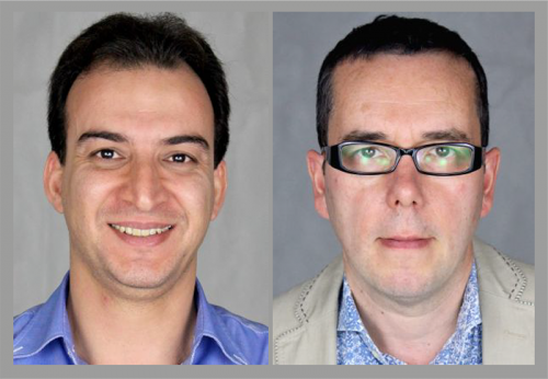
Embryogenesis begins with fertilisation, and defective activation of the egg by the sperm is implicated in human infertility. Today’s paper, published in the most recent issue of Development, investigates the role of the sperm protein PLCζ in egg activation and the calcium oscillations that accompany it. We caught up with co-first author Alaa Hachem and his PI John Parrington, Associate Professor in Cellular and Molecular Pharmacology at the University of Oxford and Fellow of Worcester College, to hear more.
Alaa and John
John, can you give us your scientific biography and the main questions your lab is trying to answer?
JP I studied at the University of Cambridge where I took Natural Sciences. An important inspiration for me was doing my third year studies at the Department of Zoology where scientists like John Gurdon, Ron Laskey, Michael Bate, and Alfonso Martinez-Arias, were harnessing the power of molecular biology to address some key questions in developmental biology. I then did my PhD at the Imperial Cancer Research Fund with Ian Kerr investigating how transcription factors regulate the activities of cells in the immune system.
My first post-doc saw a change of direction. I worked under the supervision of Karl Swann and Tony Lai at the National Institute of Medical Research trying to identify the molecular basis of the process whereby an egg is induced to develop into an embryo. These studies continued at University College London where I was able to continue my research into egg activation thanks to an MRC Career Development Award and an MRC Senior Research Fellowship. These studies culminated in the discovery – jointly with Karl Swann, Tony Lai, and Keith Jones – that the physiological agent of egg activation appears to be a novel sperm-specific protein called PLCζ. Subsequently, at the University of Oxford, where I moved to begin a lectureship at the Department of Pharmacology, my group were the first to show that mutations in PLCζ are associated with certain types of human infertility. PLCζ triggers egg activation by inducing Ca2+ signals in the egg.
Over the last decade, my group have been studying more generally the role of calcium signals in a variety of important pathophysiological processes
Over the last decade, my group have been studying more generally the role of calcium signals in a variety of important pathophysiological processes, in particular the role of the intracellular signalling molecule NAADP as a calcium mobilising messenger. These studies culminated in my discovery, in collaboration with Antony Galione, that two-pore channel, or TPC, proteins, are endolysosomal calcium channels regulated by NAADP. To study the mechanism of action and role of NAADP and TPC proteins, we generated knockout mice with loss of expression of TPC1 and/or TPC2. Our studies of these mice have identified important roles for TPCs in processes as diverse as smooth muscle and cardiac contraction, insulin secretion and sensing, autophagy and skeletal muscle function, intracellular trafficking, secretion of enzymes by the pancreas, brown adipose tissue thermogenesis, muscle development, and neo-angiogenesis.
Most recently, I became interested in using the new CRISPR/Cas9 gene editing technology to create a PLCζ knockout mouse. The reason for this was that despite numerous studies pointing to an important role for PLCζ in the egg activation process, no one had provided the definitive evidence in the form of gene knockout that PLCζ was the physiological agent of egg activation. In fact, we had previously tried using the standard embryonic stem cell approach to make a PLCζ knockout but for some reason we never managed to get germline transmission. With only a limited budget for the project, when we heard about CRISPR/Cas9 gene editing, this seemed to offer a rapid and economic way to make a PLCζ knockout. And indeed we did, in a single generation, although from this we then generated two knockout lines, our studies of which are described in the Development paper.
As well as running a lab, you have been involved in public science communication and writing books on the genome and gene editing. How did you get in to this side of science, and how important is the public understanding of science to you?
JP I’ve been interested in the communication of science to the public ever since the start of my career as a PhD student. I’ve written about new scientific discoveries and the ethics and politics of science for various publications since that time and also taken part in a variety of science communication initiatives with young people. I have debated the science and ethics of cloning and assisted reproductive technologies with sixth form students, and even took part in a project with the Northern Ballet to teach cosmology to 14-15 year olds using dance and drama. For several years now, my wife Margarida Ruas – who is also a joint first author on the Development paper – and I have taken over the classroom of a year 6 (10-11 year old) primary school classroom, to give a lesson on ‘Sex, Reproduction, and DNA’. As well as a short Powerpoint presentation, this involves us showing the children fertilization of a sea urchin egg under the microscope, freezing objects in liquid nitrogen to mimic the freezing of human embryos in clinical ART labs, and the children extracting DNA from a strawberry. The popularity of this lesson was demonstrated by comments from the children such as ‘science is wicked!’ and ‘this is the best lesson I’ve ever had!’
You can learn more about the British Science Assication Media Fellows in this video.
To further my skills in science communication, I have also completed part-time diploma courses in science communication at Birkbeck College, London, and in creative writing at the University of Oxford. Although I’ve been involved in many science communication initiatives over the years, I think a crucial moment was being awarded a British Science Association Media Fellowship. This involved me working as a science journalist at The Times in London for six weeks in the summer of 2012. I really enjoyed covering all manner of different science topics during this period, and I was very pleased that 22 of my articles were published. This gave me confidence and helped develop my writing and communication skills.
One of the stories I published concerned ENCODE – the project whose aim is to map all the functional activity in the genome. This later became the subject of my first book – The Deeper Genome, published by Oxford University Press in 2015 – which tackles the controversial question of how much of the genome is junk and how much is important. My second book – Redesigning Life, published by OUP in 2016 – is about gene editing, but also other new approaches such as optogenetics and stem cell technology. I have just finished writing a book about genetics for secondary school students, and I’m working on one about the biology of human consciousness for OUP. So you could say I have caught the book-writing bug. It’s definitely a juggling act finding the time to write popular science books as well as doing my university teaching, research and writing papers, reviews, and grants. But I have really enjoyed being able to tackle broader scientific topics than I can in my research, and the extra readership that this has brought. It has also been fun to talk about the subjects covered by the books at science and literary festivals and have a debate with the audience, who can be of a variety of ages and backgrounds. All of which should make it clear that I take communication of science to the public, including the debate about its direction and ethics, very seriously.
Alaa – how did you come to join John’s lab? I understand your PhD is funded by the Iraqi government?
AH The first time I met John was during a fertility conference in Yazd, Iran, where he presented his latest studies of PLCζ’s role during fertilization and its link with human infertility. His talk caught my attention and I decided to approach him to ask him more about his future projects, and also about the chance of doing a DPhil in Oxford under his supervision. I found him very keen in extending my knowledge of the process of mammalian egg activation, and he was also very happy to help me with my application to Oxford. In 2013, I joined his lab after successfully securing a generous grant from the Higher Committee For Education Development in Iraq (HCED-Iraq). The prime minister’s initiative provided me with sufficient funds for covering the cost of college and university fees, accommodation and living expenses, as well as some money for the research itself.
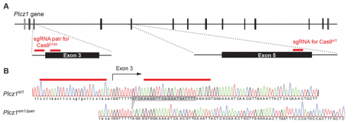
The CRISPR deletion, from Figure 1, Hachem, et al. 2017
Can you give us key results of the paper in a paragraph?
AH&JP Activation of the egg by the sperm is the first, vital stage of embryogenesis. The sperm protein PLCζ has been proposed as the physiological agent that triggers the Ca2+ oscillations that normally initiate embryogenesis. However, there has been no evidence that gene knockout of PLCζ abolishes the ability of sperm to induce Ca2+ oscillations in eggs. We used CRISPR/Cas9 gene editing to generate PLCζ knockout mice. Our studies of sperm from PLCζ knockout males showed that these fail to trigger Ca2+ oscillations in eggs. This therefore provides the first definitive evidence that PLCζ is the physiological trigger of these Ca2+ oscillations. Remarkably, however, some eggs fertilized by PLCζ-null sperm can develop, albeit at greatly reduced efficiency, and after a significant time-delay. In addition, PLCζ knockout males are subfertile but not sterile, suggesting that in PLCζ’s absence, eventually egg activation can occur via an alternative, although much less efficient route. This is the first demonstration that in vivo fertilization without the normal physiological trigger of egg activation can result in offspring.
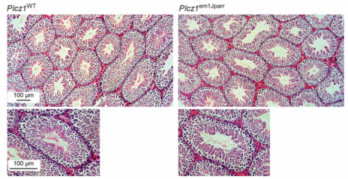
Histological analysis of testes, from Figure 3, Hachem, et al. 2017
In the absence of Ca2+ oscillations, how do you think egg activation occurs?
AH&JP The unfertilized egg is kept in a state of meiotic arrest by the maturation promotion factor, or MPF. A later decline in MAPK activity correlates with the formation of pronuclei and entry into interphase of the first cell cycle. Previous studies have shown that MPF levels can be reduced artificially in various ways, even without a Ca2+ stimulus, triggering egg activation and development to blastocyst in vitro. This mode of egg activation, without Ca2+ release, may explain the spontaneous activation observed by previous studies in ovulated, hamster and mouse eggs left to reside in the oviduct for extended periods of time in the absence of fertilization. Moreover, eggs from the C57BL/6 strain, the strain used in our studies, have been shown to have a particularly high susceptibility to spontaneous activation during in vitro maturation. Therefore a related mechanism might be responsible for the activation we observe, with the self-activation that aging, unfertilized eggs experience that normally results in fragmentation or embryonic arrest, being rescued by the PLCζ knockout sperm. A more intriguing possibility is that some stimulus supplied by the sperm, either a soluble factor, or the product of sperm-egg binding, is triggering egg activation in the absence of PLCζ. Although we failed to observe the Ca2+ oscillations typical of fertilization following ICSI or IVF with PLCζ knockout sperm, it remains possible that PLCζ knockout sperm are inducing an atypical Ca2+ signal in the egg that somehow we failed to detect in the current study. Examining this possibility will be an important focus for future studies.
Ca2+ oscillations in egg activation, from Figure S2, Hachem, et al. 2017
How might your PLCζ mice be useful for studying male infertility and its treatment?
AH&JP Currently infertility in men whose sperm fail to induce egg activation, can be treated by inducing egg activation artificially with mechanical, electrical, or chemical stimuli. However, the long term effects of such treatments on human development remain far from clear, an issue of some concern given that the Ca2+ signals induced by these treatments are highly non-physiological. The availability of PLCζ knockout sperm now makes it possible to assess in a mouse model, how artificial egg activation stimuli, used in the clinic, might affect embryonic gene expression and offspring growth, metabolism and behaviour. Importantly, it will provide a way to test the efficacy and safety of recombinant PLCζ protein as an alternative therapeutic agent to treat infertility caused by egg activation deficiency.
When doing the research, did you have any particular result or eureka moment that has stuck with you? And what about the flipside: any moments of frustration or despair?
AH Eureka moments are always a joy to share with friends and colleagues. However, such moments don’t come easily as they are usually preceded by great amounts of frustration and despair. I remember the moment when we observed the genotyping results of the mice produced after performing the CRISPR-Cas9 gene targeting of the PLCζ gene. The results showed us for the first time that mutation by out-of-frame deletion had occurred, and all the guide-RNAs had worked successfully with both wildtype Cas9 and Cas9 nickase. That was a big moment of relief because previously we tried to mutate the coding sequence of the PLCζgene in mouse zygotes, however we weren’t successful due to a one nucleotide difference between the guide-RNA we had designed based on the standard mouse genome sequence and the genomic DNA of the strain of mice used for the gene targeting. At the time, I felt extremely unlucky for this rare mismatch to have occurred, as it ruined five months of my work. However, a friend told me that it doesn’t show how unlucky I am, but rather how ‘special’ I am for this rare event to occur during my scientific career.
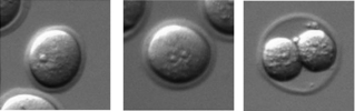
Eggs post-fertilisation, from Figure 6, Hachem, et al. 2017
Another moment that really pleased me was when I collected the epididymis from the first homozygous knockout male at the age of 8 weeks. A previous unpublished conference report had claimed that knockout of the PLCζ gene was detrimental to the spermatogenic process, leading to no sperm being produced, which would have made it impossible to use gene knockout to test the physiological importance of PLCζ in the egg activation process. Thankfully, these claims didn’t hold true as I discovered that male PLCζ knockout mice were able to produce sperm with normal parameters of viability, motility, and capacity to undergo capacitation and the acrosome reaction. This allowed us to confirm PLCζ as the source of the Ca2+ oscillations at fertilization which otherwise would not have been possible.
The PLCζ knockout mice allowed us to begin unravelling long-awaited questions regarding the identity of the ‘sperm factor’ that triggers embryogenesis and its role in the egg activation process. Initially, I assumed that the males we had created would be totally infertile, given that their sperm lacked the capacity to trigger the normal signal that induces embryogenesis. Yet, one day we received an email from our co-author Jonathan Godwin saying that some of the mutant males had managed to impregnate a few of the wildtype females. This was puzzling, and I initially assumed it was due to a mistake in labelling the homozygous mutants. However, the genotyping results of the new offspring confirmed they were heterozygous animals and that the knockout males had not been mislabelled. Therefore, in order to address this unexpected finding, we cultured zygotes generated with KO sperm by in vitro fertilisation and in vivo mating, which confirmed occurrence of activation in mice eggs in the absence of the normal Ca2+ oscillations, albeit with greatly reduced efficiency and after a significant delay. Importantly, the findings showed for the first time birth of individuals of a mammalian species in the absence of the normal physiological stimulus, and we have also witnessed the importance of PLCζ for achieving the normal physiological levels of fertility in mammals.
What are your career plans following this work?
AH I am an aspiring academic and so inherently I am drawn to teaching and research. Therefore, I plan to go back to Iraq where I hope to teach university students in embryology along with setting up my own lab where I will continue to study aspects of mammalian reproduction and infertility. My goal is to help develop the scientific community in Iraq. I believe that my studies at the University of Oxford has equipped me with the knowledge and skills to have a positive impact on both students and research. In the future, I hope to translate the findings clinically by working with infertility clinics in Iraq.
My goal is to help develop the scientific community in Iraq
And what next for the Parrington lab?
JP Providing the definitive evidence by gene knockout that PLCζ is the physiological trigger of the Ca2+ oscillations that initiate embryogenesis has been very exciting for me, since I have been on the search for the identity of the ‘sperm factor’ that kick-starts life ever since my first post-doctoral post. However, our findings raise as many questions as they answer, and I’m particularly keen now to study how egg activation and even development to term can occur in the absence of the physiological agent of egg activation. Given that some previous studies have shown that embryo development is sensitive to differences in the pattern of Ca2+ signals in the egg, there is also the interesting question of whether the offspring conceived by PLCζ knockout males differ in important respects, e.g. in their growth, metabolism, behaviour, lifespan, etc. compared to offspring conceived in the normal fashion. This issue also has clinical importance given that artificial egg activation stimuli are now being used to treat certain types of human infertility in which egg activation fails to take place without assistance. Given that such stimuli generate a Ca2+ signal that is quite different from the normal sperm-induced Ca2+ oscillations, we would like to use the PLCζ knockout sperm as a null background to test the efficacy and safety of these clinically used artificial egg activation stimuli in an animal model. In addition to these PLCζ-focused studies that I would like to carry out in the future, I am also very interested to continue my studies into the mechanism of action and pathophysiological role of the TPC endolysosomal Ca2+ channels that we are also investigating using gene knockout approaches.
Finally, what do you like to do when you are not in the lab?
AH I usually enjoy watching sci-fi movies and some sport activities when I have time.
JP As well as writing popular science articles and books, I read a lot of popular science. I also read books about history, politics, and a fair number of novels. I also like to travel and I usually try and learn a bit of the language of the country I’m visiting. I’m a political activist, and I’m particularly interested in the ethics and politics of science. For relaxation I like walking, running, exercising at the gym, cooking, listening to music, and watching the occasional film. Otherwise, my two children keep me pretty busy!
The three-day conference takes place in the University of Dundee and the University of St Andrews on 13-15 October and features speakers from around the world, exploring the many aspects and influences of D’Arcy Thompson’s landmark book – including presentations of fascinating developmental biology research following in D’Arcy’s footsteps. Don’t miss it!
Here are the highlights from the current issue of Development:
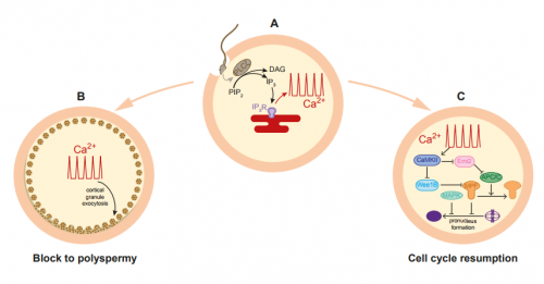
PLCζ ‘waves in’ mammalian oocyte activation
At fertilisation, fusion of the sperm with the oocyte activates a slew of downstream processes to kick-start embryogenesis. This ‘oocyte activation’ event induces the cortical reaction to prevent polyspermy, triggers oocyte metabolic and DNA synthesis pathways, and reactivates meiosis. In mammals, there is evidence to suggest that a phospholipase C isoform, PLCζ, initiates calcium oscillations associated with oocyte activation when delivered from sperm to egg. Hence, PLCζ is thought to be crucial for the activation process. Here, on p. 2914, John Parrington and colleagues directly test this hypothesis using CRISPR/Cas9 to generate a PLCζ knockout mouse. They report that while the production and quality of sperm are unaffected in knockouts, the sperm fail to induce calcium waves at fertilisation, confirming that PLCζ is required for this event. Intriguingly, although the majority of eggs fertilised by the knockout sperm do not develop, some are able to initiate embryogenesis, albeit in a delayed manner. Remarkably, PLCζ knockout mice could father a small number of offspring that develop to term, suggesting that oocyte activation can occur via an alternative route when PLCζ-triggered calcium oscillations fail. These findings will provoke further investigation into what this alternative mechanism might be, and the PLCζ knockout will be a useful tool to study infertility in mammals.
HIFs help make two halves
Just as in adulthood, an organism must respond to changes in its external environment during embryogenesis. Oxygen levels can fluctuate within a tissue, and animals have evolved a conserved signalling pathway to orchestrate a cell’s response to low oxygen levels (hypoxia). Critical to this pathway is the transcription factor hypoxia-inducible factor α (HIFα), which is degraded in conditions of normoxia. However, in low oxygen, it binds with its partner HIFβ to hypoxia-response elements and activates downstream genes that are important for a cell to cope with oxygen depletion. On p. 2940, Yi-Hsien Su and colleagues demonstrate that the hypoxia signalling pathway is active in the early sea urchin embryo, in a graded manner that mirrors the emerging dorsoventral axis. They report that while hifα mRNA is distributed uniformly throughout the embryo, the protein is stabilised at the dorsal side, and degraded more ventrally. They found that HIFα protein restricts nodaltranscripts to the ventral ectoderm only, and that the dorsoventral axis is affected by artificial perturbation of HIFα levels. Interestingly, they also found evidence for an intrinsic hypoxia gradient in embryos, which may be a forerunner to dorsoventral patterning. Together, these results provide a fascinating insight into the question of how environmental signals can impact early development.
A fishy response of transposons to demethylation
DNA methylation is an epigenetic mechanism that promotes heterochromatin formation, silences imprinted loci, the X chromosome, repeats and transposable elements. The idea that DNA methylation also represses differentially expressed genes has been challenged by experiments showing that loss of genomic methylation, either during early embryonic development or in mutants, does not result in a burst of gene activation. On p. 2925, work by Kirsten Sadler and co-workers confirms and extends the model that DNA methylation functions primarily as a gatekeeper for transposons. They report that mutants with a hypomethylated genome upregulate interferons, leading to the recruitment and expansion of immune cells in the developing larva. Rather than being directly due to derepression of interferon genes in the demethylated state, interferon production is stimulated by the detection of nucleic acids in the cytosol of a cell. This normally indicates the presence of a virus, but the mutants used in the study were not infected. So, what elicits this response? The authors reveal that the aberrant transcription of transposable elements caused by demethylation results in the production of cytosolic DNA that triggers the antiviral response. This mechanism could act as an early-warning system, allowing cells in the developing embryo with widespread epigenetic abnormalities to be put under immune surveillance so they can be rapidly eliminated if necessary.
The 2017 BSDB Cheryll Tickle Award winner talks to us about her career in science, the importance of collaboration, and the similarities between playing musical instruments and manipulating embryos.
This Primer summarises our current understanding of the diversity of WT1 functions in mammalian tissues and organs and how WT1 mutations can lead to disease.
This Review describes the gene regulatory networks, signaling pathways, morphogenetic movements and cell dynamics underlying organogenesis of the mammalian pancreas.
 (No Ratings Yet)
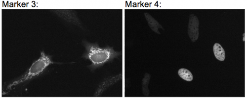
 (6 votes)


 (2 votes)
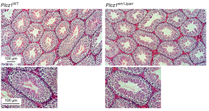 Here, on p.
 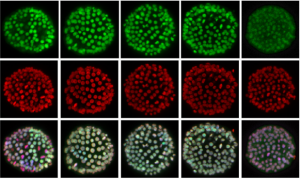 On p.
On p. 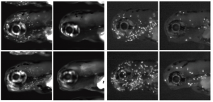 On p.
On p.