ROLE of THYROID HORMONE in MOUSE INTESTINAL DEVELOPMENT and REGENERATION.
Thyroid hormone (T3) is known to be critical for postembryonic development in mammals (around birth). This laboratory has been taking a multi-faceted approach to investigate the function of T3 and T3 receptors (TRs) in vivo by using Xenopus and mouse as models. A major recent focus is on how T3 regulates adult stem cell function during mouse postembryonic intestinal maturation and regeneration. We have shown earlier that System L amino acid transporter can influence gene regulation by TR through cellular uptake of T3 in a cell line and frog occytes. In addition, the TR coactivator PRMT1 is upregulated during mouse intestinal maturation. Yun-Bo Shi is recruiting two postdoctoral fellows to use knockout mice to study whether they play a role in the function of T3 in the intestinal maturation and regeneration.
Ritchie, J.W.A., Shi, Y.-B. , Hayashi, Y., Baird, F.E., Muchekehu, R.W., Christie, G.R., and Taylor, P.M. (2003). A role for thyroid hormone transporters in transcriptional regulation by thyroid hormone receptors. Mol. Endocrinol. 17, 653-661.
Matsuda, H., Paul, B. D., Choi, C. Y., Hasebe, T., and Shi, Y.-B. (2009) Novel functions of protein arginine methyltransferase 1 in thyroid hormone receptor-mediated transcription and in the regulation of metamorphic rate in Xenopus laevis. Mol. Cell. Biol. 29, 745–757.
Sato, Y., Heimeier, R.A., Li, C., Deng, C., and Shi, Y.-B. (2011) Extracellular domain of CD98hc is required for early murine development. Cell & Bioscience 1:7, 1-12.
Sinclair, L. V., Rolf, J., Emslie, E., Shi, Y.-B., Taylor, P. M., and Cantrell, D. A. (2013) Control of amino-acid transport by antigen receptors coordinates the metabolic reprogramming essential for T cell differentiation. Nature Immunology 14, 500-8.
Poncet, N., Mitchell, F.E., Ibrahim, A.F.M., McGuire, V.A., English, G., Arthur, S.C., and Shi, Y.-B*., and Taylor, P.M*. (2014) The catalytic subunit of the System L1 amino acid transporter (Slc7a5) facilitates nutrient signaling in mouse skeletal muscle. PLoS One 9(2): e89547,1-14.
The positions are open to all candidates within 4 years of MD/PhD degree and with
experience in mouse research. Please contact: YUN-BO SHI at shi@helix.nih.gov, NICHD/NIH, Bethesda, MD 20892, USA. (http://smm.nichd.nih.gov/)
So Adam, can you tell us a little about your previous research and university life leading up to the paper?
You could say that I have always been interested in tissue regeneration and have explored this in a variety of systems. Prior to joining Freda’s lab, my work at York University with Dr. Tom Hawke and McMaster University with Dr. Gianni Parise was largely focused on skeletal muscle physiology, muscle stem cell activity and factors that regulate muscle repair. During this time, I also became very interested in cellular cross-talk mechanisms and how the many cell and tissue types found within skeletal muscle function in a coordinated manor to achieve tissue repair and homeostasis. In a general sense, this type of thinking is ultimately what we applied when approaching the digit regeneration experiments.
“Freda Miller has a talent for recruiting individuals with unique backgrounds and skill sets in a melting pot of talent.”
Your paper was published when you were in Freda Miller’s lab in Toronto. What are the broad aims of the Miller lab and how did you fit into it?
The Miller lab is an incredibly dynamic place to do research and I consider myself privileged to have had the opportunity to do so. Freda’s research program primarily concentrates on developmental neurobiology as well as tissue regeneration/stem cell biology with many projects drawing parallels between the two fields. In addition, our group worked very closely with Dr. David Kaplan who focuses on cancers of the nervous system in addition to development. Freda has a talent for recruiting individuals with unique backgrounds and skill sets in a melting pot of talent. Being able to collaborate with my coauthors (i.e. Dr. Scott Yuzwa, Dr. Matt Krause, Matt Carr) was fundamental to this project and made for a great lab environment. Personally, I have considerable experience on IHC, cell sorting and animal work which fit well with many of the approaches utilised in the investigation.
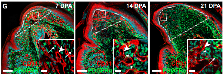
Confocal images of distal digits of at 7, 14 and 21 days post-amputation, from Fig. 1, Johnston, et al. 2016. Cell Stem Cell
And how was this particular project conceived?
Prior to this work, we published a manuscript that described the function of Schwann cell precursors in skin regeneration; a project I proudly say we stumbled into looking for something else altogether different. Based on these findings, we had an inkling that these cells may play a broad role in tissue repair which led us to investigate digit regeneration, a structure with robust regenerative capacity which is poorly understood. Freda always says “It is just as difficult to ask a big question, as it is to ask a small one” and I think that “big picture” approach resonated throughout the development of this project.
What was known about the nervous system in regeneration prior to your work?
The concept that nerve innervation is important for tissue repair is actually not a new one. The best described work to date focuses on nerve derived signals in the incredible regeneration observed in newts and salamanders. In part, these fascinating studies sparked our interest in regeneration as they actually demonstrated a functional role for Schwann cells as well as nerve axon –derived signals in limb regeneration. In mammals, it is also appreciated that nerves are necessary for the repair of many tissues (i.e. skeletal muscle) and regulates the activity stem cell populations (i.e. epidermal stem cells, HSCs). What was less understood is the mechanisms by which nerves play such an important role.
“Digit tip regeneration is one of the only examples of true multi-tissue regeneration that is possible in mammals (and even humans), and is contingent upon nerve innervation.”
Could you sum up the key results of your paper in a few sentences?
Our manuscript focused on the mammalian digit tip, a structure that has an incredible ability to regenerate following amputation. In fact, it is one of the only examples of true multi-tissue regeneration that is possible in mammals (and even humans) and is contingent upon nerve innervation. We demonstrated that cells which normally function to support nerve axons undergo de-differentiation into a precursor state (Schwann cell precursors) in response to digit amputation and dissociate from the axons. Surprisingly, these cells move into the regenerating “blastema” (area of regeneration) and intermix with the resident mesenchymal precursors where they secrete paracrine factors to enhance cell proliferation and subsequent digit regeneration. Not surprisingly, one of these factors was PDGF-AA, however, we also identified oncostatin M as a major regulator or regeneration which has not been shown before.
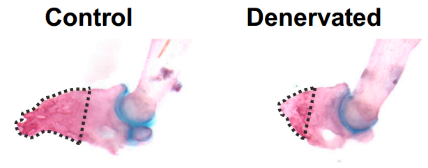
Resection of the sciatic nerve impedes digit regeneration, from Fig. 2, Johnston, et al. 2016, Cell Stem Cell
When doing this research was there a particularly exciting result or eureka moment that stayed with you?
I think this happened on a monthly basis and none of us thought the experiments would actually pan out! All kidding aside, one of the final experiments that we completed for the study was to exogenously transplant cultured Schwann cell precursors into the regenerating digits of mice that were deneravated (and regenerate poorly) in an attempt to rescue the associated defects. We all thought it was a long shot due to the technical nature of the experiments, however, due to the efforts of a very talented MD/PhD student (Matt Carr) the cells engrafted, rescued the regenerative defects, and thus, we were able to establish a definitive role for Schwann cell precursors in this process.
And what about the flipside: any particular moments of frustration or despair?
In the investigation we utilised over a dozen transgenic mouse strains, some of which required double or triple backcrossing to generate. At times it made me wonder how many manuscripts were delayed due to the reproductive habits of mice!
“Our long term aspirations are focused on identifying what is special about the blastema or “regenerative environment,” with the goal of using this information to improve tissue repair”
Does your work have any implications for human regeneration?
Interesting question; in fact, human digits also possess the ability for regeneration (if amputated distal to nail bed) and without embarrassing any of my former lab mates, we know this information through first-hand experience (pun intended!). However, our long term aspirations are more focused on identifying what is special about the blastema or “regenerative environment” with the goal of using this information to improve tissue repair.
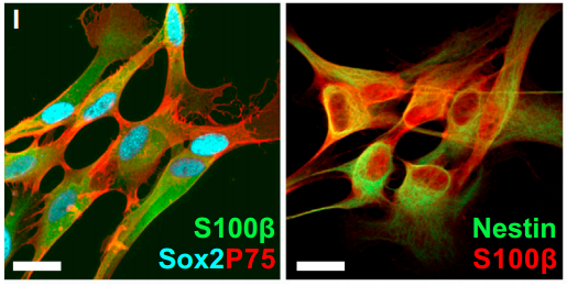
Cultured rat neonatal Schwann precursor cells, from Fig. 4, Johnston, et al. 2016. Cell Stem Cell
You’ve just started your own lab at the University of Prince Edward Island in Canada: how are you settling in?
I am beginning to settle in nicely and learn the ropes of becoming a “PI”. By making the jump to an academic position, I have lots of different responsibilities compared to being a postdoc, but I also really enjoy mentoring trainees and get on the bench as much as possible. The University of Prince Edward Island is a smaller school but still has a dedicated research community and lots of opportunity to collaborate with both academic and industrial partners.
What can we look forward to hearing about in the upcoming years?
My goal is a build a laboratory that leverages all the unique training experiences I have encountered through my graduate studies and postdoctoral work. We will still focus our efforts on understanding mechanisms of tissue repair and regeneration but my lab is also actively engaged in delineating how we can utilise exercise as a modality to enhance the stem cell niche to improve repair.
“If you have never been to Prince Edward Island you are missing out on fine seafood, beaches and very kind residents”
And life on Prince Edward Island?
If you have never been to PEI you are missing out on fine seafood, beaches and very kind residents. I am originally from the Island of Cape Breton in Nova Scotia, so PEI was not a big adjustment for me and it is great to be closer to friends and family.
And finally, what do you like to do when you are not in the lab?
My wife and I really enjoy outdoor activities such as hiking and running and I have also been an avid guitar player for a number of years.
Dear Colleagues
Washington State University’s Vancouver campus is seeking an adjunct faculty member to teach Principles of Animal Development during the Spring 2017 semester (Jan. 9-May 9). We are also looking for an instructor for Immunology.
For the complete advertisement and application instructions see:
https://admin.vancouver.wsu.edu/human-resources/employment/jobs/adjunct-faculty-developmental-biology-and-immunology
Vancouver, WA is part of the Portland, OR metro area.
Review of applications begins Oct. 10, 2016.
Thank you,
John
John Bishop, Ph.D.
Professor, School of Biological Sciences
Program Leader, Vancouver Biological Sciences
Washington State University
Vancouver, WA 98686
The 5th of July 1996 marked the birth of the world’s most famous sheep, Dolly. A scientific revelation, she was the first mammal to be cloned from an adult somatic cell through nuclear transfer. Earlier this month, scientists from far and wide gathered at the Roslin Institute in Edinburgh for a special one-day symposium dedicated to Dolly’s legacy.
The significance of the day was underlined by the opening of the symposium by the Principal of the University of Edinburgh, Prof Sir Tim O’Shea. The initial welcome was followed by a short speech on how scientific discoveries can change the world outside of the sphere of research.
The first session of the day was the keynote lecture from Professor Sir Ian Wilmut, who led the team that produced Dolly. His talk was a tour de force of the scientific thinking behind cloning and scientific work that preceded Dolly. It was surprising to hear that only a few years before the Dolly experiments, the prevailing thinking was that cells lose genes as they differentiate. Transcription factors were still a relatively new discovery, making the re-programming of a somatic cell even more revolutionary. It was a pleasure to hear Sir Ian’s anecdotes about the international reaction that followed Dolly’s birth. A short coffee break followed the talk, and after this pick-me-up, we were ready to hear how Dolly’s legacy is still creating breakthroughs.
The second session of the day was titled ‘From Dolly to Engineered Farm Animals’. This session was focussed on how cloning and genetic engineering are being used in farm animals for the benefit of human and animal health. Prof Goetz Laible opened the session with his talk on the production of transgenic cattle via siRNA insertion into the cow’s genome. The cows are engineered to alter their milk composition so it is more beneficial for human consumption. The first of the Roslin Institute’s own speakers to talk was Dr Chris Proudfoot, who also spoke about altering livestock animals, but for a different purpose. Dr Proudfoot described the use of CRISPR/Cas9 technology to produce livestock that are less prone to contracting disease, benefitting farmers.
The second of the Roslin-based speakers to talk was Dr Lissa Heron whose questionable pun title (Eggcellent therapeutics) was nonetheless followed by a discussion of great research. She spoke of genetically engeneered chickens that were made to produce high levels of pharmaceutical proteins in their eggs whites. This is a process that has commercial benefits over the way drug proteins are produced currently. The final speaker of the session, Prof Angelika Schnieke differed from the others and spoke about genetically engineered pigs as a model for human disease. After a brief tribute to Dolly, she quickly moved to the central tenet of her talk: the sheep got all the glory but now the pigs are doing the work. She spoke about how research using pigs is helping us to understand the genetic basis of cancer and how pigs should be more widely used in research, especially in pre-clinical drug testing.
The third session, titled ‘Alternatives to cloning for altering cell identity’, included talks that moved away from cloning to the area of science concerned with cell identity, cell development and how understanding these processes can be used to tackle human disease. The first speaker was Prof Shinya Yamanaka, Nobel Prize Laureate Physiology or Medicine 2012. Whilst Sir Ian’s was the highlight of my day, this talk was a close second. Prof Yamanaka spoke about his Nobel -prize-winning research in inducing pluripotency in differentiated cells – creating induced pluripotent stem cells (iPSCs). He also updated the audience on his current research, centered around creating a bank of iPSCs from ‘super donors’ that could be used in the clinic to treat disorders such as age-related macular degeneration and achondroplasia. Prof Shimoyama celebrated the 10th anniversary of his groundbreaking work this year, and his talk provided the backbone for a number of talks that followed.
The next two speakers were Dr Abdenour Soufi and Dr Sally Lowell, who are based at the University of Edinburgh. They spoke about their work on cell fate decisions made by pluripotent cells. First up, Dr Soufi detailed his work on pioneer transcription factors that pave the way during reprogramming of iPSCs. He went on to speak about how chromatin structure may hold the key to cellular identity. Following his talk, Dr Lowell spoke about her findings into how transcription factors can control the first steps towards differentiation. A new angle was presented on how cell morphology and adhesion may be responsible for the developmental cues cells receive, providing insight into why cells often differentiate in an unpredictable fashion. Prof Marius Wernig was the final speaker of the session, and he presented his work on the direct reprogramming of somatic lineages. Building on the work of Prof Yamanaka, Prof Wenig showed that mouse fibroblasts could be reprogrammed to produce functional neural cells. He spoke of his aim to improve gene targeting in iPSCs in order to correct disease-causing mutations.
Another chance to grab a coffee preceded the final session: ‘Taking stem cell science towards the clinic’. The first to talk was Prof Paul Tesar who spoke about his work studying iPSCs to analyse the molecular mechanisms of myelin disorders. He spoke about his experiments with oligodendrocyte precursor cells and how they contribute to multiple sclerosis. Using high-throughput techniques, he is using these cells to test a variety of compounds as a platform for discovering new therapeutics. Prof Stuart Forbes (also the session chair) stepped in to talk about his work on hepatocyte regenerator cells and how these could be used in cirrhosis to regenerate the liver.
The next speaker was Prof Andrew Jackson, who spoke of how microcephaly could represent a disorder of the neural stem cells. Prof Jackson explained how altered cell machinery could disrupt their normal turnover depriving the brain of neural stem cells. He described how patient skin cells can be reprogrammed to pluripotent stem cells and used to produce a 3D cortical organoid – reported by the media as ‘mini-brains’. This technique allows the cell machinery to be studied in a representative 3D environment. The next speaker Prof Mar van de Wetering gave the final talk of the day, speaking about his use of patient-derived organoids to study the crypts of the small intestine and how these may give rise to cancer.
Prof David Hume, director of the Roslin institute whose short speech was followed by a wine reception, closed the day. The symposium excellently highlighted and celebrated the legacy that Dolly left, not only on the world of research but on society in general. Rather than being a significant part of any one particular strand of science, the team behind Dolly the sheep birthed a new area of science altogether.
I would like to thank the sponsors of the symposium Disease Models & Mechanisms (DMM) for enabling me to attend by funding my registration fee. For more information about the journal, visit: http://dmm.biologists.org/
“…had you told me when I was 25 years old that I would be the director of a cancer center I would have been incredulous, given that I was totally into chemistry”
This is an extract from an interview that originally appeared in Disease Models and Mechanisms (available here Open Access)
Paraminder Dhillon, DMM Scientific Editor
Lewis C. Cantley, Director of the Sandra and Edward Meyer Cancer Center at Weill Cornell Medicine, is a world leader in cancer and metabolic disease research. His seminal discoveries have shed light on the regulation of ion pumps and other transport proteins, insulin-mediated regulation of glucose metabolism and the role of signal transduction networks in cell transformation.
In this interview, he documents his journey from serendipitous discovery of the pathway to determining its diverse physiological functions and role in cancer – an incredible odyssey that has laid the groundwork for clinical trials based on PI3K inhibitors.
I once read that, as a child, you worked out how to build fireworks. Did you always know that you would end up as a scientist?
Firecrackers were illegal where I grew up in West Virginia. When I was around 10-11 years old, I wanted to buy some, so my dad said “why don’t you make your own?” He wasn’t a chemist, but he was a very smart man and he figured out from reading the encyclopedia that we needed just three ingredients. I went to the drug store, bought the ingredients and made my own gunpowder. It wasn’t very effective in blowing up, but it burned very well. I started using my homemade gunpowder as a fuel to try to launch rockets – and it worked, although I didn’t get them very high. Today I would probably be arrested based on the buying pattern I had at the local drug store!
I did know I wanted to be a scientist from very early on. I was largely influenced by my father who, whenever I asked a question about how things work, would always come up with a logical explanation rather than resort to saying “because God made it that way”. If he didn’t know the answer, he would refer to the encyclopedia. By the time I started school I realized that I actually knew a lot more science than most of my teachers. I had a talent for it, particularly chemistry. One of my presents growing up was a chemistry set, and I loved mixing things together and seeing colors change and causing occasional explosions. I just found that amazingly fun.
When did you start using your talent for chemistry to answer biological questions?
I was always interested in biology, too. I grew up on a farm and loved growing tomato plants, which made me wonder how photosynthesis works. I found it a magical thing – that the sun shines on a plant and it takes off growing. I thought if we could figure out how this works, light energy could be captured to do all kinds of things. But I found high school biology very boring – we memorized a whole lot of things without getting any real mechanistic insight into the processes. I swore at that time that I would never take another course in biology. I decided it would be much better to learn chemistry and, ultimately, I suspected that there would be nothing in biology that couldn’t be explained by chemistry. I liked to understand kinetics and how reactions happen, so I decided to go into biophysical chemistry. In the end, that was the right path to take, although had you told me when I was 25 years old that I would be the director of a cancer center I would have been incredulous, given that I was totally into chemistry.
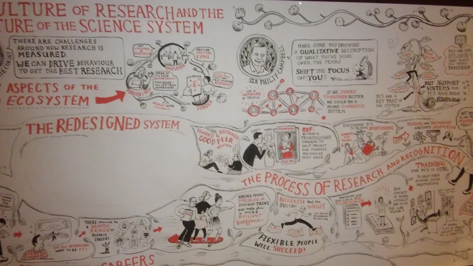
On Monday, September 19th, a group of early career researchers from across the UK and across the sciences gathered at the Royal Society in London to discuss the future of research at a one day conference.
As the Node’s Community Manager, I was lucky enough to be allowed to join in. I’ve only recently left research, and many of the issues raised were all too familiar, from publishing to career angst to representation and participation. What was encouraging was the number and diversity of ideas that came out of the debates, a lot of hope rather than despondency.
As the conference progressed, the ideas were recorded on this epic wall drawing.
As a record of what came out of the day, I collated some of the hundreds of tweets that came out of the day (using #RSsciculture; there really must have been some sore thumbs by the end of the day).
It’s a rough and necessarily selective approximation of a conference report, but I think it covers most of what was discussed on the day.
Technology is quickly changing many parts of medicine, giving people more power to take charge of their health care. Taking isotope labeled peptides as an example, stable isotope labeled peptides have been widely applied in the nuclear magnetic resonance (NMR) spectroscopy and mass spectrometry (MS). The combination of SIL peptides with NMR spectroscopy allow for the incorporation of NMR active nuclei which can reduce the complexity of spectra, and then help researchers to obtain novel correlations between atoms for more structural information.
Technology has changed people’s daily life to a large degree. In current days, people can use smartphones to track their blood sugar. And in future days, apps and accessories may be available to check cholesterol or track the heart’s electrical activity. Instead of the doctor’s office or lab, people could soon be showing up for checkups with the info already in hand, gathering information about their health. In addition to the use of new technologies, there are also some other parts of health care where doctors and patients agreed, such as the use of genetic testing for diagnose problems.
Against the backdrop of health care reform and a controversial medical device tax, medical technology companies are focusing more than ever on products that deliver cheaper, faster, more efficient patient care. Many in the industry like Isotope Labeling process have long felt overly burdened by what they consider to be an unnecessarily complex approval process. In fact, modern medical technology has achieved great success in a variety of fields, including cancer diagnosis, clinical application, experimental advance and the like, which could bring safe and effective medical devices to market more quickly and at a lower cost. For example, Melanoma Biopsies, as a huge number of dangerous-looking mole, it has been used as a handheld tool for multispectral analysis of tissue morphology.
Clearly, there are also some other aspects should be considered, such as the efficiency, side effects, etc. Investigation and research of modern medical technology ethics under the guidance of the concept of scientific development could be extremely necessary. Medical and health services to achieve long-term development, health care facilities, medical technology and health security has been significantly improved. While the development of healthcare facility is always in a dynamic process because of the influence of medical needs, medical technology, healthcare system, and other unpredictable factors. And for companies that sell medical technology, the arrival of the digital hospital has signalled a new era. Thus, maybe Integrilin can attract increasing attention in life sciences groups.
Along with the social economy prosperity and medical technology advances, medical ethics has made brilliant achievements, but currently still face a predicament in the course of development. In other words, medical technology advance not only means the medicine but also the medical spirit.
As you may have seen, we at Development have recently announced a change to our peer review process, introducing a cross-referee commenting step. This should be in place within the next week or two, and we’re hoping it will help us to make better decisions on papers, and to make the revision process easier for authors.
We were approached by Retraction Watch, who regularly run features on various aspects of publishing, about this initiative, and they’ve just posted my interview with them. So if you’re interested in finding out more about what we’re doing and why, or want to see how I’ve managed to squeeze chocolate and wine into a Q&A about peer review, please head on over to Retraction Watch for the full interview! Obviously I’m happy to answer any other questions you might have about this process, so just leave any comments or queries below, or in the RW comments feed.
And we’ll be monitoring how well this initiative works, so I expect you’ll be hearing more from me about this once we’ve had a chance to review the process and look at its impact on the papers we handle…
Changes in body organ morphology have allowed animals to better exploit diverse habitats. As organ morphology is under genetic control, genetic modifications provide the basis for the wide range of morphologies. However, as our knowledge of the genetic basis of phenotypic diversification in evolution has focused mostly on quantitative traits, it is not clear how simple genetic changes can give rise to modified functional organs. The paper by the groups of Jordi Casanova at IRB Barcelona and IBMB, and Xavier Franch-Marro at IBE and has addressed this issue by analysing the expression and function of regulatory genes in the developing tracheal systems of two insect species.
The larval tracheal system of Drosophila can be distinguished from the less derived tracheal system of the beetle Tribolium by two main features. First, Tribolium has lateral spiracles connecting the trachea to the exterior in each segment, while Drosophila has only one pair of posterior spiracles. Second, Drosophila, but not Tribolium, has two prominent longitudinal branches that distribute air from the posterior spiracles. Both innovations, although affecting different structures, are functionally dependent on each other and linked to habitat occupancy. Thus, Drosophila larvae buried in semi-liquid environments keep their posterior spiracles above the surface and distribute the air along the body via the dorsal trunks. Conversely, the lateral spiracles of free-living Tribolium larvae provide sufficient airflow to all segments, thereby making the formation of thick dorsal trunks unnecessary.
The work published in Development shows that the acquisition of each innovation is associated with change in the expression domains of individual transcription factors, spalt and cut, respectively. However, the two genetic modifications are connected both functionally and genetically, thus providing an evolutionary scenario to account for the coordinated evolution of functionally interrelated structures.
Department/Location: Wellcome Trust – Medical Research Council Cambridge Stem Cell Institute, University of Cambridge, UK.
Salary: £25,023-£28,982
Reference: PS10184
Closing date: 17 October 2016
Fixed-term: The funds for this post are available until 30 June 2017 in the first instance.
The Wellcome Trust-Medical Research Council Cambridge Stem Cell Institute is an international centre of excellence for stem cell research and regenerative medicine. Scientists in the Institute collaborate to advance our knowledge of various stem cell types and to perform pioneering work in translational research areas, providing the foundation for new medical treatments. The Institute currently comprises 29 research groups based across 6 sites in Cambridge. In 2018 all researchers will move to a new building on the Cambridge Biomedical Campus.
Applications are invited for a computational biologist to join the SCI’s bioinformatics group. We apply state-of-the-art experimental and computational methods toward understanding the biological properties and biomedical potential of stem cells.
The vacant post is at Research Assistant level and would be suitable for individuals with either a computational or biological background. The post holder will bridge the research groups within the SCI and will analyse next generation sequencing data using cutting edge software tools on internally (SCI)-produced data. He/she will work in a team of bioinformaticians dedicated to the application of modern bioinformatics techniques to stem cell research.
Candidates should be able to work in a UNIX/Linux environment. Proficiency with a scripting language (e.g. Perl/Python) and statistical data analysis tools (R, Matlab) would be a strong advantage. Additional experience with analysis of high-throughput sequencing data is desirable. The post holder will be involved in the development and interpretation of multilayer genomic, transcriptomic and epigenomic data. Necessary training in specialist computational tools will be provided; the main criterion is an enthusiasm to use bioinformatic approaches to advance stem cell research.
To apply online for this vacancy and to view further information about the role, please visit: http://www.jobs.cam.ac.uk/job/11530. This will take you to the role on the University’s Job Opportunities pages. There you will need to click on the ‘Apply online’ button and register an account with the University’s Web Recruitment System (if you have not already) and log in before completing the online application form.
Please upload your Curriculum Vitae (CV) and a covering letter in the Upload section of the online application to supplement your application. If you upload any additional documents which have not been requested, we will not be able to consider these as part of your application.
The closing date for all applications is the Monday 17 October 2016.
Informal enquiries are also welcome via email to: jobs@stemcells.cam.ac.uk.
Interviews will be held towards the end of October 2016.
Please quote reference PS10184 on your application and in any correspondence about this vacancy.
The University values diversity and is committed to equality of opportunity.
The University has a responsibility to ensure that all employees are eligible to live and work in the UK.
 (No Ratings Yet)
 (4 votes)