The community site for and by
developmental and stem cell biologists

In Development this week (Vol. 143, Issue 6)

Posted by , on 15 March 2016

Here are the highlights from the current issue of Development:


Knocking the SOX off obesity

Embedded Image

Growth restriction in utero is associated with increased risk of obesity in later life. Recently, epigenetic inheritance was identified as an important component of this phenomenon, but the precise molecular mechanisms that underpin the association between growth restriction and obesity remain unknown. Now, on p. 950, Walter Stünkel and colleagues report a role for SOX6 in adipocyte differentiation, and suggest that SOX6 may be a key player in the association between growth restriction and obesity. By comparing adipocytes differentiated from mesenchymal stem cells from normal and growth-restricted newborn umbilical cords, the authors show that SOX6 is upregulated in growth-restricted adipocytes and that it activates key adipogenic players including PPARγ, C/EBPα and MEST. The authors also show that SOX6 interacts with β-catenin, possibly inhibiting WNT/β-catenin signalling to promote adipogenesis. Importantly, the role of SOX6 in regulating adipogenesis was also demonstrated in vivo, using Sox6 antisense oligonucleotides to target white adipose tissue in mice. Taken together, these data demonstrate a clear role for SOX6 in regulating adipocyte differentiation and adipogenesis in vivo, and provide a possible mechanistic link between growth restriction in utero and obesity in later life.


New player in neonatal heart repair

Embedded Image

The mammalian heart has a transient regenerative ability during the neonatal stage. This ability depends on the replicative potential of endogenous cardiomyocytes; however, the underlying transcriptional network that controls cardiomyocyte replication during neonatal heart regeneration remains poorly understood. In this issue (see p. 936), Bin Zhou and colleagues investigate the role of GATA4 – a transcription factor that is crucial for cardiac specification and development – in cardiomyocyte turnover and neonatal heart repair. The authors utilised cryoinjury and apex resection models in a neonatal transgenic mouse in which they could control expression of GATA4 specifically in the cardiomyocytes. Following injury, the authors observed severely compromised ventricular function in Gata4-ablated mice, which was accompanied by reduced cardiomyocyte replication and hypertrophy. Importantly, the authors identified FGF16 as a downstream effector of the Gata4-ablated phenotype, and showed that cardiac-specific overexpression of FGF16 promoted cardiomyocyte replication and improved heart function after injury. These data identify GATA4 and FGF16 as important mediators of neonatal heart repair and bring hope for the possibility of paracrine-mediated repair in the adult heart.


A better MAP(K) for tubule elongation

Embedded Image

Branching morphogenesis is fundamental to the development of multiple organs, including the lungs, kidneys, vasculature and mammary glands. Tubule elongation is a crucial part of branching morphogenesis and relies on the balance between cellular proliferation and migration to achieve appropriate growth. In the developing mammary gland, receptor tyrosine kinases (RTKs) regulate tubule elongation, but the relative contribution of proliferation and migration is largely unknown. Now, on p. 983, Andrew Ewald and colleagues use fluorescent reporters and real-time imaging to investigate the role of RTK signalling in tubule elongation in a 3D mouse mammary tissue culture system. The authors show that ERK signalling is required for cell migration and elongation of budding branches, but that cell proliferation is not acutely required for branch elongation. Importantly, the authors show that mosaic expression of MEK, which activates MAPK, is sufficient to induce initiation and elongation of mammary branches. This study provides a fundamental advance in our understanding of the cellular mechanism and molecular control of tube elongation in the developing mammary gland.


PLUS…


Closing the circle: from organoids back to development

Organoid imageThis Editorial, from our Guest Editor Melissa Little,  looks at the emerging field of in vitro organogenesis and discusses how organoid technology can be applied to better understand developmental processes. Read the Spotlight on p. 905


An interview with Melissa Little

MelissaMelissa Little chats about her research and career, the potential of the organoid and stem cell fields, and what she hopes to achieve during her guest editorship with Development. Read the Spotlight on p. 907


***  find out more about our upcoming Special Issue on Organoids ***


From single genes to entire genomes: the search for a function of nuclear organization

DEV910Here, Ringo Pueschel, Francesca Coraggio and Peter Meister highlight the genome-wide techniques that have been used to shed light on the mechanisms of genome folding and unravel the regulatory functions of nuclear organization. Read the Review on p. 910


Development of the lymphatic system: new questions and paradigms
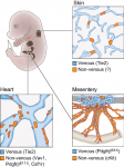
DEV924Jonathan Semo, Julian Nicenboim and Karina Yaniv discuss the molecular mechanisms controlling lymphatic system development and highlight recent findings that shed light on previously uncharacterised sources of lymphatic endothelial cells. Read the Review on p. 924


Thumbs up (No Ratings Yet)
Loading...

Categories: Research

Navigate the archive

Use our Advanced Search tool to search and filter posts by date, category, tags and authors.

Editorial- Closing the circle: from organoids back to development

Posted by , on 15 March 2016

This editorial was written by Melissa Little and first appeared in Development.


Organogenesis is an inherently fascinating developmental process. It requires the creation of complex form and function from a collection of distinct cell types, all of which come together without a template. To achieve this, cells within the developing organ undergo differentiation, migration, proliferation and appropriate growth and scaling in a spatially and temporally controlled manner. Moreover, many organs retain throughout life a capacity to regenerate or repair in response to injury in order to maintain function. Studies on both embryonic organogenesis and postnatal tissue homeostasis and repair have been essential foundations to the field of stem cell biology, as it is the stem/progenitor populations involved in these processes that are key. To date, our understanding of organogenesis, and to a lesser degree stem cell biology, has largely been based on animal models. The conservation of genes across organisms perhaps emboldened the view that we can infer knowledge of human development from animal models, but the lack of parallels shows that this is not the case. Instead, it is our capacity to examine stem cells from humans that is filling this gap. It is now possible to recreate miniature approximations of many human organs, referred to as organoids, entirely in vitro. Generated from either human pluripotent stem cells (PSCs) or human adult epithelial stem cells, organoids offer unprecedented insights into human developmental processes and can also be used to model postnatal repair and disease. As a result, organoid biology is at the forefront of our developmental studies in the human. For this reason, I am honoured to have been appointed Guest Editor at Development, with a particular focus on the emerging field of in vitro organogenesis, and with the aim of publishing a Special Issue on the topic (for more details, see below).

Two of the most seminal breakthroughs in recent decades have been the derivation of the first human embryonic stem cells (Thomson et al., 1998) and the reprogramming of human somatic cells to pluripotency (Takahashi et al., 2007). The development of protocols to direct the differentiation of human PSCs towards specific endpoints has resulted in the generation of derivatives of all three germ layers, including nerves, cardiac muscle, pancreatic beta-cells, hepatocytes and blood. Most of these studies have drawn heavily on our underlying knowledge of the morphogens involved in patterning the same cell types during embryogenesis. While initial focus was placed on generating specific target cell types, the cultures themselves began to reveal order amongst the complexity, with the formation of highly patterned multicellular structures in vitro. This occurred most notably when cultures were provided with some degree of three-dimensional space within which to interact; for example, as part of an embryoid body or with the support of an extracellular matrix such as Matrigel. The first study to recognise the emergence of a self-organising but recognisable tissue from human PSCs was less than five years ago, with the generation of a patterned optic cup from the group of Yoshiki Sasai (Eiraku et al., 2011). What followed this seminal study was a growing number of reports describing organotypic morphogenesis as a result of the directed differentiation of human PSCs. Human PSC-derived organoids of the cerebral cortex, adenohypophysis, small intestine, stomach, lung, liver and kidney have now been reported (reviewed by Huch and Koo, 2015). In all cases, these organoids contain distinct cellular components patterned in a morphologically appropriate fashion – a level of cellular complexity previously thought unattainable in vitro.

These advances are exciting not only because they represent the long-term possibility of organ regeneration, but also because they provide a new approach to studying human morphogenesis and development. But the challenges here are immense. Are we sure that a cerebral organoid that appears to show evidence of an appropriate multicellular composition and gene expression profile is a sufficiently accurate model of human brain development? Can we reliably sustain such tissue long enough under appropriate conditions to allow it to mature as it would in vivo? If these questions are not appropriately addressed, there is a danger that the field will become absorbed by the characterisation of what are no more than in vitro dysmorphologies or models of abnormal patterning. This will require disciplined and thorough analyses that take into account what the stem cell field, particularly the induced PSC field, already appreciates is substantial experimental variability. Access to human fetal tissue signatures has been, and will continue to be, essential to ensure that we are on the right track. Indeed, single-cell profiling data comparing human cerebral organoids with developing human fetal brain show remarkable congruence, which is very encouraging (Camp et al., 2015). Based on the assumption that PSC-derived organoids do indeed represent a model of human development, we can be sure that their application will extend beyond developmental biology to personalised drug development, disease modelling, functional genomics and even bioengineering.

Organoids can also be generated from adult stem cells, such as those first reported by the group of Hans Clevers from the intestinal epithelium (Sato et al., 2009). This capitalises on the presence of an epithelial stem cell population in many postnatal organs. Organoids have now been derived from the epithelial stem cell populations of a wide variety of postnatal tissues, including lung, colon, prostate and liver (reviewed by Huch and Koo, 2015). In all instances, these organoids are composed exclusively of epithelial cell types supported by the provision of an extracellular matrix surrogate for surrounding mesenchymal tissues. Adult stem cell organoids have taught us much about the regenerative populations present in many postnatal tissues and the mechanisms by which these populations maintain homeostasis. Their applications in the understanding of disease and in personalised drug screening are now coming to the fore.

Organoids will not only advance our understanding of human development and tissue turnover, but also of morphogenetic principles. A major paradigm in morphogenesis has been the concept of positional information, initially described by Wolpert (1969). That concept proposed that morphogenesis during embryonic development results from the relative position of cells with respect to each other and the secreted morphogen gradients they experience. Revised views of this concept have taken into account a capacity for heterogeneous populations to create form without a predefined pattern – a phenomenon known as self-organisation. This has never been more evident than in what is now being observed in organoid studies, where complicated, multicellular form is created from a single starting cell type, and without the complete set of spatial cues normally present in an embryo. The robustness with which a complex structure can arise in vitro is surprising. More importantly, it provides the field with a capacity to re-evaluate the underlying principles governing morphogenesis and tissue homeostasis. Coupled with continuous advances in our capacity to image developing tissues at the resolution of individual cells across time and space, investigations into how organoids arise could well lead to rapid advances in our understanding of fundamental developmental principles.

The excitement of what organoid biology can add to our understanding of development has grown exponentially over the last few years, with several scientific meetings dedicated to discussing progress in this area. Development recognises that this is an exciting and rapidly expanding area, which is why we are announcing a Special Issue on organoids, to be published in early 2017. This will include Review articles from the pioneers and leaders of the field, as well as primary papers on topics ranging from methods and techniques for organoid formation to modelling developmental processes and diseases using organoids, in both human and non-human systems. As Development’s Guest Editor, I will be taking a lead on coordinating this Special Issue, and invite interested readers to find out more about it – including further details on the issue’s scope and how to submit your work for consideration – on our website (http://dev.biologists.org/content/special-issue-organoids). We look forward to receiving your submissions.

In summary, the past decades have seen developmental biology guide us into stem cell biology, with developmental principles continuing to underpin this field. This is perhaps nowhere as compelling as in the generation of organoids. In turn, the generation of organoids is beginning to teach us not only about human development, but also about general developmental principles, helping to build an information flow in the reverse direction in a way that was previously impossible. The challenge now is to close the circle completely using organoid systems to reinforce the link between stem cells and developmental biology, which will bring these two fields closer together than ever before.


References

Camp, J. G., Badsha, F., Florio, M., Kanton, S., Gerber, T., Wilsch-Bräuninger, M., Lewitus, E., Sykes, A., Hevers, W., Lancaster, M. et al. (2015). Human cerebral organoids recapitulate gene expression programs of fetal neocortex development. Proc. Natl. Acad. Sci. USA 112, 15672-15677.
Eiraku, M., Takata, N., Ishibashi, H., Kawada, M., Sakakura, E., Okuda, S., Sekiguchi, K.,  Adachi, T. and Sasai, Y. (2011). Self-organizing optic-cup morphogenesis in three-dimensional culture. Nature 472, 51-56.
Huch, M. and Koo, B.-K. (2015). Modeling mouse and human development using organoid cultures. Development 142, 3113-3125.
Sato, T., Vries, R. G., Snippert, H. J., van de Wetering, M., Barker, N., Stange, D. E., van Es, J. H., Abo, A., Kujala, P., Peters,  P. J. et al. (2009). Single Lgr5 stem cells build crypt-villus structures in vitro without a mesenchymal niche. Nature 459, 262-265.
Takahashi, K., Tanabe, K., Ohnuki, M., Narita, M., Ichisaka, T., Tomoda, K. and Yamanaka, S. (2007). Induction of pluripotent stem cells from adult human fibroblasts by defined factors. Cell 131, 861-872.
Thomson, J. A., Itskovitz-Eldor, J., Shapiro, S. S., Waknitz, M. A., Swiergiel, J. J., Marshall, V. S. and Jones, J. M. (1998). Embryonic stem cell lines derived from human blastocysts. Science 282, 1145-1147.
Thumbs up (No Ratings Yet)
Loading...

Tags: , ,
Categories: Discussion, News

What would you ask Nobel Prize Laureate Shinya Yamanaka?

Posted by , on 14 March 2016

It’s been 10 years since induced pluripotent stem cells (iPSCs) were first reported from the lab of Prof. Shinya Yamanaka. Since then, the field of direct reprogramming has grown immensely, and iPSCs have proved themselves to be an extremely useful and versatile tool, enabling research into basic developmental biology, the mechanism of reprogramming itself, as well as translational avenues of drug discovery and cell therapies.

Next week, I’ll be heading to Kyoto to interview Prof. Shinya Yamanka who, together with Sir John Gurdon, was jointly awarded the Nobel Prize for Physiology or Medicine in 2012 for the discovery that mature cells can be reprogrammed to become pluripotent.

As part of my interview, I’m going to ask Shinya the best, most interesting question put forward by the community – that’s YOU!

So: what would you ask Shinya? Now’s your chance. Submit your questions in the comments below, via our facebook page or on twitter using #askshinya.

The video forms part of Development’s interview series. Watch my interview with Prof. Austin Smith, where he chats about his role as an Editor at Development, and what it takes to be a good scientist. Plus, keep watching as Austin tries to identify stem cell scientists from photos taken before they were famous. Can you tell who they are? Watch the video and see for yourself.


Thumbs up (1 votes)
Loading...

Tags:
Categories: Discussion, News

Science consulting: helping out a scriptwriter

Posted by , on 13 March 2016

During my years in New York, I unexpectedly experienced an interesting form of science outreach. I randomly met a film director at a party, Sasha Collington. This director explained to me that she needed the help a biologist to work on her new script. I accepted right away for three reasons. First, curiosity. Second, because just like many other experimental scientists, I get very frustrated with dead-end experiments and I regularly start questioning the meaning of my life. I liked the idea of being directly and immediately helpful to someone. Third I get angry a lot when I hear all the scientific none-sense or the misleading and condescending over-simplification of science in articles or movies. I was therefore tempted by this opportunity to get the science as accurate as possible.


Consequently, we exchanged several emails and met a few times. She began by explaining the story she was working on. The film, in production, is called “Love Type D”:


In her film, the outcome of relationships is governed by biology. We are either dumpers or dumpees. Therefore she was looking for ideas about what kind of biological concept could explain this, how could the characters experimentally test it and whether or not they could overrule it. I understood that she was not looking to double check the science in her script. She was not looking for technical or procedural details either. I rather felt that she was looking for general biological knowledge to nourish her artistic creativity. It was thus a broad discussion, very upstream in the film making process. After her description of her project, I started talking a lot. She had in mind to go for some king of genetic phenomenon, so I explained her the basics of genetics and epigenetics. What is a gene? What is the difference between a gene and an allele? What is chromatin? How is gene expression regulated? How are genes related to phenotypes and why is it much more complicated that we once thought? What are the classic techniques used to identify genes or to assess their level of expression? She was very active in the discussion. I could tell that she was trying to see how she could use those pieces of information to help her story. Indeed, she was asking a lot of questions about practical aspects: what kind of experiments could we use? Could a child perform that experiment? Would the reagents be accessible to a non-scientist? This is where it became really interesting because I perceived a bit better what it means to talk about science with non-researchers.


When it comes to discussions within the scientific community, I am personally very opposed to the storytelling approach. I feel that science is turning into a marketing activity and I really dislike that. It shifts the focus away from data and logic toward fancy tales. However, in this context of communication between an artist and a scientist, and between this artist and her public, I realized that storytelling was not hurting and, on the contrary, was necessary to convey the message properly. First, I understood that aesthetic is important. As an artist using a visual medium, her recurring worry was to know how a given experiment would look like. Would the result appear on a screen? On a paper? In a test tube? Does it have colours? Does it move? Does it make a sound? Does it have to be in the dark? Questions I never even thought of. We had to forget “unfilmable” or visually unexciting experiments such as western blots or ELISA. We were rather trying to imagine microscopy-based results that would visually make sense on the screen. I perceived the benefit of taking into account the visual content and not just the conceptual content. That is something I will keep in mind whenever I have to communicate with people from outside the academia.

Second, I understood the importance of analogy. Among the various scientific concepts we explored, she particularly liked the notion of chromatin compaction and decompaction during the epigenetic regulation. She liked it because it spontaneously evoked the idea of a book that someone closes or opens. It is something the public unconsciously knows very well and therefore they would almost emotionally connect with this scientific concept. I realized that making something “understandable by the public” did not mean “absence of complicated words”, or “not too many parameters”, it rather meant using concept people could relate to. Next time I have to interact with non-scientific people I will try to appeal to those “cultural images” they have instead of falling into the (condescending) trap of over-simplification.

Finally, I had expected that the biggest obstacle would be the knowledge gap between us. But it rather turned out that the main difficulty is the divergence of objectives. I wanted to get the script scientifically accurate whereas her prime goal was, understandably, to have a scientific element that would serve the story: something clear, coherent and pretty. Because of this divergence, we spent quite some time trying to come up with ideas that could be both scientifically and artistically satisfying. I felt a bit like a very annoying person, repeating things such as “well, not exactly…”, “no it’s more complicated”, “I guess we could say that only if…” and often “well, we don’t really know.” I guess that this is a common feeling among people who happened to serve as consultants. We may come off as dull, picky mood-killers. But that’s ok. We are here to give a scientific point of view to something. That is, to provide logical, accurate, fact-driven interpretations or ideas. When advising a film scriptwriter or director, it is important to keep acting as scientists, and not trying to anticipate or satisfy the artist expectation or vision. Even if we reinforce the image of nerdy researchers by doing so.


Overall, this experience was a lot of fun. I loved this opportunity to see how a script was being made and to experience the different perception of what is biology. I would gladly do it again, especially if it is at a downstream step, such as technical coaching during the shooting. Was my help useful? I like to think it was. At least to get her started on the scientific part of her script, to set some landmarks in her mind that would help her come up with that part of the story. Regarding the scientific accuracy, which was one of my objectives, I think it did not work out as well as I thought. There are other limitations in a film production (time, money, space, material, actors…). Seeing that your advice is not always followed must probably be frustrating for scientific consultants, but that’s the way it is. After all, it remains a piece of fiction.


Outreach logo new squareThis post is part of a series on science outreach. You can read the introduction to the series here and read other posts in this series here.


Thumbs up (2 votes)
Loading...

Tags:
Categories: Outreach

PhD position opened in Switzerland for a developmental / cell biologist

Posted by , on 11 March 2016

Closing Date: 15 March 2021

Evolutionary developmental genetics of reptile skin colour

In the context of evolutionary developmental biology analyses of reptilian skin colours and colour patterns, we offer one PhD position for an outstanding, highly motivated, and creative experimental wet-lab biologist with strong skills in developmental biology and cell biology.

Michel Milinkovitch’s group at the University of Geneva (UNIGE) integrates the expertise of developmental biologists, evolutionary biologists, computer scientists and physicists for an improved understanding of the mechanisms generating a diversity of skin colours and colour patterns in reptiles.

We have recently shown (Saenko et al. 2013) that the extensive variation of skin colours and patterns in Sauropsida reptiles is generated by precise co-localisation of interacting pigmentary and nano-structural elements. In this framework, we have also shown (Teyssier et al. 2015) that chameleons shift colour through active tuning of a lattice of guanine nanocrystals, which photonic effect is filtered by a layer of pigments. In addition, we have built extensive transcriptomic and genomic resources (Ullate-Agote et al. 2014; Tzika et al. 2015) for mapping colour mutations in our new model species of snakes and lizards. For example, we recently mapped and identified the mutation responsible for the amelanistic mutation in corn snakes (Saenko et al. 2015).

The successful candidate will (i) participate to linkage mapping of multiple colour and colour pattern mutations in snakes and lizards and (ii) use molecular/cell/developmental biology methods (microscopy, immuno-histochemistry, in-situ hybridisation, transcriptomics, in-vivo assays, ex-vivo cultures, etc.) to characterise the effects of these mutations on neural-crest cell migration, as well as on the physiology of pigmentary and structural-colour cells. The new PhD student will also interact with a physicist PhD student who is mathematically modelling the reaction-diffusion processes that generate colour patterns in snakes and lizards.

Candidates must have a Master in biology or biochemistry. Skills and experience with developmental biology and/or cell biology are mandatory. Skills in biophysics are useful. The successful candidate will have a genuine interest for organismal biology and will appreciate interactions with physicists and computer scientists.

The University of Geneva (UNIGE) is world-renowned for its research in Biology and Physics.  UNIGE is among the top 1% best universities in the world and the Faculty of Sciences is ranked 32th world best (Shangai Academic Ranking of World Universities).

PhD students are remunerated according to the standards of UNIGE, which are very generous when compared to other international programs.

Geneva is an international city occupying a privileged geographical situation.

Candidates must send their application – in the form of a single PDF file including a brief letter of interest, a CV, as well as contact information (not support letters) of two persons of reference – to:

Prof. Michel Milinkovitch (Michel.Milinkovitch@unige.ch), Laboratory of Artificial & Natural Evolution (www.lanevol.org), University of Geneva, Switzerland.

Refs: Saenko, Teyssier, van der Marel & Milinkovitch. Precise colocalization of interacting structural and pigmentary elements generates extensive color pattern variation in Phelsuma lizards. BMC Biology 2013, 11: 105; Teyssier Saenko van der Marel & Milinkovitch. Photonic Crystals Cause Active Colour Change in Chameleons. Nature Communications 6: 6368 (2015); Tzika, Ullate-Agote, Grbic & Milinkovitch. Reptilian Transcriptomes v2.0: An Extensive Resource for Sauropsida Genomics and Transcriptomics. Genome Biol. Evol.  7: 1827-1841 (2015); Ullate-Agote, Milinkovitch & Tzika. The genome sequence of the corn snake (Pantherophis guttatus), a valuable resource for EvoDevo studies in squamates. Int. J. Dev. Biol. 58: 881-888 (2014); Saenko, Lamichhaney, Martinez Barrio, Rafati, Andersson & Milinkovitch. Amelanism in the corn snake is associated with the insertion of an LTR-retrotransposon in the OCA2 gene. Scientific Reports 5, 17118 (2015) .

Thumbs up (No Ratings Yet)
Loading...

Categories: Jobs

Postdoc position to study Mechanisms of neural progenitor specification

Posted by , on 11 March 2016

Closing Date: 15 March 2021

A full 3-year ANR funded postdoctoral position is available at the Centre de Biologie Intégrative Toulouse to study mechanisms by which time is translated into a precise developmental sequence in the context of neocortex development. This position is part of the postdoctoral program of CBI Toulouse (http://cbi-toulouse.fr/eng/). Our research focuses on the role of local cell-to-cell communication via Eph:ephrin signaling in the specification of neural progenitors in the mouse. The specific aim of the proposed project is to identify the cellular and molecular mechanisms linking Eph:ephrin signaling to neural progenitor fate decisions, concentrating on the transcriptional response elicited downstream of Eph activation in neural progenitors.

The project includes genome-wide gene expression analyses (RNA-Seq and ChIP-Seq) to fully characterize the repertoire of genes downstream of Eph:ephrin signaling in cultured progenitors and the assessment of candidate genes in regulating the production of projection neurons in vitro and in vivo. The succesful candidate should have a PhD and a strong background in genome-wide gene expression analyses. Expertise in cell culture and/or mouse genetics would be a plus. The position is available from october 2016.

Highly motivated candidates should send a brief description of their research interests and career goals, their CV, and contact information for three references to Alice Davy (alice.davyatuniv-tlse3.fr)

Thumbs up (No Ratings Yet)
Loading...

Categories: Jobs

Researchers dig up new molecular details on “the other type” of stem cells

Posted by , on 11 March 2016

  • Scientists at IRB Barcelona and CSIC reveal that the combination of two molecular signals determines which cells that have already differentiated can regain their stem cell properties.
  • Their studies on fruit flies allow for advancements in the field of regenerative medicine and a better understanding of processes involved in cancer.

In a study published this week in PLos Genetics, scientists Nareg J. Djabrayan and Jordi Casanova from the Institute for Research in Biomedicine (IRB Barcelona) and CSIC, have identified two molecular signals and the pathway of

Nuclei of Drosophila trachea cells during the transition from larvae to an adult fly. The cells in the Tr2 segment are dividing; they are the facultative stem cells. (N.J. Djabrayan, IRBBarcelona)

events that allows cells in a tissue that are already specialized to regain their behaviour as stem cells. The study offers new information about how cells become differentiated and how “this other type” of stem cells, called facultative, get activated, which is of particular interest in cell reprogramming, regenerative medicine, and in understanding cancer.

Facultative stem cells are being identified more and more often in human tissues and organs, but much less is known about them compared to typical stem cells, which have distinct morphological traits. “The existence of facultative stem cells indicates that many differentiatied cells continue to have great plasticity and are able to respond in situations of stress, such as in wounds or in the regeneration of worn out tissues,” explains Jordi Casanova, head of the Development and Morphogenesis in Drosophila group at IRB Barcelona.

“This plasticity is good if it is well regulated. If any of the mechanisms involved becomes unbalanced, it becomes dangerous,” adds Casanova. “In fact, cellular plasticity in tissues that regenerate often, are associated with organs that have a high incidence of cancer, such as the colon or blood,” he explains.

A combination of signals in space and time

The researchers looked at the moment when a Drosophila larva changes into an adult fly. “This model is a good one to identify and study facultative stem cells because there are some cells in the larva that will give rise to the adult fly and they get activated during the switch from larva to adult, a transition which could be considered a condition of stress,” explains Casanova.

Concretely, the researchers studied cells with facultative stem cell traits in Drosophila trachea. They showed that only some cells in the tissue receive a spatial signal that marks them. Then, when the transition occurs, a second signal is sent, triggered by hormones, that prompts the reactivation of the stem cell programme. This signal reaches all cells in the tissue, but only those that have been marked previously with the spatial signal are reactivated as stem cells.

“Understanding these signal regulation mechanisms is fundamental in order to control, for  example, the reprogramming of cells,” indicates Casanova. Using the same model, the scientists now want to investigate which mechanisms allow differentiated cells to maintain their plasticity and how they go from being a differentiated cell to a stem cell and vice versa.


Reference article:

Snoo and Dpp act as spatial and temporal regulators respectively of adult progenitor cells in the Drosophila trachea

Nareg J-V Djabrayan and Jordi Casanova

PLoS Genet. (2016 Mar 4) DOI: 10.1371/journal.pgen.1005909


This article was first published on the 11th March 2016 in the news section of the IRB Barcelona website

Thumbs up (1 votes)
Loading...

Tags: , , ,
Categories: Research

1-2 postdoc positions to study neuromuscular patterning during development & regeneration

Posted by , on 10 March 2016

Closing Date: 15 March 2021


5.cover
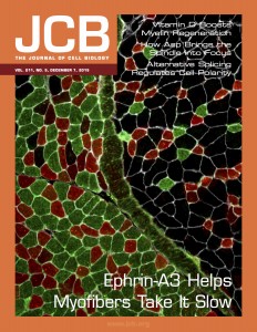
Each adult mammalian skeletal muscle has a unique complement of fast and slow myofibers, a consequence of cell-autonomous and non-cell-autonomous patterning decisions during development. Intriguingly, following either acute muscle regeneration or deinnervation/reinnervation, the proportionality and patterning of muscle fibers is largely preserved, suggesting a mechanism for ongoing maintenance of fiber type specification during homeostasis and regeneration/repair. We have recently reported that interactions between the repulsive guidance ligand ephrin-A3 [which is expressed on all and only slow (Type I) myofibers] and EphA8 [an ephrin receptor expressed by terminal Schwann cells at all and only neuromuscular junctions of fast (Type II) myofibers] promote and preserve slow myofiber identity by preventing stable innervation of slow muscle fibers by fast motor neurons [Stark et al, JCB 211:1077–91 (2015)]. Additional data from our lab suggest that variations on this mechanism may also direct slow fiber type-specific interactions with muscle satellite (stem) cells, and/or promote and preserve fast (Type IIb) myofiber identity during development and regeneration. The future potential of this work includes not only extending our basic understanding of muscle patterning during development, regeneration, neuromuscular disease, and aging but also has implications for targeted gene or cell transplantation therapies.


The Cornelison lab at the University of Missouri is recruiting 1-2 postdoctoral fellows to work on this NIH-funded project, addressing the significant unsolved question of how muscle fiber type is specified, maintained, and recapitulated in each muscle in that muscle’s unique pattern. The project will involve analysis of muscle fiber patterning in wild type, loss-of-function and gain-of-function mouse models during development and following muscle or nerve damage, using section immunohistochemistry, RNA in situ hybridization, timelapse videomicroscopy, flow cytometry, tissue culture and coculture, cell transplantation, and global gene expression and epigenetic profile analysis.


We are looking for highly motivated colleagues, ideally with a background in muscle and/or nerve development and regeneration, who are able to work both independently and collaboratively. A doctoral degree, strong background in cell and molecular biology, and proficiency in spoken and written English are required.


Columbia is a vibrant college town with a low cost of living that consistently appears on ‘Best Places to Live’ lists (ForbesLivabilityOutside). It is only two hours away from either St. Louis or Kansas City, and also has easy access to outdoor activities in-town (biking on the Katy Trail, rock climbing, camping) or nearby (climbing or canoeing in SW Missouri, boating on Lake of the Ozarks). The University of Missouri is a top-ranked public research university with the main undergraduate campus, the medical school, and the veterinary school all on the same campus, facilitating active collaborations in the life sciences. The Bond Life Sciences Center is a new multidisciplinary facility housing 38 labs from 14 different departments, as well as multiple University core facilities.


To apply, please send a CV, cover letter, and the names of three references to cornelisond@missouri.edu.

Thumbs up (No Ratings Yet)
Loading...

Categories: Jobs

Forgotten classics- Principles of morphogenesis

Posted by , on 10 March 2016


Gustafson, T., Wolpert, L. (1967) Cellular movement and contact in sea urchin morphogenesis. Biological reviews of the Cambridge Philosophical Society 42, 442-498.

Recommended by Thomas Lecuit (IBDM- Developmental Biology Institute of Marseille)


In the beginning of his famous 1969 paper on positional information, Lewis Wolpert states that “the central problem of the development of form and pattern is how genetic information can be translated in a reliable manner to give specific and complex multicellular forms and varying spatial patterns of cellular differentiation”. In considering this problem, Wolpert continues, it is important to distinguish between three different, but interdependent, aspects: molecular differentiation (i.e cell fate choice), spatial differentiation and morphogenesis. Most biologists’ first (and maybe only) encounter with Wolpert’s work will be his conceptual contributions to our understanding of spatial differentiation, i.e. “the process by which the individual cells within a population are specified to undergo a particular molecular differentiation, which results in a characteristic spatial pattern”. His solution was positional information, the idea that a cell will differentiate in a specific way by interpreting its position, most famously applied to the French Flag problem.

Wolpert did not centre his attentions exclusively on spatial control of differentiation though. In fact, just two years earlier, he published another work, in collaboration with Tryggve Gustafsson, where he focuses on morphogenesis. At 57 pages, one would assume this to be a long review on morphogenesis (and this is indeed how it is classified on PubMed). In reality, the article is somewhere between a review, a paper and a hypothesis. It is an overview of Gustafson and Wolpert’s thoughts on the mechanisms underlying morphogenesis, in the context of other work published in the field and their own careful and detailed observations of sea urchin development.

Gustafson and Wolpert explain their motivation in the introduction. By the late 60s much was already known about how information encoded in DNA is translated into proteins, as well as other biochemical processes in cells. But how do these processes ultimately determine and influence morphogenesis? Part of the problem was that both biochemistry and genetics were seen as separate fields from development. This paper attempted to bridge the gap. As the authors state “our purpose is to reduce the complex processes at the organ level to activities of the individual cells […] that may be more meaningful for the biochemist than concepts such as gastrulation, mesenchymal patterns and coelom”. To do this, they “reduce the complex morphogenic events […] into a question about the cellular forces involved.” They argue that much of early morphogenesis depends on only two cellular activities: adhesiveness and motility. They show how these determine each morphogenetic step of early development, using the sea urchin as their model.


Wolpert image

Reproduced with permission of Wiley


By today’s standards this paper might be considered ‘too descriptive’. Gustafson and Wolpert are basically just using a microscope to observe sea urchin development. But this paper shows that you don’t necessarily need fancy techniques to provide ‘mechanistic insight’. With detailed observations and clear thinking, the authors argue that most of morphogenesis, regardless of organ or developmental stage, can be explained by a few simple mechanisms. In an age when labs dedicate decades of work to a single gene in a specific tissue system, it is surprising and refreshing to read a paper that tries to identify overall principles and puts the big picture at the forefront. Of course, this paper was just the beginning and, as the authors admit themselves, further work was necessary, e.g. more direct measurements of the forces involved and additional ultrastructural information. Maybe your own research is trying to fill some of these gaps? Either way, next time you come across Wolpert’s positional information paper, spare a thought (and some reading time) for his 1967 paper on sea urchin morphogenesis. As Thomas Lecuit put it, this largely unknown paper is “a must read […] [which] lays down the foundation of tissue morphogenesis in all animals”. In fact, why not read both? “Together these two papers cover most of the important concepts in morphogenesis, including cellular behaviour, physical forces and regulatory information.”


Further thoughts from the field


Gustafson and Wolpert’s 1967 paper was a landmark in the field of developmental biology. The paper summarized many of the cell behaviors that these two biologists described in a series of earlier publications (published in the 1950s and 1960s), all of which were based on time-lapse, light microscopic observations of transparent sea urchin embryos. The 1967 paper had a tremendous impact when it was published because it revealed a whole new world of remarkable cell behaviors that accompany embryonic development. The paper remains highly relevant today. It highlights the power of observing the behavior of living cells in intact embryos by time-lapse imaging. In fact, many of the cell behaviors Gustafson and Wolpert described are still being deciphered at the molecular/genetic levels. The paper also described simple mechanical models of tissue morphogenesis based on a small number of cell-level properties, such as cell adhesion, cell division, and cell contractility. These models remain relevant today, but more importantly they highlight the critical importance of integrating information at multiple levels of biological organization in order to understand morphogenesis.

Chuck Ettensohn (Carnegie Mellon University)


Wiley has kindly provided free access to this paper until  June 2016!


—————————————–
by Cat Vicente


Forgotten classics doodle squareThis post is part of a series on forgotten classics of developmental biology. You can read the introduction to the series here and read other posts in this series here.


Thumbs up (10 votes)
Loading...

Tags: , , ,
Categories: Education, Research

Postdoc Position Available

Posted by , on 9 March 2016

Closing Date: 15 March 2021

The laboratory of Jianjun Sun at the University of Connecticut Department of Physiology and Neurobiology is inviting applications for a postdoctoral researcher to work on a NIH-funded project.


Our group studies female reproductive organ formation and function. We recently developed a Drosophila model to investigate the conserved mechanism of ovulation and its contribution to ovarian cancer (Sun and Spradling, Curr Biol, 2012; Sun and Spradling, eLife, 2013; Deady et al., PLoS Genet, 2015; Deady and Sun PLoS Genet, 2015). We are working to identify the signaling network that control the spatiotemporal activation of matrix metalloproteinase in Drosophila ovary, which is essential for ovulation and cancer metastasis. Please see our website (sunlab.pnb.uconn.edu) for more information.


Ideal candidates will be accomplished, highly motivated, and creative scientists with a recent Ph.D. in the life sciences (less than 2 years of postdoctoral experience is strongly preferred), or who anticipate completion of their degree prior to starting the position. Previous experience in Drosophila genetics, cell biology, and/or developmental biology is desirable.


Applicants should email a brief cover letter describing research accomplishments and future research goals, current CV with list of publications, and contact information for 3 professional references to: Jianjun.sun[at]uconn.edu in one PDF file. Salary will be commensurate with appropriate experience according to NIH scale.


The University of Connecticut is an EEO/AA employer.

Thumbs up (No Ratings Yet)
Loading...

Categories: Jobs