The community site for and by
developmental and stem cell biologists

Biocurator positions at Xenbase, the Xenopus Model Organism database

Posted by , on 13 October 2015

Closing Date: 15 March 2021

Xenbase (www.xenbase.org) is the Xenopus bioinformatics and genomics resource. Xenopus is a major model for fundamental cell and developmental biology and a model for human disease. Xenbase is a totally free, and globally accessible database, used by Xenopus researchers worldwide, and is funded by the National Institute of Child Health and Human Development. Xenbase has two performance sites: the curation team is based in Cincinnati, OH (PI: Dr. Aaron Zorn) and the developer/database management team is based at the University of Calgary in Canada (PI: Dr. Peter Vize).

Xenbase is seeking to fill 2 full time Curation positions to join the curation team at the Division of Developmental Biology, Cincinnati Children’s Hospital, Cincinnati, OH, USA. Curation positions offer a challenging job away from the wet-lab and research bench, where interpreting, annotating and displaying complex data is our main task. Curators also develop strategies to improve data curation; work to improve data display/querying on the website; interact with our user community at research conferences; develop programming skills; and contribute to Xenbase publications.

Job Description:

  • Curation and annotation of published Xenopus scientific literature, focusing on gene expression and the extraction of other research data: genes, transgenics, antibodies, morpholinos, phenotypes, genetic interactions, gene product functions and models of human disease.
  • Import and annotate data from large-scale screens (e.g., loss-of-function morpholino screens, gain-of-function mRNA screens).
  • Help develop new features: curation and processing of public and directly submitted RNA-seq and ChIP-seq NGS data from Xenopus experiments, curation of mutant phenotypes and transgenics; expanding our an anatomy atlas; implementing GO annotations.
  • Co-author reports and publications, and give presentations at national and international meetings and workshops.

Qualifications:

  • MSc or PhD degree in bioinformatics and/or developmental biology, genomics, genetics, molecular biology, zoology, anatomy or related field.
  • Demonstrated ability to produce scientific papers, reports and presentations
  • Demonstrated ability to work in a team as well as independently, efficiently (i.e both quickly and accurately) and be self-motivated
  • Strong interpersonal and communication skills, including excellent written and spoken English

Preference will be given to applicants with:

  • Experience with a bioinformatics, genomics or model organism database
  • Experience in data annotation/biocuration, knowledge of relational databases, and familiarity with ontologies.
  • Experience in a Xenopus or other vertebrate (mouse, zebrafish or chick) developmental biology lab.
  • Experience in analyzing genomics data, using GRN software, genome browsers and common bioinformatics tools.

How to Apply:

Please submit your application, to: aaron.zorn[at]cchmc.org with the following information:

  • A cover letter , including a statement of interest/purpose
  • CV/Resume.
  • Copy of your degree(s).
  • List 3 references/referees whom we may contact (please include their postal address, email and phone number).

Salary and Start Date:

Salary will be commensurate with qualifications and experience. Start date is negotiable, but expected to be in early 2016.

The successful applicants will be employees of Cincinnati Children’s Hospital and will undergo background checks, orientation and a 3-month probationary period. Employees are required to receive an annual flu vaccination.

More information about working at Cincinnati Children’s Hospital and living in Cincinnati can be found here: http://www.cincinnatichildrens.org/careers/working/default/

Thumbs up (No Ratings Yet)
Loading...

Categories: Jobs

Navigate the archive

Use our Advanced Search tool to search and filter posts by date, category, tags and authors.

DTP studentships on the Norwich Research Park

Posted by , on 13 October 2015

Closing Date: 15 March 2021

PhD opportunities available here:

http://biodtp.norwichresearchpark.ac.uk/projects

Use the search functions for particular topics or partner institutions, which include the JIC, TGAC, IFR, TSL and the UEA.


Thumbs up (No Ratings Yet)
Loading...

Categories: Jobs

Postdoctoral Researcher position with Andrea Münsterberg, UEA Norwich

Posted by , on 13 October 2015

Closing Date: 15 March 2021

Using chick embryos we recently identified BMP/Smad and Wnt/GSK3b signals as extrinsic cues that control the migration of prospective cardiac cells. We showed that BMP and Wnt pathways converge on a common effector: the transcription factor Smad1 (Song, McColl et al., PNAS 2014). We will use systematic approaches to determine the targets of BMP/Smad1 and Wnt/GSK3b signalling in early mesoderm cells. Identified candidate genes will be assessed for their function in controlling cell migration and fate choice.

More information can be found here http://www.jobs.ac.uk/job/AMD289/senior-research-associate/


Thumbs up (No Ratings Yet)
Loading...

Categories: Jobs

DTP studentships available at Oxford Brookes University

Posted by , on 12 October 2015

Closing Date: 15 March 2021

New call for applications for DTP studentships in the Department of Biological and Medical Sciences at Oxford Brookes University:

http://bms.brookes.ac.uk/research/studentships

The application deadline is 12 noon on November 20th.

Thumbs up (No Ratings Yet)
Loading...

Categories: Jobs

Postdoctoral Fellowship in Neurodevelopmental Disorders

Posted by , on 9 October 2015

Closing Date: 15 March 2021

NIH-funded postdoctoral position is available in the laboratory of Dr. Kevin Francis at The Children’s Health Research Center at Sanford Research (Sioux Falls, SD) to study the consequences of altered cholesterol metabolism on neurodevelopment and neuronal function.  The successful applicant will utilize a wide range of experimental approaches, including iPS cell models, electrophysiology, whole genome sequencing, confocal imaging, and genome editing, to answer questions of biological and clinical significance.  Current research projects are exploring lipid-protein interactions critical for neurodevelopment, identifying mechanisms underlying neuronal fate choice, and developing iPS cell-based assays for high-throughput small molecule screens.

Candidates with established expertise in electrophysiology are particularly encouraged to apply.  Experience in neurobiology, molecular biology, and stem cell biology is also desirable.

Additional information about the Children’s Health Research Center can be found on the center’s website here.

Contact:  Candidates should submit a cover letter (including career goals), curriculum vitae, and contact information for three or more professional references to Dr. Kevin Francis at:  kevin.francis@sanfordhealth.org.
Thumbs up (No Ratings Yet)
Loading...

Categories: Jobs

2016 Workshop – Tissue mechanics in morphogenesis: Focus on theoretical modeling – May 9-13, 2016, Paris

Posted by , on 9 October 2015

We are happy to announce the forthcoming workshop on « Tissue mechanics in morphogenesis: Focus on theoretical modeling ». This is an informal workshop open to anyone, theorist or experimentalist, who is developing or is using theoretical models to understand tissue mechanics during morphogenesis.

How tissues acquire and maintain their shape is a crucial question at the crossroad between developmental biology and physics, which requires the joint efforts of experimentalists and theorists. Theoretical modeling plays different roles in this context: it guides experiments, helps data analysis and provides conceptual and predictive frameworks, which are instrumental to understand tissue morphogenesis and homeostasis.

Several models have been developed over the past years, some are published, and some others are in preparation.
This workshop aims at encouraging:
– discussions between researchers in the field
– exchange of published and unpublished information
– discussion of regime of applicability of theoretical frameworks
– link between different scales: molecule, cell, tissue
– studies of dynamical processes in morphogenesis
– new approaches to data analysis
– improvement of existing models
– development of new theory/models
– new collaborations

Date and place : May 9-13, 2016, Université Paris 7 Diderot
For application and details see : http://www.msc.univ-paris-diderot.fr/tissue-mechanics

Looking forward to seeing you in Paris,

François Graner, Paris, France
Pierre-François Lenne, Marseille, France
Guillaume Salbreux, London, UK

contact: tissue.mechanics@univ-paris-diderot.fr

Thumbs up (No Ratings Yet)
Loading...

Tags:
Categories: Events, News

Postdoc position in Jim Smith’s lab at the Crick

Posted by , on 8 October 2015

Closing Date: 15 March 2021

A postdoc position is available in Jim Smith’s lab at the Francis Crick Institute to study the role of PAWS1 during embryonic development and in cancer. PAWS1 regulates signalling by bone morphogenetic proteins (Vogt et al., Open Biology 4, 130210; 2014), and we have recently demonstrated that it influences the activity of additional signalling pathways and modulates a range of different cellular functions, including ciliogenesis. See www.crick.ac.uk/jim-smith.

Thumbs up (No Ratings Yet)
Loading...

Categories: Jobs

Postdoctoral position at Sars Centre in Bergen/Norway

Posted by , on 8 October 2015

Closing Date: 15 March 2021

A 3-year postdoc position is available in the group ‘Evolution of nutrient and growth homeostasis in animals’ at the Sars Centre in Bergen/Norway. The successful candidate will identify novel genes involved in the molecular and cellular regulation of feeding and fasting using a tissue-specific transcriptome analysis in the cnidarian Nematostella vectensis. Expertise in the analysis of next-generation sequencing data is particularly appreciated.

Further details on the position, group and application procedure are available here:
http://www.sars.no/jobs/2015-10580_postdocFWF_steinmetz.php

Contact details:

Dr Patrick Steinmetz

e-mail:patrick.steinmetz@uib.no

Thumbs up (No Ratings Yet)
Loading...

Categories: Jobs

EDRC 2015 Meeting report: “Ich hab mein Herz in Heidelberg verloren”

Posted by , on 8 October 2015

Hundreds of fly researchers from Europe and around the world gathered in the picturesque German city of Heidelberg to attend the European Drosophila Research Conference or EDRC. The local organizers held the meeting at the beautiful historic “Stadthalle”(City Hall) of Heidelberg. I will briefly highlight some of the work that was presented at this meeting, although I will never be able to grasp the full width of topics. From neurobiology to population genetics, the program covered almost all disciplines in which this little fly has shown its strength over decades of research.


Before the official kickoff, the meeting had several interesting workshops. My personal favorite was the gut workshop, which covered the Drosophila intestine as a model for adult stem cell biology and immunity. Talks by Saskia Suijkerbuijk on cell competition in Apc mutant tumors in the gut, Maria Dominguez on the transcription factor Escargot and the micro-RNA miR-8 in stem cell identity and motility and beautiful live imaging of midgut stem cells from Lucy O’ Brien’s lab were but a few of the highlights from this session.


The meeting officially started with an EMBO plenary talk by Nobel-prize winner Eric Wieschaus about his long-time passion: embryonic gastrulation. A talk with beautiful movies and modeling approaches showed just how multidisciplinary the field of developmental biology has become. Next, Linda Partridge talked about the mechanisms of aging in a way that crossed organism boundaries to highlight both the history and the future development of this field. The meeting had a very strong plenary lecture program with Gero Miesenboeck, Andrea Brand, Herbert Jaeckle, Trudy Mackay and Norbert Perrimon highlighting Drosophila’s strength in their respective fields.


The Drosophila field keeps renewing due to the rapid development and adaptation of novel techniques. The low cost of Drosophila maintenance allows for genome-wide transgenic resources being available to the community. A great example at the EDRC was the collaborative effort to tag all protein-coding genes with GFP from the Schnorrer, Tomancak,Vijayraghavan, Sarov and Knust labs. Furthermore, Filip Port from the Bullock lab reported on various efforts to optimize CRISPR/Cas9 in Drosophila.


Obviously, it was evident for most of the audience that Drosophila is a great model organism. However, Andreas Prokop from the Manchester Fly Facility reminded us that it is of crucial importance to communicate Drosophila research to a wider audience as well. He highlighted several excellent outreach activities organized at schools as well as many resources generated by the Fly Facility that are made available to use as teaching tools. Check out his presentation on F1000Research Slides.


Meeting co-organizer Bruce Edgar reveals the location of the next EDRC 2017 in London
EDRC 2015 co-organizer Bruce Edgar reveals the location of the next EDRC 2017 in London

Poster sessions and lunches with rich German cuisine provided ample opportunity to talk science and catch up with colleagues. The meeting ended with the announcement where the next EDRC will be held: in 2 years time it will be London Calling to all Drosophilists!

Thumbs up (No Ratings Yet)
Loading...

Tags: , ,
Categories: Events, Research

The Hippo effector YAP in retinal stem cells

Posted by , on 7 October 2015

Xenopus represents a prime model for dissecting in vivo the signalling network that controls retinal stem cell behaviour. Its retina indeed retains a reservoir of active neural stem cells in a peripheral region, the ciliary marginal zone (CMZ), that sustains continuous eye growth throughout life. To identify regulatory mechanisms underlying retinal stem cell activity in this model system, we recently focussed our interest on the terminal effector of the Hippo pathway, the co-transcriptional factor YAP (Cabochette et al., 2015). The Hippo pathway acts as a global regulator of organ size during development (Lian et al., 2010; Ramos and Camargo, 2012). We were thus curious to question its function in the context of Xenopus indefinite retinal tissue growth.


Not surprisingly, overexpressing Yap by the mean of blastomere mRNA injections resulted in eye overgrowth, while knocking it down by Morpholino injections led to microphthalmia. In order to know whether the latter phenotype was due to embryonic or post-embryonic growth defects, we adapted in Xenopus the use of photo-cleavable Morpholinos (photo-MO). This technology, previously set up in zebrafish (Tallafuss et al., 2012), allows for inducible or reversible gene knockdowns through UV-induced cleavage of either sense or antisense photo-MOs. Our data support the conclusion that the small eye phenotype rather results from defective post-embryonic CMZ-dependent growth. This raised the hypothesis that YAP may play a specific role in the homeostatic control of post-embryonic retinal stem/precursor cell activity (Figure 1).


fig1
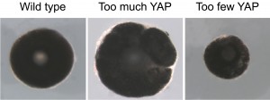
Figure 1. YAP controls post-embryonic eye growth. Dissected eyes from stage 40 Xenopus tadpoles following blastomere microinjection of Yap mRNA or photo-Morpholinos (that allow knocking-down Yap only at post-embryonic stages). Compared to a control situation (on the left) Yap overexpression leads to eye overgrowth (in the middle) while post-embryonic Yap knock-down leads to a microphthalmic phenotype (on the right).

In the post-embryonic retina, Yap expression is restricted to the tip of the CMZ where stem cells reside. We therefore anticipated that Yap knock-down may have exhausted the stem cell pool. But surprisingly stem cells were still present and analysis of their proliferation showed that they were still dividing as well. However, a severe reduction in EdU incorporation was observed, suggesting that something was wrong in their cell cycle progression. We thus employed a variety of approaches dedicated to in vivo analysis of cell cycle phase duration. We very unexpectedly found that although the cell cycle of Yap-morphant retinal stem cells lasts longer, their S-phase length is severely reduced from 17 to 4 hours. How come the S-phase of a stem cell can decrease to such extent?

During S-phase, an eukaryote cell replicates its DNA, starting from multiple replication origins scattered on the genome. This tightly regulated process follows a strict temporal program. The genome is indeed partitioned into early and late replication domains, such that some origins fire during early S-phase while others fire during late S-phase. We found that the precise choreography of the DNA replication program was altered upon Yap knock-down, with a decreased proportion of stem cells exhibiting late S-phase patterns. Late origin may thus have fired prematurely or may have not fired at all. In any cases, this likely explains the shortening in S-phase duration. Among rare factors known to produce such phenotype is c-Myc, whose expression was interestingly found to be increased in Yap morphant CMZ. Although not formally demonstrated, we thus propose that YAP may control S-phase progression through direct or indirect control of c-Myc expression.

Deregulation of DNA replication timing is known to be a source of genomic instability. In line with this, we observed an increased occurrence of DNA damage, enhanced p21 and p53 expression and increased cell death among progenitor cells derived from Yap-depleted stem cells. This ultimately leads to a failure in producing new neurons, which provides an explanation for the reduced post-embryonic growth of Yap morphant retina.

Finally, we also showed that in this temporal control of S-phase progression, YAP physically and functionally interacts with a novel partner, PKNOX1, a mammalian Homothorax ortholog belonging to the Meis/Prep homeodomain factor family, involved in the maintenance of genomic stability (Iotti et al., 2011).

Although relatively young, the research field on Hippo signalling has raised incredibly fast (Lin et al., 2013; Yu and Guan, 2013), with recent interest in the field of stem cell biology (Hiemer and Varelas, 2013; Mo et al., 2014; Piccolo et al., 2014). However, little mechanistic insights are known into how this pathway regulates stem cell properties. Our work revealed a novel role for this factor in the control of the temporal program of DNA replication (Figure 2). We propose a model where this YAP function would protect neural stem cells of the retina from experiencing genomic instability.

figures_230715.ppt
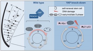
Figure 2: Model illustrating YAP function in retinal stem cells. We found that YAP is expressed in CMZ retinal stem cells (left panel). The middle panel shows the cell cycle of wild type retinal stem cells and the putative role of the YAP/PKNOX1 complex in the control of S-phase temporal progression (represented by the distinct patterns of DNA replication foci). YAP knock-down (right panel) leads to a dramatic reduction of S-phase length likely due to c-Myc-dependent premature firing of late replication origins. This results in genomic instability (increased occurrence of DNA damage, enhanced p21 and p53 expression and eventually cell death).

S-phase duration recently emerged as a major target of cell cycle regulation in different neural progenitor types during cortical development. Neural stem cells exhibit a substantially longer S-phase than progenitors committed to neuron production (Arai et al., 2011; Turrero García et al., 2015), a feature proposed to be dedicated to high quality control of replicated DNA, as errors would be inherited by all the progeny (Arai et al., 2011). Unique mechanisms may therefore operate in neural stem cells to control S-phase duration and ensure genomic integrity. We propose that YAP is part of the genetic network involved in this stem cell-specific regulation of the replication temporal program.


Main paper:

Cabochette, P., Vega-Lopez, G., Bitard, J., Parain, K., Chemouny, R., Masson, C., Borday, C., Hedderich, M., Henningfeld, K. A., Locker, M., et al. (2015). YAP controls retinal stem cell DNA replication timing and genomic stability. eLife 4, e08488.

References

Arai, Y., Pulvers, J. N., Haffner, C., Schilling, B., Nüsslein, I., Calegari, F. and Huttner, W. B. (2011). Neural stem and progenitor cells shorten S-phase on commitment to neuron production. Nat. Commun. 2, 154.

Hiemer, S. E. and Varelas, X. (2013). Stem cell regulation by the Hippo pathway. Biochim. Biophys. Acta 1830, 2323–34.

Iotti, G., Longobardi, E., Masella, S., Dardaei, L., De Santis, F., Micali, N. and Blasi, F. (2011). Homeodomain transcription factor and tumor suppressor Prep1 is required to maintain genomic stability. Proc. Natl. Acad. Sci. 108, E314.

Lian, I., Kim, J., Okazawa, H., Zhao, J., Zhao, B., Yu, J., Chinnaiyan, A., Israel, M. a, Goldstein, L. S. B., Abujarour, R., et al. (2010). The role of YAP transcription coactivator in regulating stem cell self-renewal and differentiation. Genes Dev. 24, 1106–18.

Lin, J. I., Poon, C. L. C. and Harvey, K. F. (2013). The hippo size control pathway–ever expanding. Sci. Signal. 6, pe4.

Mo, J.-S., Park, H. W. and Guan, K.-L. (2014). The Hippo signaling pathway in stem cell biology and cancer. EMBO Rep. 15, 642–56.

Piccolo, S., Dupont, S. and Cordenonsi, M. (2014). The Biology of YAP/TAZ: Hippo Signaling and Beyond. Physiol. Rev. 94, 1287–1312.

Ramos, A. and Camargo, F. D. (2012). The Hippo signaling pathway and stem cell biology. Trends Cell Biol. 1–8.

Tallafuss, a., Gibson, D., Morcos, P., Li, Y., Seredick, S., Eisen, J. and Washbourne, P. (2012). Turning gene function ON and OFF using sense and antisense photo-morpholinos in zebrafish. Development 139, 1691–1699.

Turrero García, M., Chang, Y., Arai, Y. and Huttner, W. B. (2015). S-phase duration is the main target of cell cycle regulation in neural progenitors of developing ferret neocortex. J. Comp. Neurol. [Epub ahead of print].

Yu, F.-X. and Guan, K.-L. (2013). The Hippo pathway: regulators and regulations. Genes Dev. 27, 355–371.


Thumbs up (No Ratings Yet)
Loading...

Tags: , ,
Categories: Research