The community site for and by
developmental and stem cell biologists

A day in the life of a skate lab

Posted by , on 7 October 2015

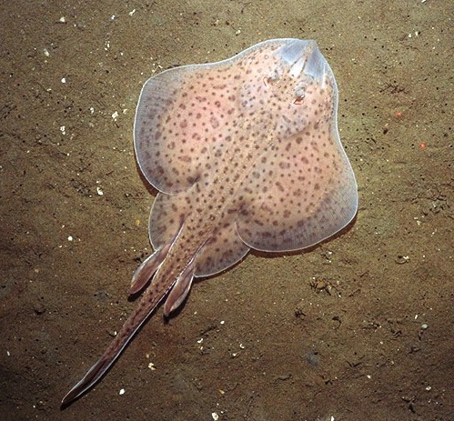
Greetings! My name is Kate (but you can call me skate) Criswell and I am a Ph.D. student in the Department of Organismal Biology and Anatomy at the University of Chicago. I study axial column evolution and development in fishes, and my developmental study organism is the little skate, Leucoraja erinacea. Little skates are little known fish with a cartilaginous skeleton, and they live in the northwestern Atlantic Ocean, from North Carolina up to Nova Scotia.


little_skate1_fullsize
An adult male little skate, Leucoraja erinacea. Photo from NEFSC/NOAA, taken by Page Valentine, USGS.


What’s so great about skates?

Skates are dorsoventrally flattened and have broad, flat pectoral fins and long, slender tails. They are closely related to sharks, rays, and chimaeras, and together these groups make up the chondrichthyans, or cartilaginous fishes. Apart from being elegant and charismatic (and for some, toothy and aggressive), chondrichthyans can provide valuable information on the broad range of developmental mechanisms at work across vertebrates. By comparing the development of cartilaginous fishes with more established organisms like zebrafish, chickens, and mice, we can tell which processes or structures are derived (specialized and more recently evolved) and which are ancestral to all vertebrates. As the sister group to all other jawed vertebrates, cartilaginous fishes exhibit many characteristics that may have been lost in their bony counterparts, or they might display features that are unique and are not found in any other vertebrates.


How do we raise skate embryos?

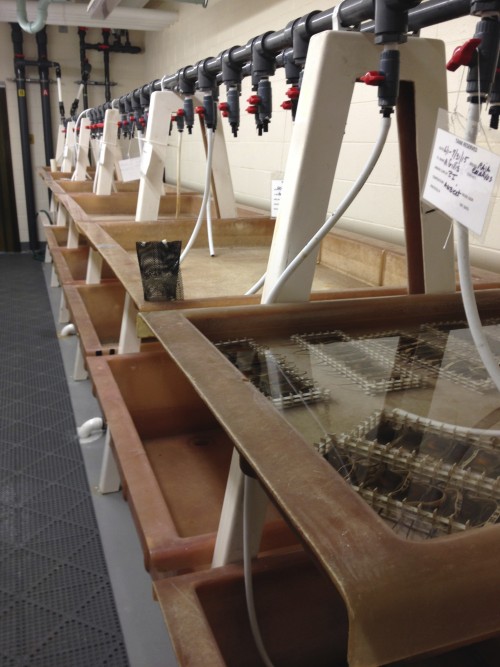
Skates are relatively easy to keep in a lab, and the embryos are very robust, with no need for filtered seawater or antibiotics (as long as you keep them under 18 degrees!). I do most of my live-animal manipulations in the summers at the Marine Biological Laboratory (MBL) in Woods Hole, Massachusetts. The MBL has a fantastic Marine Resources Center (MRC) with large skate brood stock tanks that they use to house adult skates (they also care for many other marine organisms, like snails, bony fish, squid, and sea urchins; a walk through the MRC is better than a trip to the aquarium!). The adult skates mate in the wild and are then collected and housed in the MRC. Females can store sperm for up to 9 months, enabling them to lay eggs throughout the year, which makes it easy to for me to acquire embryos in the winter months. Indeed, there are a number of different labs across the United States that obtain skate embryos from MBL.


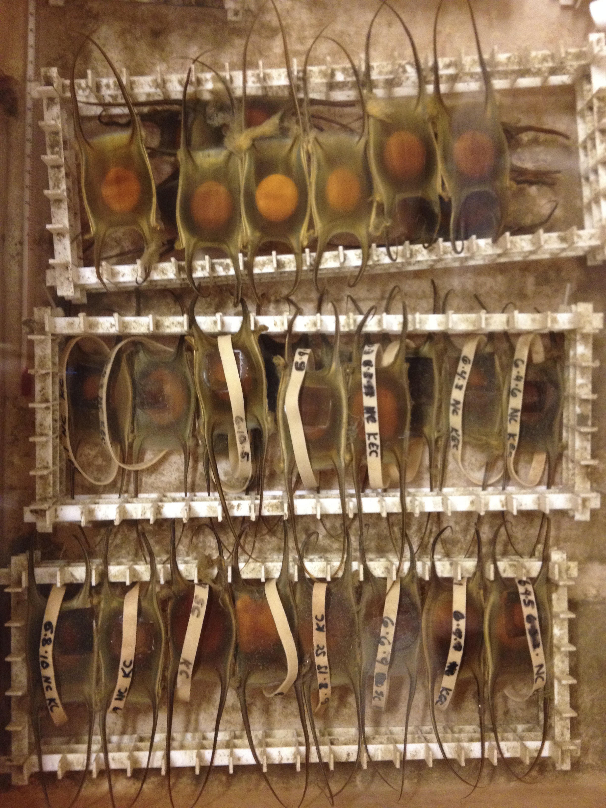
Each egg contains one embryo, which is enclosed in a capsule made of collagen fibers called a mermaid’s purse. These egg cases are rectangular in shape, with two long tendrils at each end. Once laid, staff at the MRC sort the egg cases by week and keep them in 16 degree circulating seawater that is pumped in from the nearby Great Harbor. The egg cases are closed to the environment for the first six or so weeks of development, and then small slits open at the ends of the tendrils to let water flow through. The skates take approximately 5-6 months to hatch into fully-formed, miniature adults, which gives me plenty of time (sometimes too much time) to study their development.


This video was made by my friend and collaborator, Andrew Gillis, who recorded video of the major stages of skate development this past summer at MBL. He’s got a bunch of other great skate videos over on his YouTube channel.


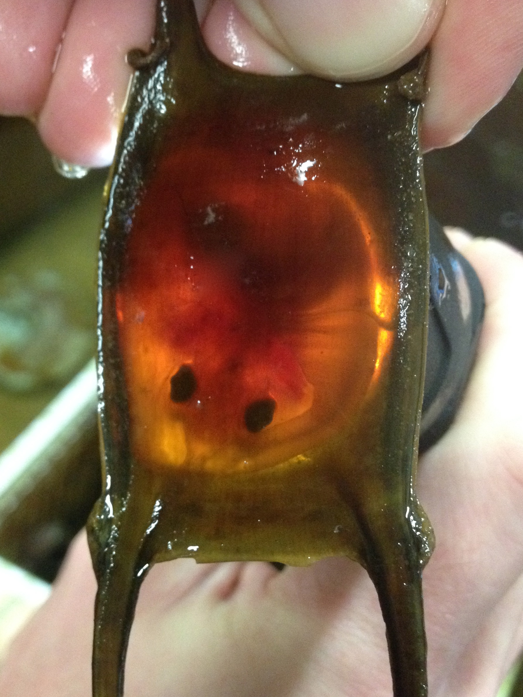
During the summers, while I am doing manipulations of live skate embryos at MBL, I make weekly trips to the MRC to go “shopping” for eggs. I collect eggs of approximately the right age, bring them to the lab space in the Loeb building, and keep them in a sea table until they are at exactly the right stage for experiments. Determining the embryonic stage of skate embryos is actually quite easy, despite their thick egg cases, because the eggs are translucent, and shining a flashlight underneath reveals the embryo curled up inside.


IMG_5609
A late-stage skate embryo almost ready to hatch.


What kinds of experiments can we do with skates?

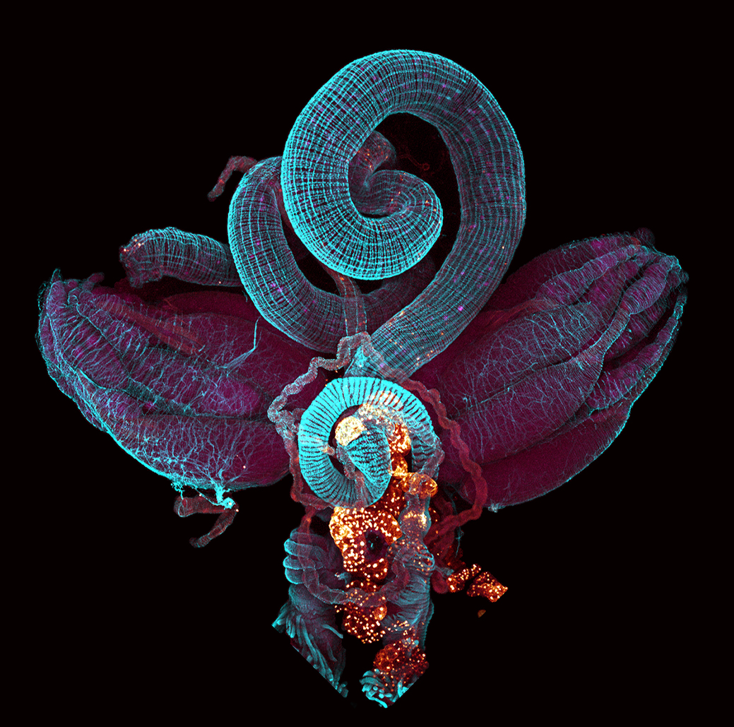
Their hardy nature and (relatively) short developmental period makes skates ideal chondrichthyans in which to do fate-mapping experiments. To study the embryonic origins of different parts of the axial skeleton using young embryos I cut a small window in the egg case, inject a fluorescent dye into the embryonic tissue of interest, and then glue a piece of donor eggshell over the hole (Krazy glue works best; all other glues are subpar). Skates develop a bit like chickens, with the embryo sitting atop a large yolk. When the embryos are young the yolk is extremely fragile and can’t be removed from the egg case. Slightly older embryos (stage 24 and above) have a tougher yolk, and to work with them I can simply cut around the edges of the egg case to make a large window, deposit the entire embryo + yolk in a petri dish with seawater and the anesthetic tricaine, and inject the embryo while it is in the dish. I then scoop the embryo up (using a very sophisticated tool – a plastic soup spoon from the Woods Hole Market) and place it back in the egg case for continued development. There is no need at this stage to glue the egg case shut.


shellgraft
In this manipulated embryo you can see the window I cut into the original eggshell, the graft of donor eggshell that I krazy-glued on after injecting the dye, and the outline of the large yolk within the egg.


After dye injection I let the embryos develop for several months in a sea table, checking the temperature every morning and removing any dead embryos. This means constantly having wet and cold hands, and having chilly seawater drip down my back when I’m not expecting it. By the end of the summer (or several weeks post-injection) I can be sure that most of the remaining embryos will survive into old age. When the axial skeleton is well developed, which takes about 3-4 months, I return to the MBL to fix the embryos and bring them back to Chicago. I spend the rest of the year analyzing experiments from the summer, using paraffin histology to cut thin sections of the axial column and then looking for dye-labeled cells in different parts of the vertebrae.


IMG_4178
This is the sea table room in the Loeb building at the MBL, where I keep skate embryos throughout the summer. The sea tables have both ambient and chilled seawater lines, enabling me to adjust the temperature to the embryos’ liking (as long as there are no power outages!).


In addition to fate-mapping, skates respond well to other experimental manipulations, like drug treatments and bead implantations. For drug treatments, the drug of choice can either be injected into the egg case before it opens to let seawater flow through or, for later stages, the embryos can be removed from their egg cases and placed in baths for several days. Once returned to circulating seawater the embryos can continue developing perfectly well sitting in glass bowls in the sea table, outside of their egg cases.


IMG_4239
I keep skate embryos organized in racks in the sea table. The top row houses unmanipulated embryos, while the bottom two rows contain experimental fate-mapped embryos after dye injection. Rubber bands are great for labeling.


Even though studying skate development can be difficult (especially when your experiments take four months!) it is extremely rewarding. I am excited to continue working in this system and to expand upon the tools currently available to learn as much as I can about the evolution of vertebrate development.


Node day in the life new doodle squareThis post is part of a series on a day in the life of developmental biology labs working on different model organisms. You can read the introduction to the series here and read other posts in this series here.


Thumbs up (16 votes)
Loading...

Tags: ,
Categories: Lab Life

Navigate the archive

Use our Advanced Search tool to search and filter posts by date, category, tags and authors.

An interview with Irene Miguel-Aliaga

Posted by , on 6 October 2015

This interview first featured in the Journal of Cell Science and is part of their interview series Cell Scientists to Watch


Irene Miguel-Aliaga left sunny Barcelona to pursue a PhD at Oxford University in the laboratory of Kay Davies. She did her postdoctorate at Harvard University in Stefan Thor’s lab, and briefly relocated with him to Sweden before coming back to the UK. She continued her postdoctoral work and developed her current research interests as a Marie Curie Fellow in the lab of Alex Gould at the National Institute for Medical Research in London. She established her own lab at the University of Cambridge in 2008, thanks to a Wellcome Trust Career Development Fellowship. In 2012 she moved to London and is currently a Programme Leader at the Medical Research Council Clinical Sciences Centre and a Reader at Imperial College London. Irene was elected to the EMBO Young Investigator programme and holds an ERC Starting Grant. Her lab investigates how gut cells maintain homeostasis and communicate with cells of other tissues and organs.


What motivated you to become a scientist?

I wasn’t one of those children who dissected bugs in their back garden at the age of two. Depending on the day, I wanted to be a vet, an astronaut or a travel agent like my parents. But somebody gave me Carl Sagan’s ‘Cosmos’ for Christmas, and then there was this TV series with lizard aliens that invaded the Earth. They persecuted scientists and the scientists organised themselves against the lizards and overcame the occupation by studying the lizards’ biology and working out their weaknesses. I think maybe the two of them synergised and turned me into a biologist rather than into Brian Cox.


Your research focuses on the brain–gut interface and the cross-talk between the different organs. What are the specific questions that your group is currently trying to answer?

We’re interested in exploring ‘gut intelligence’. The specific questions are about how the gut senses and responds to nutrients or signals from other cells, and about the plasticity of the gut itself – what it responds to and why it matters.


And now you’re also looking at differences between the sexes?

Yes, we’ve been working on sex differences in the intestinal epithelium. Flies can have male or female guts, and the difference is not only developmental – it can be reversed in adult flies. I think it is really fascinating that an adult somatic organ knows its sex and that this matters to the fly!


Of the cell biology methods that you use, which one would you say is the trickiest?

We used to tackle very developmental questions. I found that in development, everything was more black and white – you mutate a gene and end up with no wings. But as soon as you start addressing metabolic or behavioural questions, your n numbers have to become much larger, and you need to control for all sorts of variables such as background effects. I think that has been the challenge for us: these days, it’s harder to persuade ourselves that we’re seeing a real phenotype and to distinguish cause from effect.


Are there any new techniques that you’re thinking of adapting for your research?

I try to think of questions first, and then think of what techniques I need to address them. Otherwise it’s easy to get distracted and end up doing things because you can, rather than because they are important.
JCS178962F2

The digestive and reproductive systems of an adult female Drosophila. Image taken by Paola Cognigni.


What were the biggest experimental roadblocks that you faced and how did you deal with them?

I think sometimes the roadblocks are your own set of preconceptions. Human nature means that, even if we try not to be, we tend to be too hypothesis driven. You have this preconception of how things are going to work. And then sometimes what happens is you do the experiments and you think the experiments are not working, but in fact they are working, they’re just telling you that what you were thinking was wrong. When that happens you just need to take a step back and say “look, I was probably too dogmatic or too one-dimensional in my approach”. We underestimate the value of negative results.


How do you establish your collaborations?

By getting overexcited about new results, talking to everyone willing to listen about them and often presenting unpublished work in progress at meetings. I like to meet scientists with different backgrounds, think of problems together and collaborate to see what we can do about them.


So you would say it’s good to be open?

I hope so. It is true that there is increasing competition and I know that some people in my lab don’t feel particularly comfortable presenting unpublished data. Perhaps we have been lucky, but so far it has definitely been beneficial.


What challenges did you face when starting your own lab that you didn’t expect?

There wasn’t really a very sharp change in any way, because my lab was pretty small until fairly recently, so it was a natural continuation of a postdoc. I’ve got children now, and I probably underestimated the amount of work that it would be. I always thought I’m very good at multitasking, so a couple of children are just a couple more things on your to-do list, when they’re in fact a (welcome) life changer. The challenge now is to get to do everything, including getting a few hours’ sleep a day. But we’re all still alive [laughs], so it seems doable.


Do you think taking part in science outreach events should be more of a priority for scientists?

From a social perspective, it’s our responsibility, but I also do it because it’s fun. It can also be extremely useful for your research: there’s nothing like talking to lay people to help you see the wood for the trees. Children are a great source of scientific questions too – my three-year-old daughter has been wondering why we stop growing, and what the differences between being dead and alive are. I may let her write my next grant proposal!


Since you left Spain, you have worked in the UK, US and Sweden. What challenges have you faced as a scientist when working in different countries?

I found it very stimulating and the experience made me more adaptable, both personally and scientifically. There are some differences, but at the end of the day, the scientific method is the scientific method and what changes is what different scientific environments perceive as interesting or important. So the only challenges are logistics. I remember moving CDs and books around. These days it’s easier because you just take your iPad.


I asked earlier what motivated you to become a scientist. What is your motivation now?

Surprise. A taste for the unexpected. I like it when we get weird results, and when, two years down the line, you find yourself working on something that you never thought you’d be working on. A passion for scientific pursuit often provides consistent yet intermittent reward – probably a good recipe for long-term happiness.


Could you tell us something about yourself that people wouldn’t know just by looking at your CV?

I failed my driving test nine times and I regularly scratch my car, so I guess I live up to the stereotype of an absent-minded scientist!
Also watch this additional short clip:

Thumbs up (7 votes)
Loading...

Tags: , ,
Categories: Interview

In Development this week (Vol. 142, Issue 19)

Posted by , on 6 October 2015

Here are the highlights from the new issue of Development:


Hormone-mediated flower development: a HEC of a job

Graphic

Fruits originate from the female reproductive part of the flower, the gynoecium, the development of which is controlled by the phytohormones auxin and cytokinin, with evidence for the latter just emerging. HECATE bHLH transcription factors are required for gynoecium development and are thought to coordinate auxin signalling, although direct evidence for this is still lacking. Now, on p. 3343, Jan Lohmann and co-workers investigate the function of HECATE 1 (HEC1) in the development of the female reproductive tissue in Arabidopsis. They show that, as in the shoot apical meristem (SAM), which houses the stem cells that generate all the above-ground parts of a plant, HEC1 interacts with SPATULA (SPT) to modulate cytokinin signalling. Furthermore, the authors report that HEC1 impinges on auxin transport by directly regulating the expression of the auxin transporters PIN-FORMED (PIN) 1 and PIN3, a mechanism not at play in the SAM and thus specific to the gynoecium. This study suggests a model in which HEC1 and SPT orchestrate auxin and cytokinin crosstalk during reproductive organ development.


Making blood cells: a FOXy affair

Graphic

The mesoderm, which is specified during gastrulation, generates diverse cell lineages such as endothelial, blood and muscle cells. However, the transcriptional network that orchestrates this process is largely unknown. FOXF1, a forkhead box transcription factor expressed in the extra-embryonic and lateral plate mesoderm, is known to be essential for specifying mesoderm cells to a cardiovascular fate but how it functions is unclear. Here, Valerie Kouskoff and colleagues (p. 3307) generated embryonic stem cells (ESCs) and transgenic mouse lines carrying a Foxf1-venus knock-in allele to study the expression of FOXF1 and its contribution to early mesoderm specification. During ESC commitment to a mesodermal fate, FOXF1 is first expressed after FLK1, a protein essential for endothelial and hematopoietic specification. In the embryo, FOXF1 is highly expressed in all extra-embryonic mesodermal derivatives with the notable exception of the blood islands, the source of blood cells, and increased FOXF1 expression levels correlate with decreased hematopoietic potential. Indeed, using an inducible FOXF1 ESC line, the authors show that FOXF1 is sufficient to irreversibly impair the hematopoietic potential of mesodermal precursors while maintaining their endothelial potential and enhancing smooth muscle fate. These findings shed light on the molecular mechanisms governing hematopoietic specification and are likely to facilitate the derivation of specific lineages from ESCs in vitrofor therapeutic applications.


CO(CO)nverting stem cells into photoreceptors

Graphic

The death of cone and rod cells – the photoreceptors that mediate phototransduction – causes visual loss in millions of people worldwide. Currently, human embryonic stem cells (hESCs) can be differentiated into photoreceptors but the process is inefficient and long. Now, Gilbert Bernier and colleagues (see p. 3294) report that the exposure of hESCs to COCO, a member of the Cerberus gene family, and insulin growth factor 1 (IGF1) in a feeder- and serum-free culture system efficiently differentiates them into functional cone photoreceptors. Such cells express cone-specific genes and key phototransduction proteins, and degrade cGMP when exposed to light – a unique property of photoreceptors. COCO-induced retinal progenitors can also self-organise into polarised sheets of morphologically differentiated cone photoreceptors that show evidence of connecting cilium and outer segment formation and adopt a cone photoreceptor fate in vivo upon injection into the mouse eye. Mechanistically, COCO acts as a potent neural and photoreceptor inducer by simultaneously inhibiting BMP, TGFβ and Wnt signalling, which suggests that cones are formed by default, and this inhibitory activity is potentiated by IGF1. This study provides an efficient and rapid means to generate cone photoreceptors and opens the way to biochemical and genetic studies of photoreceptor development and pathology for regenerative purposes.


PLUS:


Spreading the word: non-autonomous effects of apoptosis during development, regeneration and disease

DEV3253Apoptosis was originally regarded as a ‘silent’ mechanism of cell elimination designed to degrade the contents of doomed cells. However, during the past decade it has become clear that apoptotic cells can produce diverse signals that have a profound impact on neighboring cells and tissues. Here, Pérez-Garijo and Steller discuss how these findings reveal unexpected roles for apoptosis in tissue remodeling during development, as well as in regeneration and cancer. See the Review on p. 3253


Photoreceptor cell fate specification in vertebrates

DEV3263Photoreceptors – the light-sensitive cells in the vertebrate retina – have been extremely well-characterized with regards to their biochemistry, cell biology and physiology. They therefore provide an excellent model for exploring the factors and mechanisms that drive neural progenitors into a differentiated cell fate in the nervous system. Here, Brzezinski and Reh outline the signaling and transcription factors that drive vertebrate photoreceptor development and discuss how these function together in gene regulatory networks to control photoreceptor cell fate specification. See the Review article on p. 3263


A developmental framework for induced pluripotency

DEV3274
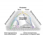
During development, cells transition from a pluripotent to a differentiated state, generating all the different types of cells in the body. Although development is generally considered an irreversible process, it is now possible to reprogram mature cells to pluripotency. Here, Takahashi and Yamanaka discuss the connections and disparities between differentiation and reprogramming, and assess the degree to which reprogramming can be considered as a simple reversal of development. See the Review article on p. 3274


Featured movie

Thumbs up (No Ratings Yet)
Loading...

Categories: Research

A new Special Collection on ‘Model Systems in Drug Discovery’

Posted by , on 6 October 2015

drugdiscoveryissueModel systems, including laboratory animals, microorganisms, and cell- and tissue-based systems, are central to the discovery and development of new and better drugs for the treatment of human disease. In the latest issue, Disease Models & Mechanisms (DMM) launches a Special Collection that illustrates the contribution of model systems to drug discovery and optimisation across multiple disease areas. This collection includes reviews, Editorials, interviews with leading scientists with a foot in both academia and industry, and original research articles reporting new and important insights into disease therapeutics.

The Special Collection Editorial provides a summary of the collection’s current contents, highlighting the impact of multiple model systems in moving new discoveries from the laboratory bench to the patients’ bedsides. The launch issue can be accessed in full here.

To read and sign up for updates on the full Collection, which also includes key drug discovery research and review articles published earlier in DMM, go to the Collection page at http://dmm.biologists.org/cgi/collection/drugdiscovery

Thumbs up (1 votes)
Loading...

Tags:
Categories: News

BSDB student and postdoc survey

Posted by , on 6 October 2015

BSDBlogoThis is a news item which was first posted on the bsdb.org site. Please, note that not all items will be duplicated on The Node. To ensure you stay informed, please, take a minute to subscribe for email notifications on the bsdb.org site: simply enter your email address in the 3rd item of the side bar. Be ensured that the amount of emails sent to you will usually not exceed one per fortnight or month.

—————————–—————————–

A message from Alex Ashcroft (BSDB student rep) and Michelle Ware (BSDB postdoc rep):

The British Society for Developmental Biology (BSDB) has elected us as your student and postdoc reps. We now have the opportunity to give students and postdocs a voice in moving the BSDB forward.

Therefore, we are trying to gauge what you want to see included as part of the society and the meetings. We have lots of ideas and have created a short survey for students and postdocs who are BSDB members (LINK). Your feedback will be key to determine what will be worthwhile in pursuing.

As a thanks we are offering four kindles and two £30 Amazon vouchers as prizes. We’d be grateful if you could complete it by October 26th.

Many thanks,
Alex Ashcroft and Michelle Ware

Thumbs up (1 votes)
Loading...

Tags:
Categories: News, Societies

Postdoctoral position: Formation and Roles of Drosophila Axonal Endoplasmic Reticulum

Posted by , on 6 October 2015

Closing Date: 15 March 2021

A vacancy for a postdoctoral research associate is available for 2 years from 1 Jan 2016 in the first instance. Axonal endoplasmic reticulum is a poorly characterised compartment that is probably ubiquitous in axons, may mediate local and long-distance communication, and probably underlies a number of neurodegenerative mechanisms.

We have developed ways to visualise axonal ER in Drosophila, and identified mutations affecting its organisation. We now wish to understand more of its mechanisms of formation, and its physiological role.

For details of the post and how to apply, see: www.jobs.cam.ac.uk/job/7054/

Informal enquiries to Dr. Cahir O’Kane, Department of Genetics, University of Cambridge (c.okane@gen.cam.ac.uk)

Thumbs up (No Ratings Yet)
Loading...

Categories: Jobs

The Node in Portugal

Posted by , on 5 October 2015

This week the Node will be in Portugal! We are first attending the joint meeting of the Portuguese, Spanish and British Developmental Societies, that is taking place in the Algarve. Let us know if you are attending as well, as we would love to meet you and hear your thoughts about the Node. If you are not attending, do check our twitter account. We will be tweeting from the meeting if the internet connection is good enough.


We are also making a scheduled stop in Lisbon. If you are in or around Oeiras on Monday the 12th of October, pop by the Institute Gulbenkian de Ciência at midday. Cat, our community manager, will give a talk about how to use social media to communicate your science (Ionians seminar room). We look forward to meeting you!
Node Portugal

Thumbs up (2 votes)
Loading...

Tags:
Categories: News

EmbryoMaker: a general modeling framework to simulate developing systems and perform experiments in silico.

Posted by , on 4 October 2015

One of the main challenges of Developmental Biology is to understand the complex developmental mechanisms giving rise to different organs or whole organisms. In most cases, these involve the interplay between cell-cell signalling and cell and tissue movements driven by one or several cell behaviours (such as cell proliferation, cell migration, etc.). Cell signalling will affect how surrounding tissues grow or change in shape, which in turn will change the spatial context in which signalling is taking place. Such complexity in the developmental dynamics can account for the formation of quite complex organs, such as mammalian teeth1 or vertebrate limbs2, but understanding how perturbations on those developmental processes will affect the resulting phenotype is not trivial.

Multi-scale computational models can help in better understanding the dynamics of developmental processes both qualitatively and quantitatively. They should be built upon explicit mechanistic hypotheses about how development takes place. Computational models can then provide explicit quantitative predictions on how the morphology of the organ or embryo or the expression pattern of certain gene products change during development (Figure 1).

Mathematical models need to be based on experimental observations. These are interpreted and a mechanistic hypothesis is formulated (i.e. how the system is supposed to work). The model is implemented by translating the hypothesis into a mathematical formulation and solved through computational methods. Model validation consists on testing how accurately the model can reproduce experimental observations. A validated model can then be used to predict the behaviour of the system under conditions that haven't been yet reproduced experimentally.
Figure 1. Model design and validation. Mathematical models need to be based on experimental observations. These are interpreted and a mechanistic hypothesis is formulated (i.e. how the system is supposed to work). The model is implemented by translating the hypothesis into a mathematical formulation and solved through computational methods. Model validation consists on testing how accurately the model can reproduce experimental observations. A validated model can then be used to predict the behaviour of the system under conditions that haven’t been yet reproduced experimentally.


In the Salazar-Ciudad lab we work on the design of computational models of development in order to study how the complexity of developmental dynamics may give rise to complex structures and how the presence of different types of developmental mechanisms in different lineages may affect their evolution. For some time we have been using a tooth development model1 in order to approach those questions. However, if one wants to tackle those questions from a more general point of view, organ-specific models of development are not enough. For that purpose we need a modelling framework that implements a general developmental toolkit, that is: 1) the ensemble of cell behaviours known to happen in animal development (cell growth, division, death, cell migration, adhesion, epithelial-mesenchymal transition, cell signalling and extracellular matrix modification), 2) the basic mechanical properties of epithelial and mesenchymal cells and extracellular matrix and 3) the freedom to design gene regulatory networks (GRN) that dynamically control the mechanical properties of cells and their behaviours. Such a modelling tool should be able to simulate the development of, say a tooth or a limb, given that we correctly choose the gene networks and initial conditions in each case. Most interestingly, this would allow to study in silico how to transform one organ into the other by replacing, for example, the tooth forming GRN by the limb forming GRN and vice versa. In a similar way evolutionary transitions between different organs or structures could be inferred by rewiring the GRN step-wise.

Although there already exist modelling frameworks of development3,4,5 none explicitly implements the differential mechanical properties of both epithelial and mesenchymal cells or the whole range of cell behaviours we enumerated above. Thus, we decided to develop the most general developmental modelling framework up to date with all the elements described above.

In a paper recently published in Bioinformatics we present this new modelling framework implemented in the open source software EmbryoMaker, freely available for download at our lab’s website. The software provides a graphical interface to visualize the progress of a simulation in real time. In addition, it comes with a user-friendly editing tools in order to design the spatial initial conditions of any developmental system made of either epithelia, mesenchyme and/or extracellular matrix (the size and shape of the tissues at time 0 and their mechanical properties and gene expression profiles) and the GRN (that is defining the number of genes that will participate, their regulatory interactions and their regulation of different cell behaviours). The editing tools can be further used during the simulation of development in order to manipulate the system in real time. For instance, groups of cells can be removed or replaced in the fashion of a graft experiment, or growth factor releasing beads can be placed in any point in the developing system (Figure 2A). Thus, EmbryoMaker may work as an in silico wet lab that allows to predict the outcome of possible experiments on the system of study before carrying them out in vivo or in vitro (Figure 1).

We also show how the modelling framework is able generate complex morphologies from rather simple initial conditions by combining different cell behaviours in a dynamic way (Figure 2B). In this case, the joint action of localized cell contraction, cell polarization and polarized cell division drive the invagination of a spheric epithelium by epiboly.

Overall we expect this new modelling framework to contribute positively to the advance of the Developmental Biology and Evolution fields by providing powerful predictive tools to aid experimental design but also as a means to systematically study the capacity of Development to generate complex and disparate structures and how those might evolve.


A, Simulation of a growth factor releasing bead experiment in which. The first row shows a developmental sequence in which an epithelial bud (blue and purple) grows over a mesenchymal condensate (pink). The second row shows the same process, but in this case a growth factor releasing bead is place at a certain time during the simulation. The growth factor diffuses towards the epithelial cells and increases their proliferation rate, thus making the left side grow larger than the right side. B, Simulation of a complex developmental system by combining several cell behaviours on a hollow spheric epithelium. The contraction of a localized group of cells causes a shallow invagination of the epithelium. Meanwhile, a molecular gradient is being formed towards the invaginated region, and cells across the embryo are instructed to proliferate in the direction of the gradient. The directional growth of the embryo pushes cells deeper into the embryo in a fashion reminiscent of the process of gastrulation by epiboly.
Figure2. Examples of simulations performed with the EmbryoMaker software. A, Simulation of a growth factor releasing bead experiment. The first row shows a developmental sequence in which an epithelial bud (blue and purple) grows over a mesenchymal condensate (pink). The second row shows the same process, but in this case a growth factor releasing bead is placed at a certain time during the simulation. The diffusible growth factor will only reach the cells closest to the bead and will increases their proliferation rate, thus making the left side grow larger than the right side. B, Simulation of a complex developmental system by combining several cell behaviours on a hollow spheric epithelium. The contraction of a localized group of cells causes a shallow invagination of the epithelium. Meanwhile, a molecular gradient is being formed towards the invaginated region (colors depict the concentration of the molecule: yellow is high and blue is low), and cells across the embryo are instructed to proliferate in the direction of the gradient. The directional growth of the embryo pushes cells deeper into the embryo in a fashion reminiscent of the process of gastrulation by epiboly.


Main reference: 

Marin-Riera M, Brun-Usan M, Zimm R, Välikangas T, Salazar-Ciudad I (2015) Computational modeling of development by epithelia, mesenchyme and their interactions: a unified model. Bioinformatics


Other references:

1- Salazar-Ciudad I and Jernvall J (2010) A computational model of teeth and the developmental origins of morphological variation. Nature 464, 583-586.

2- Hentschel HGE et al. (2004) Dynamical mechanisms for skeletal pattern formation in the vertebrate limb. Proc. Royal Soc. B. 271, 1713-1722.

3- Pitt-Francis J et al. (2009) Chaste: a test-driven approach to software development for biological modeling. Comput Phys Commun. 180: 2452-2471

4- Izaguirre JA et al. (2004) CompuCell, a multi-model framework for simulation of morphogenesis. Bioinformatics 20, 1129-1137

5- Delile J et al. (2013) Computational modeling and simulation of animal early embryogenesis with the MecaGen platform. In: Kriete A, Eils R editors. Computational Systems Biology, 2nd ed. Academic Press. Elsevier pp. 359-405


Thumbs up (6 votes)
Loading...

Tags: ,
Categories: Research, Resources

DMM Travel Grants for 2015

Posted by , on 2 October 2015

Are you interested in attending a meeting or course in a DMM-relevant field during 2015? Apply for one of our new travel grants, valid for travel before the end of the year.

Applicants will usually be PhD students and postdoctoral researchers at the beginning of their research careers, who will use the funding to support their travel to relevant scientific meetings. We also welcome applications from independent group leaders and PIs with no independent funding, seeking support to attend meetings, conferences, workshops, practical courses, PI laboratory management courses and courses to re-train.

For further information, and to download an application form, go to http://dmm.biologists.org/site/misc/DMM%20travel%20grants.xhtml

Thumbs up (1 votes)
Loading...

Categories: Funding, News

Upcoming Free Webinars sponsored by Abcam

Posted by , on 1 October 2015

Save the Dates!  

Abcam is pleased to announce that our November calendar of free live webinars, presented by expert guest speakers, is now LIVE!

Register for a webinar (or three!) today and come prepared with your questions for the live Q&A session at the end of every webinar.

Can’t attend the live webinar? Not a problem! Register for the event today and simply look for an email from Abcam Events the day after the live webinar with the on-demand recording.


Developing Durable miRNA Biomarker Technologies for Microbial Carcinogenesis in Resource Poor Settings

Register Here: http://bit.ly/mirnaprofilingwebinar 


Benefits and Limitations of Primary and Secondary Antibody Conjugates

  • Thursday, November 12th
  • 15:30-16:30 CET/ 14:30-15:30 GMT/ 09:30-10:30 EST/ 06:30-07:30 PST
  • Presenter: Dr Javier Conde Vancells of Abcam

Register Here: http://bit.ly/antibodyconjugateswebinar


Intestinal Organoids as a Model System and Tools for Genetic Manipulation

Register Here: http://bit.ly/intestinalorganoidswebinar


Questions? Feel free to contact Abcam Events at events@abcam.com

Thumbs up (No Ratings Yet)
Loading...

Categories: Education, Events