An interview with Benoit Bruneau
Posted by the Node, on 5 November 2013
This interview first appeared in Development.
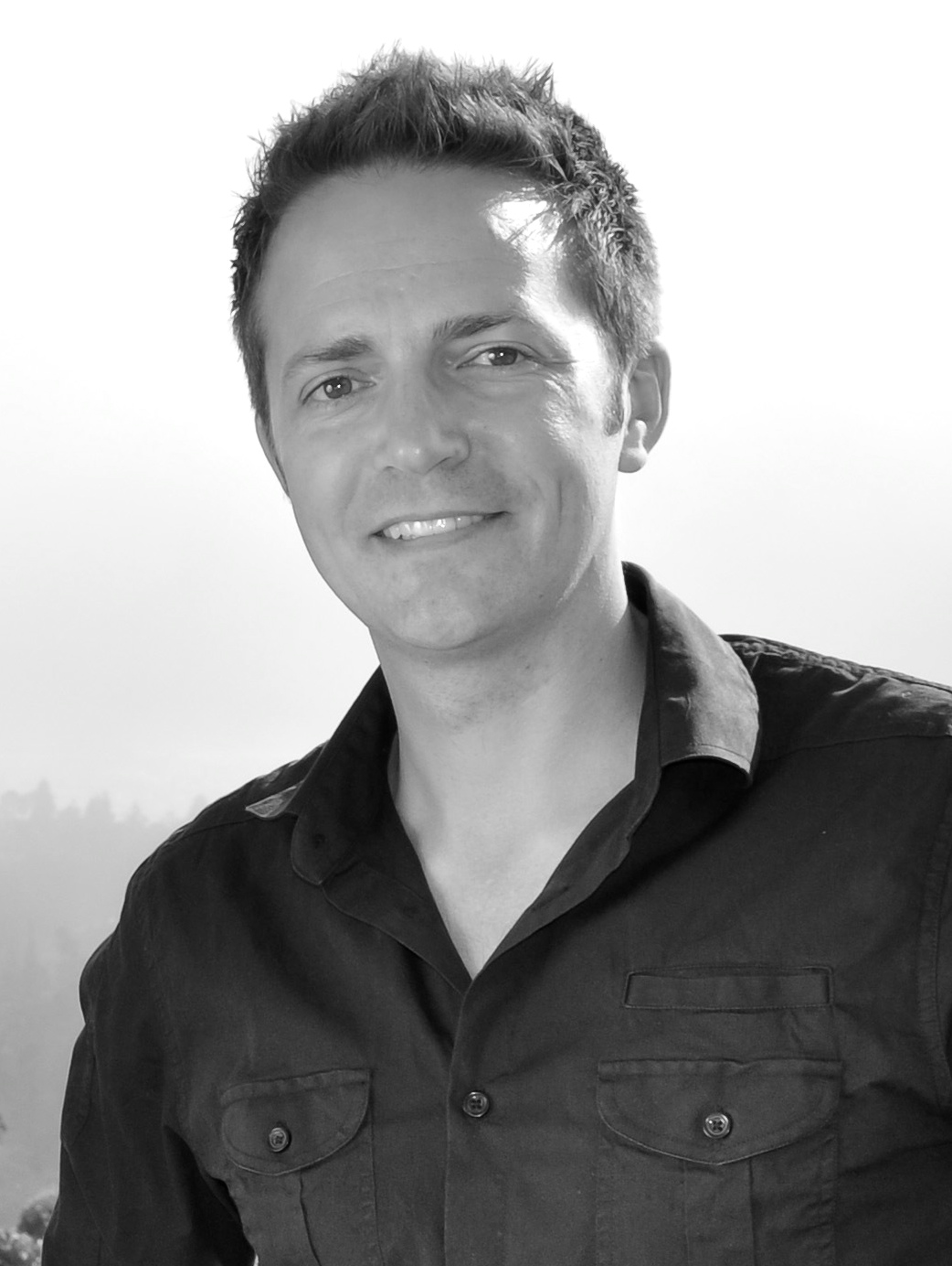
Benoit Bruneau is a developmental biologist based at the Gladstone Institutes in San Francisco. His lab studies the transcription factors and chromatin remodelling complexes that regulate cardiac organogenesis and differentiation, with the aim of uncovering the basis for congenital heart defects. Benoit has recently become an editor for Development, and we asked him about his research and career and discussed how social media can help scientific progress.
 When did you first become interested in developmental biology?
When I was in my third year of undergraduate studies at the University of Ottawa, I took a developmental biology course that had a research component. The university had an axolotl colony, so we did all sorts of classic experiments, such as grafting Spemann organisers. I totally fell in love with developmental biology. However, when I asked the teaching assistant what it took to become a developmental biologist he wasn’t very motivating. He said you had to spend 6 years in graduate school, then postdoc for a number of years, then find a job that doesn’t pay well and fight for grants; I shelved that idea and instead had thoughts of medical school. Then a plant genetics research project the following year got me interested in genetics, and I ended up doing a PhD in physiology, looking at heart gene expression.
Developmental biology took a back seat for several years, but during my PhD I found myself reading lots of developmental biology papers, as well as papers on transcriptional regulation. These got me thinking about how great it would be to understand how gene expression is regulated in development. When I was doing my postdoc with Jon and Christine Seidman they discovered that the gene encoding the T-box transcription factor TBX5 was the mutated gene in Holt-Oram syndrome, which includes congenital heart defects. They asked me whether I wanted to make the Tbx5 knockout mouse, model the disease and understand its function. Right there I saw the opportunity for putting together everything that I had always dreamed of doing in one project. I wasn’t in a developmental biology lab but it seemed like I was embarking on a project in that field, and I was fortunate to be surrounded by developmental biology labs, such as those run by Cliff Tabin, Norbert Perrimon and Connie Cepko. Simply by osmosis, by doing and talking constantly about developmental biology over the years, I immersed myself in it.
Why did you decide to focus your research on the heart?
My graduate work was on cardiac physiology, and I joined the Seidman lab so that I could make mouse models of cardiomyopathies. I always had an interest in the heart because of heart disease in the family. When I started out, one might have said that if I wanted to study heart disease I should have been studying heart attacks and atherosclerosis. Now, it is obvious that if we really want to fix hearts after a heart attack, we need to be able to build new heart cells, and that is what developmental biology is all about. This has actually become a reality, and I have been fortunate to participate in some of those discoveries.
What are the projects your lab is working on at the moment?
An important clinical motivation for our research is to understand the basis for congenital heart defects. I am really excited because we are finally able to do things in a way and on a scale that I had always dreamt of. We want to understand all the genomic switches and regulators that are involved in cardiac lineage determination and cardiac differentiation. We are taking a strategic approach by focusing on certain chromatin remodellers and DNA-binding transcription factors that we know are involved. We are also using new approaches to understand gene regulation during differentiation: for example, investigating the 3D interactions in the genome that shape or control these regulatory events.
We have really migrated from investigating a single gene at a time to addressing what is actually happening at the genome level: what is interacting with what, and how is that important? With the advent of new genomic and engineering technologies, such as TALENs (transcription activator-like effector nucleases) and CRISPRs (clustered regularly interspaced short palindromic repeats), the sky is the limit. For example, it took me 2 years of my postdoc to make the Tbx5 mutation, but a new postdoc in my lab generated the same mutation in 3 weeks. This means we can now address the function of regulatory elements not just in a heterologous context or in an artificial assay, but in a differentiating cell and, as we work on mouse, in a differentiating organism. Ultimately, the goal is to get a genomic blueprint of cardiac differentiation.
How stem cell research fits within developmental biology is a much-discussed topic. Your work is at the crossroads between these two fields: where do you stand in this discussion?
Yesterday someone referred to me as a ‘stem cell guy’, which is funny because we have really only published one and half stem cell-related papers! I’ve had this discussion with the lab recently: do we have a lab identity, and does it matter? Are we a stem cell lab now? Are we still a heart development lab? Or are we a transcription/chromatin lab that happens to be studying that molecular process in the developing heart? Our conclusion was that we are all of those things. We don’t need to pigeon-hole ourselves, because stem cells are a part of developmental biology. We want to understand progenitor allocation, morphogenesis, and how cells behave from the point of view of gene regulation. Our research topic allows us to be stem cell scientists, developmental biologists and chromatin biologists. I have had people join my lab with a background in developmental biology who are now doing primarily stem cell-based differentiation projects or chromatin-based projects, and vice versa. We incorporate all of their different skill sets and approaches, and I have fantastic people in the lab who can keep track of all the techniques and approaches. I hope we are uncategorisable!
Did you have a mentor or someone who inspired you during your career?
There are two people who have been major influences on my career. One of them is Janet Rossant and the other is Eric Olson. They have both been enthusiastic supporters of my science, which is really important. When you are starting out you don’t know if what you are doing is actually any good or if it will be appreciated, and I got really wonderful encouragement from both of them. I sought and got fantastic career advice from Janet very early on, when I was still a postdoc, and afterwards when I moved to Toronto. She has been a very good career mentor, and a generous colleague. Eric was an influence scientifically. I have got to know him over the years and the way he does things is tremendously inspiring. After I hear Eric give a talk I have two immediate reactions. The first is that I might as well quit science, because I will never be able to achieve something as impressive. But the stronger reaction is to be really motivated to go where I didn’t think I would be able to go, and go there without any fear.
How have you found your first months as a Development editor?
It is a tremendous honour. Development has always been one of my favourite journals and to be an editor among all the current and former editors who are the giants of developmental biology is humbling and a great honour. One of my goals is to try to help the journal increase its visibility and broaden its scope. In a way, the journal has already been doing this quite successfully with the recent Stem Cells and Regeneration section. I also want to encourage those colleagues in my field who are not contributing so much anymore to come back to the journal and those that are newer to consider sending their best stuff in. The history and the prestige behind Development is apparent to most but is not appreciated by many. That is what I would like to be able to bring to my role as editor.
Is there any particular type of paper, or particular topics, that you would like to see people submitting to Development?
I would like people who work on stem cell models of differentiation to think of the journal as a good place for their papers. I would also like to see more people who are doing very high quality molecular embryology to send their best work to the journal. There’s more competition now in the journal sphere, and I think Development has one of the most important places. However, we need to continue persuading people to send what they consider their best work to the journal. We will do that by accepting the best papers and by submitting our own best papers.
You are a very active user of Twitter, but many scientists see social media as a waste of time. Why do you use Twitter, and do you think scientists should be more active on social media?
Most scientists aren’t aware of the advantages of using Twitter. People wonder why it is interesting to know what someone had for lunch, or where they are going every minute, but that is not what Twitter is about. Twitter for me has been a really important source of information. I have been able to have real-time scientific discussions from my living room with people across the four corners of the world – mini conversations that I would not necessarily have otherwise had. I also get to know about some of the science that people are doing, especially in the genomics field, which has embraced Twitter as a means of communication. Of course, there are a lot of general views and amusing things that are nice to know about, but I see it more as a global science communication tool. You are also able to interact and get people’s opinions in a really immediate way. Sometimes people are overly frank on Twitter, which can be a bad thing; but it can also be nice: you really get to know what people are thinking, as opposed to just reading their work. I’m enthusiastic about Twitter, and I tell people about it, but I try not to beat them over the head with the gospel of Twitter. It’s not for everyone.
What would people be surprised to find out about you?
Despite being Canadian I didn’t live much of my childhood in Canada. My father was a diplomat, so I have lived in a number of different countries across the world. I was born in Tel Aviv, and I was baptised in the ancient monastery of Latrun, which only very rarely (every 50 years or so) has baptisms.
 (2 votes)
 In mammals, adult neurogenesis is highly restricted to the subventricular zone and to the subgranular zone (SGZ) of the hippocampal dentate gyrus. Is neurogenesis in these regions a recapitulation of developmental neural production, or does it involve distinct molecular and cellular processes? And are these processes conserved across mammalian species? To help answer these questions, Ed Lein and colleagues (
 The female gametes of flowering plants are produced within a structure known as the gametophyte, which develops inside the carpel of the flower. The female gametophyte (FG) contains several cell types, and it has been proposed that their fate is specified, according to position, by an internal auxin gradient. Ueli Grossniklaus and co-workers (
 Although much is known about the specification and differentiation of midbrain dopaminergic (mDA) neurons, the mechanisms regulating their migration within the ventral midbrain (VM) are poorly understood. Migration of several other neuronal types is under the control of CXCL12/CXCR4 signalling, which has been shown to impact on migration, neuritogenesis and axonal pathfinding. Now, Ernest Arenas and colleagues (
 Stem cell renewal in vivo often requires a specialised microenvironment, the stem cell niche. Niche cells provide self-renewal signals as well as structural and spatial cues to regulate stem cell maintenance and differentiation. Here, Pankaj Sahai-Hernandez and Todd Nystul (
 Hox genes provide positional information along both the body’s anterior-posterior and the limb’s proximal-distal axes. Analysis of Hox gene function in the limb has primarily focussed on their roles in skeletal patterning. Now, Deneen Wellik and co-workers (
 Before fertilization, animal eggs are maintained in cell cycle arrest, to prevent parthenogenetic activation. In vertebrates, this is achieved by MAPK- and Emi2-mediated inhibition of the anaphase promoting complex/cyclosome (APC/C). Sperm induce egg activation by calcium-dependent activation of CaMKII, which triggers the destruction of Emi2, activating APC/C. However, invertebrates do not possess an Emi2 homologue, raising the question of how egg activation is achieved in these species. On
  Despite numerous studies, the molecular mechanisms underpinning the fertilization event in mammals remain largely unknown. However, as summarized here by Masuru Okabe, recent work using both gene-manipulated animals and in vitro studies has begun to elucidate essential sperm and egg molecules and to establish predictive models of successful fertilization. See the Primer on p.
  The satellite symposium on ‘Making and breaking the left-right axis: implications of laterality in development and disease’ was held in June 2013 in conjunction with the 17th ISDB meeting in Cancún, Mexico. As summarized by Rebecca Burdine and Tamara Caspary, leaders in the field gathered at the symposium to discuss recent advances in understanding how left-right asymmetry is generated and utilized across the animal kingdom. See the Meeting Review on p.
  Benoit Bruneau is a developmental biologist based at the Gladstone Institutes in San Francisco. His lab studies the transcription factors and chromatin remodelling complexes that regulate cardiac organogenesis and differentiation, with the aim of uncovering the basis for congenital heart defects. Benoit has recently become an editor for Development, and we asked him about his research and career and discussed how social media can help scientific progress. See the Spotlight article on p.
  (No Ratings Yet)

 We’re excited to announce that our short film, Stem cells – the future: an introduction to iPS cells, is now available in French, German, Italian, Polish and Spanish as well as English, with a supporting quiz for the classroom. You can
 Many congratulations to our partner, Dr

 This post is part of a series on a day in the life of developmental biology labs working on different model organisms. You can read the introduction to the series
  (16 votes)