Distant Developments: In conversation with Professor Stephen Robertson, clinical geneticist and developmental biology researcher
Posted by Megan Wilson, on 15 July 2013
“We have the ability to understand human disease and deliver it back to the clinic – allowing families to understand and then move on.”
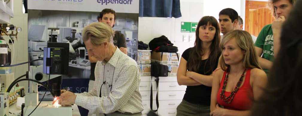
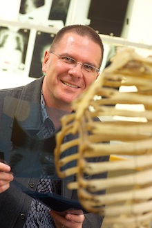
This week, I caught up with Professor Stephen Roberson to discuss how he combines his research interests in developmental biology with a position as a clinical geneticist. Stephen is a medically trained researcher based in the Department of Women’s and Children’s Health, University of Otago. He worked in Paediatrics and Clinical Genetics in both Auckland and Melbourne in the 1990s. In 1999 he changed tack to join the Institute of Molecular Medicine at the University of Oxford, working on the genetic determinants of congenital malformations. Stephen was recruited back to Otago in 2002 to take up a position as Curekids Professor of Paediatric Genetics. Stephen is located in the Department of Women’s and Children’s Health, University of Otago, where he heads the Clinical Genetics Group. His group has over 90 publications including high profile journals such as Nature Genetics, PNAS, and the American Journal of Human Genetics.
Stephen’s career highlights include identifying mutations in FLNA that caused both brain and skeleton malformations while at Oxford. Then back at Otago, his group identified mutations in a paralogue (FLN B) in individuals with a related spectrum of disorders, leading to the recognition of a group of development disorders now named filaminopathies. His team has also identified germline mutations in a tumour suppressor gene also resulting in disorders of morphogenesis characterized by a broad slew of malformations in different organ systems.
In addition to running a successful research laboratory, he still is a practicing clinical geneticist spending nearly a third of his time travelling to clinics in the South Island and Wellington. On top of this he is a popular lecturer, teaching in both Genetics and Medicine courses at Otago.
Combining clinical work with research. Early on in Stephen’s medical career, he had a strong interest not just the clinical side of medical work but also the molecular factors underlying a disorder. He was really driven by a desire to combine these two together throughout his career. As one of only 8 clinical geneticists in the NZ Genetic Health Service, this part of the job involves sitting down with the patient and family and discussing what the diagnosis means both in terms of future options but also heredity. Many great scientific opportunities have arisen through Stephen’s work in clinic. Often a patient can walk in to clinic with a rare condition and sometimes due to an understanding of the molecular pathways of involved in development it is possible to speculate that a defect in a given pathway may underpin a given clinical presentation , with the advantage of being go to the back to the lab with the patient’s DNA samples to work on identifying the gene at fault.
“NZ families are very open to research and very generous and have a desire to contribute to understanding the broader picture of a disease”.
The Major research projects in Stephen’s laboratory centre around three core questions.
They seek to further understanding of how neural cells migrate during brain development particularly in relation to brain malformations often found in rare genetic disorders. His group has just recently completed analysis of a new neurological syndrome that has identified a pathway involved in brain development that up until now was not suspected to play such a role (to be published soon –watch this space!).
His group has an ongoing interest in bone malformations including the genetic pathways responsible for gravity sensing and how the skeleton triggers an anabolic response to mechanical stress.
Stephen’s group also have a number of clinical projects running, including understanding genetic contributors to conditions characterized by atresia of gall bladder and bile ducts, a disorder which presents not long after birth with the babies becoming very jaundiced, followed by liver failure. If they can find the genetic cause, this will hopefully lead to earlier diagnosis and more successful treatment outcomes for these patients.
Stephen is fascinated, like all developmental biologists, by the whole amazing process of going from a cell to a whole organism.
“Everyone studying their own corner of a much bigger picture, how the overall process is conducted is mind blowing.”
Stephen’s group makes use of many classic developmental biology model animals to study the functional role of genes identified by mutation screening. They make use of the Otago Zebrafish Facility to perform morpholino studies to begin to characterize gene function, and transgenic mouse ‘knock-in’ studies to study the effect of the disease mutation on mouse development. One key advance his lab has adopted is the use of next generation sequencing, particularly exome sequencing, allowing much faster genetic characterization of patient DNA samples.
Working as a scientist in New Zealand. The biggest advantage is the collegiality, more group leaders have some job security that makes the environment more collegial and less combative. . Due to NZ’s size it is easier to get to know each other and also span more areas of interest such as going to journal clubs on topics far removed from your research area.
Of course, the greatest challenge for most scientists and NZ scientists in particular is Funding, limited amount of money and funded only for a short time three year time frame, when often the project is really just getting started. Stephen suggests that researchers need to rise to this challenge by being more creative and collegial with their limited resources, and to communicate their work to the public to highlight its benefits and relevance.
Giving back to the community. Stephen works closely with CureKids as one of their three Chairs of Child Health Research. CureKids’ focus is raising funds for medical research to find cures for childhood diseases. This has been so successful in NZ, CureKids will soon be starting up in the USA and Australia. This involvement with Curekids has had a stronginfluence on both Stephen’s research and the way he communicates back to the community. This has been a challenge since Stephen was previously more atune to the more traditional routes of science communication such as journal articles and research seminars. To reach the public, he now has learnt to communicate more broadly through medias like TV, radio, and talking “off the cuff” at events without prompts in order to engage with the people who are really the ones that are funding this research.
They also keep asking the hard questions, the ones the families want the answer to rather than just curiosity-driven investigation – “that’s a nice discovery, but how does that fix the disorder or improve the lot of the child or family?” This helps keep the research centred around the central purpose of helping people. Stephen is also interested in questions surrounding the ethical questions raised by the implementation of the many new genomic tools becoming available and publicised – how do we put this into context for the public?
So how can scientists help the public to understand genetic disorders? Stephen’s advice is we need to put the research into context – make more use of visual learning both print and online to help the public understand developmental biology. Key for this will be developing resources and making better use of graphic tools to help explain our research.
“We need to make the public more aware of how much is imprinted in very early development in the embryo, it really casts the mold for the adult.”
 (4 votes)
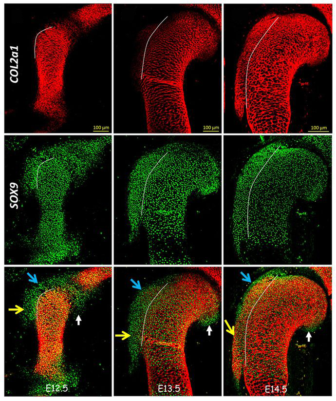
 (8 votes)
 (No Ratings Yet)