The community site for and by
developmental and stem cell biologists

Overly Honest Methods

Posted by , on 8 January 2013

Why did you incubate that sample for 16 hours? Because you wanted to go home for the day – but that much detail is not in your paper! Last night, a few scientists on Twitter started sharing their “overly honest methods”, and today the #overlyhonestmethods hashtag exploded with lots of funny and true stories about scientific experimentation. There are thousands of tweets, still coming in, so I can’t show you all of them, but here is a selection of some of my favourites:

Thumbs up (9 votes)
Loading...

Categories: Lab Life

Navigate the archive

Use our Advanced Search tool to search and filter posts by date, category, tags and authors.

In Development this week (Vol. 140, Issue 3)

Posted by , on 8 January 2013

Here are the research highlights from the current issue of Development:


Directed differentiation: Nodal steps forward

The directed differentiation of pluripotent stem cells into endodermal derivatives, including insulin-producing pancreatic β cells, has considerable clinical promise in cell replacement therapies. The first step in this process is the conversion of pluripotent stem cells into definitive endoderm (DE). Here (p. 675), Douglas Melton and colleagues investigate the endodermal populations generated from mouse embryonic stem cells treated with Nodal (which is required for in vivo development of DE) or Activin A (which is thought to mimic Nodal activity). These TGFβ family members use the same signalling pathways but, although the researchers show that Nodal- and Activin-derived DE cells have similar gene expression patterns, Nodal-derived endoderm contributes much more efficiently to embryonic endoderm upon transplantation into the gut endoderm of mouse embryos. Importantly, this functional difference between Nodal- and Activin-derived endoderm extends to the subsequent development of pancreatic progenitors in vitro and maturation into insulin/c-peptide-expressing cells in vivo. These data provide a firm basis for the derivation of insulin-producing cells for disease modelling and cell therapy.


Unpaired Sox17 models biliary atresia

Congenital biliary atresia is an incurable disease of newborn infants that is characterised by deformation of the gallbladder and biliary duct system. Yoshiakira Kanai and co-workers now report (p. 639) that haploinsufficiency of Sox17 in C57BL/6 background mice provides a genetic model for this poorly understood condition. The researchers show that SOX17, a transcription factor that is required for definitive endoderm development in various vertebrate species, is expressed at the distal edge of the gallbladder primordium during gallbladder and bile duct development. In Sox17+/− C57BL/6 embryos, cell-autonomous defects in the proliferation and maintenance of the gallbladder/bile duct epithelia lead to epithelial cell detachment from the luminal wall, bile duct atresia (blockage), bile leakage and inflammation in the bile ducts and liver at late foetal stages. These results suggest that SOX17 has a dose-dependent function in the morphogenesis and maturation of gallbladder and bile duct epithelia during late organogenesis and provide new insights into the pathogenesis of congenital biliary atresia.


Embryonic DNA methylation without Dnmt3L

During embryogenesis and gametogenesis, the DNA methyltransferases Dnmt3A and Dnmt3B establish the genome-wide methylation patterns that are essential for mammalian development and reproduction. The catalytically inert Dnmt3-like (Dnmt3L) is known to regulate de novo methylation in the germline but does it function in the early embryo? Déborah Bourc’his and colleagues have been investigating this question and, on p. 562, they report that, although mouse embryos initially contain a maternal store of Dnmt3L, the protein is rapidly degraded. The researchers show that zygotic Dnmt3L deficiency slows down the rate of de novo methylation in the embryo by affecting methylation density at some, but not all, genomic sequences. Importantly, however, Dnmt3L is not strictly required for de novo methylation in the embryo because methylation patterns are eventually established in its absence, possibly through upregulation of Dnmt3A. De novo methylation can therefore be achieved in vivo without Dnmt3L, which suggests that early mouse embryos are more plastic than the germline in terms of how they acquire de novo methylation patterns.


Mending a broken heart

Human hearts do not regenerate after a heart attack because adult mammalian cardiomyocytes proliferate poorly in response to injury. By contrast, zebrafish regenerate heart muscle after trauma by inducing cardiomyocyte proliferation. Studies of zebrafish heart regeneration might, therefore, identify ways to repair damaged human hearts. Here (p. 660), Wen-Yee Choi and co-workers develop a surrogate model for zebrafish heart regeneration that uses fluorescent ubiquitylation-based cell cycle indicator (FUCCI) technology to visualise cardiomyocyte proliferation in live zebrafish embryos. The researchers generate transgenic lines in which heart-specific promoters drive the expression of G1 and S/G2/M FUCCI probes and use these lines to identify several small molecules that alter cardiomyocyte proliferation during heart development. These molecules act via the Hedgehog, IGF or TGFβ signalling pathways, they report. Moreover, the researchers show, the same pathways are activated in regenerating zebrafish cardiomyocytes, and their pharmacological manipulation alters cardiomyocyte proliferation during adult heart regeneration. Future use of this new screening system may identify molecules with the potential to improve human heart regeneration.


Eyeing up proneural bHLH factors

During retinal development, seven retinal cell types are specified from a common pool of retina progenitor cells (RPCs). Several proneural basic helix-loop-helix (bHLH) transcriptional regulators, including Atoh7 and Neurod1, direct the intrinsic programming of RPCs but how do individual bHLH factors influence RPC fate? On p. 541, William Klein and colleagues ask whether replacing one bHLH gene with another redirects the fate of RPCs. Previously, the researchers showed that Neurod1 can replace the function of Atoh7 in specifying retinal ganglion cells (RGCs), which suggests that Atoh7-expressing RPCs are pre-programmed to produce RGCs. Now, they report that insertion of Atoh7 into the Neurod1 locus reprogrammes Neurod1-expressing RPCs, which normally produce amacrine and photoreceptor cells, into RGCs. Thus, Atoh7 acts dominantly to specify an RGC fate. The researchers also identify an Atoh7-dependent enhancer within its target gene Nrxn3 that is used by Atoh7 but not by Neurod1 in the developing retina. Together, these results provide new insights into the specification of retinal cells by proneural bHLH factors.


Body elongation goes with the flow

The tailbud is the posterior leading edge of the growing vertebrate embryo. Now, by measuring the three-dimensional cell flow field of the zebrafish tailbud, Scott Holley and co-workers reveal a posterior flow within the tailbud that reflects ordered collective cell migration (p. 573). They identify a transition in tissue fluidity at the tailbud tip where there is a decrease in the coherence of the cell flow but no alterations of cell velocities. Inhibition of Wnt or Fgf signalling reduces the coherence of the flow, but affects trunk and tail extension differently. By using computer simulations to interpret these complex phenotypes, the researchers show that a decrease in the coherence of the flow combined with a normal flow rate leads to a ‘traffic jam’ in the posterior tailbud and a severely contorted trunk, whereas decreases in both coherence and flow rate merely ‘kink’ the tip of the tail. Thus, the balance between flow rate and the coherence of collective migration within the tailbud steers zebrafish body elongation.


PLUS…

Transcriptional repressors: multifaceted regulators of gene expression

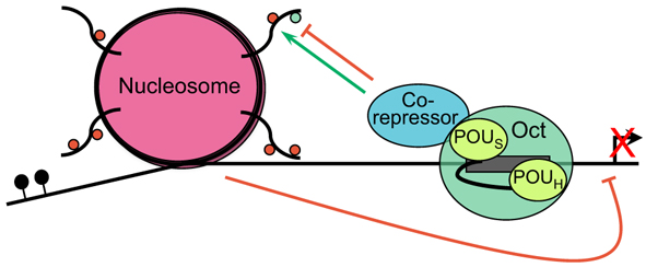
Although classic repressors undoubtedly silence transcription, genome-wide studies show that many repressors are associated with actively transcribed loci. Reynolds, O’Shaughnessy and Hendrich review this evidence and propose that the modulation of gene expression by co-repressor complexes provides transcriptional fine-tuning that drives development. See the Review on p. 505


Establishing and maintaining gene expression patterns: insights from sensory receptor patterning

Rister, Desplan and Vasiliauskas review the mechanisms that generate and maintain sensory receptor expression patterns and compare them to those that control sensory receptor expression patterns in the mouse retina and in the mouse and fly olfactory systems. See the Primer article on p. 493


Click here to see other articles in the ‘Development: The Big Picture’ series.


Radial glia – from boring cables to stem cell stars

As part of the ‘Development Classics’ series, Malatesta and Gotz look back at their 2000 Development paper, in which they showed that radial glial cells act as neural stem and progenitor cells in development – a discovery that has led to a change in the concept of neural stem cells in the adult brain. See the Spotlight on p. 483


A germline-centric view of cell fate commitment, reprogramming and immortality

At the recent EMBO/EMBL symposium ‘Germline – Immortality through Totipotency’, researchers discussed the mechanisms that establish and control totipotency, with an eye towards the mechanisms that may endow germ cells with the ability to propagate totipotency across generations. See the Meeting Report by Torres-Padilla and Ciosk on p. 487


Thumbs up (No Ratings Yet)
Loading...

Categories: Research

Postdoc position

Posted by , on 4 January 2013

Closing Date: 15 March 2021

A postdoctoral position is available in the laboratory of Dr. Sophie Astrof to study roles of cell-extracellular matrix interactions in cardiovascular development and disease using mouse model system. The research will involve investigation of the role of extracellular matrix in orchestrating signaling/communication between various progenitor cell populations during morphogenesis of the aortic arch arteries.  In our lab, we use genetics, conditional mutagenesis, and transgenic approaches to explore roles of tissue microenvironment during organogenesis and disease. Experience with genetic manipulation, embryology and cell biology is desirable.  My laboratory is a part of the Center for Translational Medicine at Jefferson Medical College (http://www.tju.edu/jmc/medicine/translational_medicine/faculty/astrof.cfm?detail=0) located in the heart of Philadelphia. To apply, send a letter of interest, CV and names and contact information of three references to sophie.astrof@gmail.com


Thumbs up (No Ratings Yet)
Loading...

Tags: , , , , , , , , , , ,
Categories: Jobs

What’s the future of peer review?

Posted by , on 3 January 2013

Jordan Raff’s recent Biology Open editorial on the future of publishing, posted on the Node, sparked quite a debate in the comments section. Much of that discussion focussed on perceived problems with the peer review system in scientific publishing. Particularly with the rise of journals like PLoS One and BiO, it seems that authors are increasingly dissatisfied with the time and effort – and with the sometimes cryptic decision-making – involved in publishing in more selective journals (journals whose selection criteria include some measure of ‘conceptual advance’ or ‘general interest’). I promised in one of my comments to Jordan’s post to write in more detail about my take on these issues, and what Development is trying to do to alleviate community concerns with the peer review process. So, here goes…

To start with what is perhaps an obvious point: one of the key aims of the peer review process is to improve the submitted paper, and in the vast majority of cases, I think it does just that – the finally accepted version of a manuscript tends to be both scientifically more sound and easier for the reader to understand than the original submission. Importantly, peer review – whether it’s of the more selective or the purely technical kind – provides some kind of quality assurance stamp on a published paper: although erroneous and fraudulent papers do end up being published, I’m sure there are far fewer of them in the public domain as a direct result of the peer review process.

However, that’s not to say that the system is perfect, because it certainly isn’t. Particularly with the rise of supplementary information, it’s all too easy for referees to ask for a ‘shopping list’ of experiments, many of which can be peripheral to the main story of the paper. And all too easy for editors to simply pass on those referee reports without comment – either because they’re too busy to go through the reports in sufficient detail to figure out what the really important points are, or because they don’t have the specialist knowledge to pass those judgments (which, after all, is why we need referees in the first place!). With a few tweaks to the system, we can do better than this.

For a selective journal, which Development unashamedly is, I think the key is to encourage referees to focus their reports on two things:

1. What’s the significance of the paper and why should it be of interest to the journal’s readership?

2. Do the data adequately support the conclusions drawn, or are there additional experiments necessary to make the paper solid?

With clear answers to those two questions in hand, it should be much easier for editors to decide firstly whether the paper is in principle suitable for the journal (spelled out in the answer to question 1), and secondly what the authors need to do for potential publication (the experiments given in response to question 2). It should get rid of that long list of ‘semi-relevant’ experiments (that aren’t really pertinent to q2), and it should make decisions much more definitive. There’s nothing worse than going through 2-3 rounds of extensive revision only for an editor to decide that the paper’s not worth publishing after all (something that, incidentally, Development is good at avoiding: around 95% of papers that receive a positive decision after the first round of review are published in the journal). Having a clearer (and shorter!) list of necessary revisions should help to avoid such situations.

I’m not a radical and I think it’s evolution not revolution of the system that’s required here. But I (and we at Development) do want to improve things. To this end, we’re looking at ways of changing our report form to reflect the aims laid out above. It might seem like a small step, but I genuinely believe that it could be a valuable one in easing the path to publication.

Moreover, I don’t think that the more radical alternatives work – various possibilities have been proposed and tested, but success is thin on the ground. Deposition in pre-publication servers and community commenting works very well in the physical sciences, but not in the biological sciences – as trials by Nature (see here and here) have demonstrated. Post-publication commenting could be a valuable addition to peer review, or even an alternative to it, but it just hasn’t taken off: I just looked at a random issue of PLoS Biology from 2012 and of the 17 papers published, only 3 had comments, none of which were particularly substantial. Open peer review – where referees sign their reports – would be great in an ideal world, but whenever I ask an audience if they’d be happy to sign their report if they were reviewing a paper for a top name in their field who might in turn be reviewing their next grant application, the vast majority opt to stay anonymous. It’s a competitive world out there, and scientists (like everyone else) hold grudges. Double-blind peer review – where the authors are also anonymous – might have some benefits in terms of reducing potential referee or editor bias, but it’s not easy to implement, and in most cases the referees will know who the authors are in any case.

So given the limitations of the alternatives, I believe that most journals will continue to operate some form of traditional peer review for the foreseeable future, and I don’t think this is a bad thing. That’s my opinion, but we also want to hear your views on this. What most frustrates you about the whole publishing process? Would a more streamlined review process like the one I’ve suggested help? What else can we do to make the system better?

Finally, though, there’s one thing that always comes to mind when I hear people complaining about the review process. You as authors are also the reviewers (or if you aren’t yet, then you one day will be) – meaning that you’re the ones giving ‘unreasonable’ lists of experiments to other people. It’s easy to pick holes in a paper, but harder to recognise when the authors have already done enough. So when you put on your reviewing hat, remember how you felt about the anonymous hyper-critical reviewer of your own paper so you don’t risk turning into one of them!

Thumbs up (6 votes)
Loading...

Tags: ,
Categories: Discussion

Top Node posts of 2012

Posted by , on 3 January 2013

Happy New Year!

We’ve looked at our stats for the past year to see which posts were the most popular. At the top of the list were the Woods Hole images and the two essay competition nominees, There’ll be dragons? and An Excitingly Predictable ‘Omic Future.

But there were many other popular posts this past year. The following list includes some of the most-viewed or top-rated posts of 2012. It’s a mixed bag of publishing discussions, research news, career features and pretty images – written by students, post-docs, lab heads, and others. Thanks for another year of great content!

Fast times at MBL (by Andrew Mathewson)

A career in science management (interview with Andrea Hutterer, by Natascha Bushati)

Colouring the mouse embryo (by Michael Wong)

Piecing together the squint puzzle (by Karuna Sampath)
See also her student Shimin’s perspective at New visions from the 3’ end of squint.

Bio Editorial – Publishing in the biochemical sciences: if it’s broken, fix it! (by Jordan Raff)
There’s a lively discussion in the comments of this post that’s also worth a read.

Freeware for scientists (by Nishal Patel)

Photo-morpholinos (by Philip Washbourne)

How obsession can fuel science blogging: the story of Retraction Watch (by Ivan Oransky)

A few older posts still received enough regular visitors to keep them among the most visited of 2012:

Hair follicle stem cells – the hairy truth (by Erin Campbell, posted in 2011)

Turtles in a nutshell (by Bruno Vellutini, posted in 2011)

7th European Zebrafish meeting (by Maria Nicolas Perez, posted in 2011)

The intestinal crypt (video from Hans Clevers’ lab, featured on the Node in 2010)

Is your favourite post missing from this list? These most-viewed and top-rated posts were generally the ones that were emailed around a lot, or shared on social media. If you see something you like on the Node, tell your friends!

Thumbs up (3 votes)
Loading...

Categories: Highlights

This month on the Node – December 2012

Posted by , on 31 December 2012

And we’ve reached the end of another full calendar year on the Node. What were your favourite posts this year? As a refresher, here is a look back on some of the content of this past month.

Advent calendar
For most of this past month, you will have been able to find a new paper each day behind the virtual doors of our advent calendar. A summary of this collection of papers was posted on Christmas morning.

Node news
The Node now has some custom tea bags, which you’ll be able to pick up at various conferences. You can drink the Node tea while reading or writing for the Node. But maybe you want to do more than read, write and drink tea? In that case: come work for us! Eva is leaving at the end of February, so we’re hiring a new community manager to run the Node. It’s a very interesting job with a lot of creative input, and you can apply until January 20.

Open Access Discussion
The post itself is from earlier this year, but in the past weeks a lot of people have been contributing to the discussion on open access publishing in the comments of Jordan Raff’s Biology Open editorial.

Research
We heard about some exciting research this last month of the year. In an interview, Roger Barker talks about an international collaboration studying the generation of medium-sized spiny neurons from stem cells.

“In this particular paper, the group in Italy, led by Elena, have such fabulous expertise in developing striatal neurons, but their lack of access to human fetal material makes it very difficult for them to do the project without collaborating with a lab like ours, in a country that does have access to fetal material. So these are truly international collaborations and without either party the project wouldn’t happen.”

Candy FISH

Elsewhere, Martin Jakt writes about his paper on a technique to estimate gene expression within single cells.

“The future brings with it hopes of understanding complex biological phenomena such as embryonic differentiation through computational modelling of the interactions between regulators and regulatees. Such models make predictions of cellular behaviour, which in the case of differentiation of multipotent cells must include the generation of diversity. Methods such as candy FISH allow not only the direct observation of the behaviour of systems at the individual cell level, but also make it possible to take into account effects of interactions between cells thus turning the problem on its head.”

Also on the Node:
Wikipedia edit-a-thon in Oxford
New Development book reviews
Don’t get rid of the middle-man

Thumbs up (1 votes)
Loading...

Categories: Highlights

Book review: Theme and variations on biology and civilisation

Posted by , on 27 December 2012

This book review originally appeared in Development. Alfonso Martinez Arias reviews “Cells to Civilizations: The Principles of Change that Shape Life ” by Enrico Coen.

Book info:
Cells to Civilizations: The Principles of Change that Shape Life. By Enrico Coen. Princeton University Press (2012) 360 pages ISBN 9780691149677 $29.95 (hardback)

In 1953, Alan Turing, a mathematician who had enabled the allies to break the Nazi communication codes – thereby making a significant contribution to ending the war in Europe – turned his attention to biology. Acknowledging the chemical make-up of living systems, he wondered what kind of reactions could generate the spatial patterns that are so pervasive in the outer layers of plants and animals. He noted that carefully coordinated interactions between an activator and an inhibitor, coupled to their diffusion, would, under certain conditions, be able to generate stable patterns of spots and stripes that resemble some of those found in nature. This simple chemical circuit had the potential to explain many phenotypes and as such has received attention over the last 50 years, although Turing himself only studied it as a proof of principle.

Biology is not just about the spots of the leopard. The field encompasses an apparently bewildering array of remarkable facts, from the ability to sense our environment to the workings of the cell cycle or the amazing periods of the cicadas. As our understanding of the elementary composition of biological systems increases, the question emerges whether there are any unifying principles to the variety that is their hallmark. Biology is not rich in laws or principles in the way that the physical sciences are. Thus, if a common thread, a principle of some sort, could be found, running from DNA to the behaviour of a population of ants, or even further to our ability to conceive and execute a painting or a symphony, this would be a remarkable observation with the power to transform our understanding of nature. In Cells to Civilizations, Enrico Coen, who stimulated us with The Art of Genes, shares his insights and solution to this puzzle. What he calls “life’s creative recipe” is claimed to be a collection of simple principles that, when applied to diverse biological systems, reveal a surprising number of similarities and relationships that can be cast into an understandable, explanatory diagram. If true, it is tantalisingly close to a unified theory for biology. Furthermore, Coen dares to extend it further and explores the possibility that his recipe applies to culture, thus claiming to establish a seamless connection from genes to the paragon of human nature – its creativity.

Understanding heredity, development, evolution and the mind are the big challenges of biology. Coen sets out in search of a general explanation that encompasses the structure and function of each challenge and brings them together. He recounts that similar principles have been dreamt of before, but hastens to add that nobody has managed to produce something lasting. However, we are told that here we might find this elusive idea. Do not be daunted by the scope of the book, which is written for a wide audience, although it contains enough science for biologists and anthropologists to ponder and argue with Coen. For the rest, it is an easy read, particularly as the biology it contains is peppered with vignettes drawn from painting and art history, which act as a guide for the more dry science that forms the meat of the argument.

Having outlined the problem, and taking evolutionary biology as a reference, Coen rephrases some of the notions associated with Darwinism to put forward seven principles that can be seen at work at many levels in biology and which are the fabric of “life’s creative recipe”: population variation, persistence, reinforcement, competition, cooperation, combinatorial richness and recurrence. These principles are then applied to several biological questions, to bring together different phenomena under a unifying umbrella. “Life’s creative recipe”, at least qualitatively, provides a common mechanism underlying diverse processes at different scales of time and space. It is a deceptively simple double-feedback loop, resembling an abstraction of Turing’s chemical machine but looking more like a Moebius strip. At the heart of the loop is a positive catalytic system (the principle of reinforcement), which is restrained by the negative effect: the principle of competition. The book unfolds as a sequential application of these principles to the major problems of biology, and shows that when these opposing forces are applied to a specific process they reveal related behaviours. The recipe is brought to bear on evolutionary theory and developmental biology in four chapters of about 50 pages each. From here the pace slows down and Coen launches into neurobiology, which is less familiar territory for him. He carries us slowly through his view of sensory neurobiology, learning and memory: a remarkable tour de force. The promise of a general recipe appears to work. As simplified examples, if the recipe has ‘reproductive success’ at the centre of the engine, ‘genes’ as activator and ‘environmental limitations’ as inhibitor, it models evolution. Plug in ‘firing’ and ‘neural inhibition’ onto ‘synaptic strength’ in the same recipe and you have learning. The patterns that shape life at different levels emerge from a regulated dialogue between opposites. Simple and effective. Interesting references to art history will retain your interest through the more challenging parts of the book. I particularly enjoyed the reference to the biologist and medieval armour expert Bashford Dean on the evolution of the helmet as a paradigm of biological evolution.

Having dealt with biology, Coen becomes ambitious and in Chapter 11 tackles culture. He acknowledges that this is a serious challenge and tells us that “we should be careful not to push (…) resemblances too far” and that “it is important to stand back to view the relationship between culture and the other processes at an appropriate level of abstraction”. I agree on both accounts, and the reader should bear Coen’s warning in mind when reading the book. At such a high level of abstraction, relationships can emerge that might not be real. To use one of the analogies in the book: there is a portrait of Ambroise Vollard by Cézanne, which was reinterpreted by Picasso. However, if we looked at the Picasso alone, without knowing about this relationship, we would very likely make different interpretations as to what the canvas represents. Sometimes we see what we want to see. In the end, the problem with abstractions in science, particularly with qualitative ones, is that they turn into metaphors of limited value. Science is about the ability of a thought to explain detail rather than to describe a loosely defined reality. Herein lies an important consideration for the still young biological sciences. When the physicists strive to find unified theories, they have numbers to aim for and experiments that they can do – experiments that have drowned more than one sublime theory. In physics, knowing whether we understand something is well defined: either you get the number or your predictions – which often consist of numbers – are wrong. At first sight, biology is not like this and determining whether or not a certain theory, principle or even idea is right or wrong requires a precise definition of what is being implied or said, as well as considerable time to investigate it. So, how can we know whether Coen’s proposed “life’s creative recipe” really tells us something about nature, or whether his book provides just another pleasant read?

Caveat lector. In physics, unifying theories have a strong quantitative basis and outlook. The theories live or die by how much of the detail, in particular of the quantitative world, they can explain and how much they can predict. Art, on the other hand, is content with the view it creates of a reality, which it captures without constraints. Enrico Coen is aware of this difference and only time will tell what exactly his effort has achieved, how much it explains and how much it predicts. It is likely that recent developments in the quantitative analysis of biological processes will lead to a rewriting of biology over the next few years. This in turn should provide some precise elements to the general argument developed in Cells to Civilizations, which is still very much grounded on the qualitative analysis of biological phenomena.

Thumbs up (2 votes)
Loading...

Categories: Book Reviews

From sharks to mammals

Posted by , on 26 December 2012

My name is Idoia, I am a PhD student within the Brainshark group at the University of Santiago de Compostela (Spain). I am currently finishing a research stay at the University of Edinburgh (Scotland) funded by the travelling fellowships offered by the Company of Biologists.

It is being four years since I started my predoctoral period. During this time, I have considered several times to carry out an international experience but never found the right moment. Now, just in the final stages of my thesis, I though “now or never”, and here I am, in a wonderful wild place called Scotland.

This stay had involved many changes for me. Language, culture but, most of all, a change in the animal model I work with: from shark to mouse. In our lab in Santiago de Compostela, we use the shark Scyliorhinus canicula (also called lesser-spotted dogfish) as a model for developmental studies of the nervous system. Yes, a shark! Ok, a small one, but still a shark. We are interested in the evolutionary changes that have occurred in the developing and adult nervous system throughout vertebrate phylogeny but also in the analysis of the conserved traits between different animal groups. Analyzing shared and derived features in our model is yielding interesting data like the presence of neuronal tangential migratory routes in the developing telencephalon of sharks homologous to those described in other vertebrates.

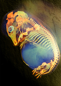
I will start by answering the first question that people used to ask me whenever I go to a conference: “why sharks?” Of course, the phylogenetic position of cartilaginous fishes is crucial to assess the ancestral condition of the vertebrate brain. Moreover, in the last decades, there has been an increasing interest in the dogfish as a model for developmental studies. This is due to it presents some advantages with respect to other vertebrate groups which allow a detailed analysis of developmental processes. For example: external egg gestation and transparent eggs (easy accessibility to the embryo to the implementation of different experimental approaches), protracted embryonic development (gestation period from 6 to 8 months), large size of the embryonic brains and availability of embryos at any time of the year. See the eggs in figure 1. Moreover, unlike teleost fish (as zebrafish, the main fish model nowadays), the telencephalon of cartilaginous fish develops by a process of evagination instead of eversion wich allows more reliable comparisons with other vertebrates. In fact, many people cannot distinguish at a glance a shark embryo from a mouse one. Do you want to try? See figure 2.

In the last years, we have been studying the expression patterns of Pax6, a well-conserved transcriptional factor, during forebrain development in sharks. Some open questions in our investigation made us to contact to Dr David Price, in the University of Edinburgh, and propose him a short collaboration. He kindly accepted to having me at least for three months in his lab to check our hypothesis in a different model as well as to help in other aspects of his ongoing project on Pax6.

At the beginning was tricky, working with mice was a big challenge for me; but at the same time was amazing to do research in a species in which several optimized techniques are available. Particularly I enjoyed learning slice culture techniques and I hope to have time to implement them in shark embryos and perform some axon guidance experiments upon my return.

On the other hand, the non-academic experience was also terrific. I loved Scotland, their people, their accent, landscapes, music and traditions. This is a real beautiful corner of the world you have to visit at least once in life. Of course the weather was not the best thing but I have to say that it was way better than I was told.

But maybe the best thing I obtained from this experience is the personal enthusiasm impulse to face the final stage of my predoctoral period. It has been one of the best decisions I have made, without a doubt whatsoever, and I have to sincerely thank to the Company of Biologists for their support.

Thumbs up (20 votes)
Loading...

Tags:
Categories: Funding, Research

December 25

Posted by , on 25 December 2012

You have reached the end of the advent calendar! Please see the round-up of all entries here.

Happy holidays!

Thumbs up (No Ratings Yet)
Loading...

Tags:
Categories: Research

Advent calendar round-up

Posted by , on 25 December 2012

If you have been following along with the advent calendar we’ve had in the sidebar for the past couple of weeks, you will have seen twenty-four papers – all selected by readers of the Node.

We’ve seen a diverse selection of papers, all describing recent work in developmental and stem cell biology. The people who suggested the papers wrote a brief description of the work, and why they chose it. Those little recommendations added an extra dimension to what otherwise would just have been a list of papers.

For example, on December 16 we featured a paper on pigmentation patterns in cats. Heather Etchevers, who selected the paper, wrote: “this paper was justifiably published in a general science journal because to some extent, everyone has asked themselves the question of how the leopard got its pattern of spots.” On December 24, Tohru Yano excitedly described a paper on limb development: “This paper shows us crazy results of extra fins/limbs at the same position!” And near the start of the month, on December 5, Bob Goldstein wrote “an important step forward, and beautiful!” about a recent paper on neural tube closure imaging.

We got a lot of great feedback about the advent calendar, but of course it was all down to your select not papers. So a big thank-you to everyone – from grad students to professors – whose suggestions were included. In order of appearance on the calendar: Nishal Patel. Tohru Yano, Rachael Inglis, Mary Todd Bergman, Bob Goldstein, Eva Amsen, Nik Papageorgiou, Katherine Brown, Heather Etchevers, Claire Cox, Barry Thompson, Heather Buschman, Andrew Renault, Seema Grewal, Benoit Bruneau, the Raff Lab, and Joanna Asprer.

Another round of special thanks goes to the journals who temporally freed access to papers so that our advent picks were available to everyone on their respective days. Thank you very much to Developmental Dynamics, Cell, Science Translational Medicine, Current Biology, Developmental Cell, Science, Genes & Development, Nature, and Development.

If you missed any of the entries, you can find the archive here. Some of the papers are no longer free to access, though.

Finally, the complete list of all the papers we’ve featured these past weeks:
(more…)

Thumbs up (4 votes)
Loading...

Tags:
Categories: News, Research