The community site for and by
developmental and stem cell biologists

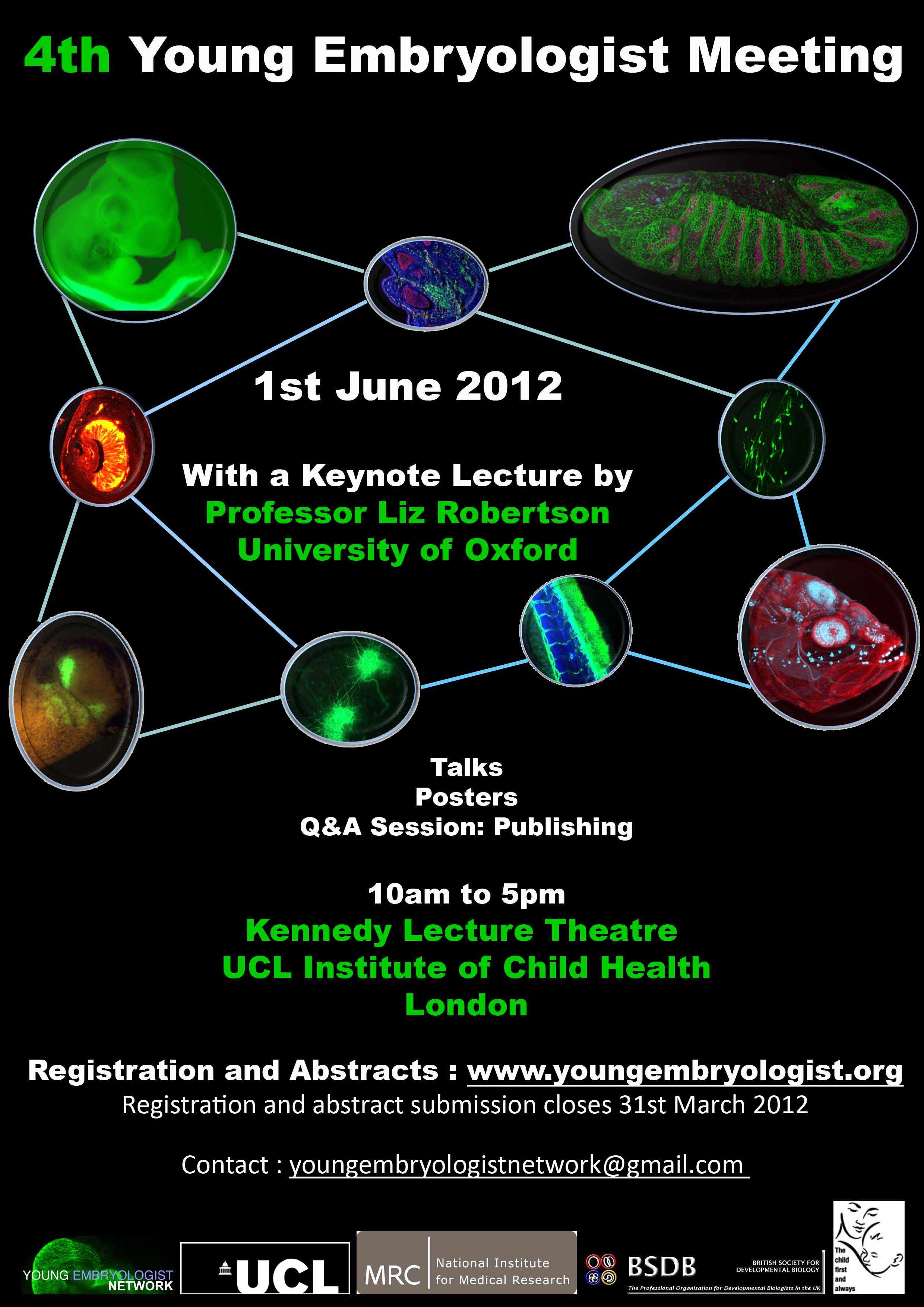
Mammalian Genetics and Development Workshop

Posted by , on 2 November 2011

THE MAMMALIAN GENETICS AND DEVELOPMENT WORKSHOP
A meeting of the Genetics Society

Venue: UCL Institute of Child Health, 30 Guilford Street, London WC1N 1EH
Date: November 17th 2011.
Organisers: Nick Greene, Andrew Ward & Andrew Copp

The Mammalian Genetics and Development Workshop is an annual meeting covering any aspects of the genetics and development of mammals. Meetings are based on the submitted abstracts, and usually include diverse topics ranging from early mammalian development (not exclusively human or mouse), imprinting and identification of disease genes to human population genetics and association studies. In recent years, presentations on other model systems (such as chick and zebrafish) have also been included where these relate to general developmental questions or disease models.

The meeting is traditionally a venue for post-docs and PhD students to talk rather than laboratory heads and so is an excellent training ground and a friendly, informal forum.

Registration
A £10 registration fee is payable by all attendees on arrival at the meeting.  This fee entitles registrants to attend all of the scientific sessions, and to receive the abstract booklet plus tea and
coffee refreshments on both days.  Speakers and chairpersons will be provided with lunch, free of charge, on the day of their presentation. Other participants will be expected to make their own arrangements for lunch. There will also be a wine reception on the first day of the meeting.

Abstract Submission
All Workshop presentations will be in lecture format .  If you would like to present a paper at the Workshop at this year’s meeting, please e-mail your abstract to the following address: mgd.workshop@ich.ucl.ac.uk by Friday 4th Nov*, specifying your preference for a 15 or 30 min slot. *Due to the late notice there will be some leeway on this closing date for readers of the Node provided you let us know your abstract is coming!

With the authors’ permission, abstracts will be published in Genetical Research.

Thumbs up (3 votes)
Loading...

Tags: ,
Categories: Events

Navigate the archive

Use our Advanced Search tool to search and filter posts by date, category, tags and authors.

Parenting in Science

Posted by , on 31 October 2011

After the interview with Ottoline Leyser was posted last week, a discussion on Twitter focussed on the last part of the interview, about parenting.

That interview question referred to a little booklet Leyser published a few years ago, after winning the Royal Society Rosalind Franklin Award in 2007. The book, called “Mothers in Science: 64 ways to have it all” features interviews with mothers who have managed to maintain a career in science while raising children. (Here is a link to the PDF on the Royal Society website.) In the booklet, all featured scientists have a page with a timeline showing the important events in their career and family life. They’re all unique stories, because every situation is different.

When I wanted to address the ensuing Twitter discussion (which you can read in the Storify embedded below) I thought I could add a poll to ask how other people have managed to combine their career with children, or perhaps to ask how others have failed to do so. But I quickly realised that there is no question I could possibly ask for which the answer can be reduced to a set of multiple choice answers. The possible answers would need to include all combinations of which family members are scientists, what the other partner’s job is, the age difference between parents (e.g. one is a postdoc, one a PI), working hours, who took the main child-rearing responsibilities or whether there is a balance, gender, which country you’re in, competition in the field of research, how close together the kids are, and much, much more.

Even a quick show of hands, just to ask who has children or not, would be meaningless if it didn’t account for gender, age, career stage, country, family situation, and desire to even have children in the first place.

So there is no poll. There is no poll because clearly there isn’t one clear-cut problem, and because there is not just one type of family unit.

What Ottoline Leyser’s book did is showcase a group of women who all managed to combine a family with a career in their own way. It’s an example to show that it can be done, but it’s not a collection of recipes for success. Each case really is different, and this Twitter discussion between @fishscientist and @David_S_Bristol tells a different story. (Text continues after the embedded file.)

So are there solutions? One promising step was made last week in the UK, when the Research Excellence Framework (REF) announced that “UK funding bodies have taken an early decision on the arrangements for taking account of maternity leave in the REF. … researchers may reduce the number of outputs in a submission by one, for each period of maternity leave taken during the REF period.”

That doesn’t help most of you, but it positively affects the career progress of a few mothers, and at least changes their stories.

If you have your own story to add, please leave a comment, as a poll was just too complicated….

Thumbs up (3 votes)
Loading...

Categories: Careers, Discussion

This month on the Node – October 2011

Posted by , on 31 October 2011

What was new on the Node this month? Here are a few of the highlights from October:

MacArthur genius grant for Yukiko Yamashita

Sasha Terashima interviewed Yukiko Yamashita, one of this year’s MacArthur Fellows. The MacArthur foundation hands out “genius grants” of half a million dollars to a select group of people in all areas of arts, science, and humanities. There is no application process, and recipients are free to choose what they do with the money. Half a million out of the blue, with no strings attached – how would you react if you got that phone call?

“When Yukiko received the phone call informing her about being selected and asking her asking her not to discuss it with anyone except her spouse until the official announcement, she had a hard time believing that it was not a scam. “I called my husband right after I hung up my phone call with the foundation and [he] seriously warned me that ‘if you get a second phone call asking your bank account and pin number, so that they can transfer the award money, don’t give it to them.’” “

Academic teaching
If you teach plant science courses, make sure to have a look at Teaching Tools in Plant Biology. In the comments, Mary Williams give some additional advice.

Meanwhile, Lucia Prieto Godino travelled to Uganda to coordinate a course on insect neuroscience and Drosophila neurogenetics. The course members are graduate students and Junior Faculty from Uganda, Tanzania, Kenya, Malawi, Nigeria, and Cameroon, and in the course they will learn how to effectively use insects to teach or do research in neuroscience. In a later post, Lucia updated us on the course progress after the first few weeks.


One student in the course explains the experiment on olfactory choice in Drosophila that his group performed the day before.

Meetings, Jobs, and more Interviews:
– The entire Company of Biologists Workshop on Growth, Division and Differentiation is covered in a series of posts from the meeting.
– Several PhD and postdoc positions have been posted on the jobs page.
– Interviews with Development Editors Gordon Keller and Ottoline Leyser were reposted on the Node. The interview with Ottoline Leyser sparked a lively discussion on Twitter – more about that later today!

See the full October archive here.

Thumbs up (1 votes)
Loading...

Categories: Highlights

November desktop calendar

Posted by , on 28 October 2011

It’s that time of the month again, when we upload the desktop calendar for next month. This time an image that you may remember from April – either from the contest on the Node or from the pub quiz at the BSDB meeting.

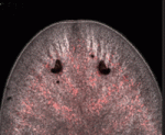
november_thumbnailIt’s a sea biscuit during metamorphosis from larval to adult stage. This image, taken by Bruno Vellutini of the Marine Biology Center of University of São Paulo, was the runner up in the Intersection Image Competition held earlier this year.

Visit the calendar page to select the resolution you need for your screen. The page will be updated at the end of each month with a new image, and all images are chosen from either the intersection image contest or from the images we’ve featured from the Woods Hole Embryology 2010 course.

Thumbs up (No Ratings Yet)
Loading...

Tags: ,
Categories: Images

Some of the things we have done during these two weeks

Posted by , on 27 October 2011

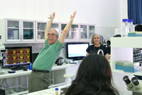
We have now finished the first two weeks of the course. Over these two weeks the students have learned about Drosophila as a model organism and how to set-up a Drosophila lab. They have learn about the genetics of Drosophila, which took quite some time, but now I am sure it is almost like their mother tongue. They have also learned basics concepts in neuroscience, such as the nature of neural impulse, and basic concepts on how neurons work to produce adaptive behaviors. During the practicals among other things they have learned  how to do muscle recordings of neuronal activity with inexpensive amplifiers. They have recorded from the legs and wing muscles of grasshoppers (see picture 1)! and observed under the microscope a multitude of different insects, which all together helped them to appreciate in the practice the nature of the neural impulse and the diversity of sensory and motor systems used by insects.

Picture 1. Improvised set-up for recording activity of flight muscles in locust

The students also now know how to collect virgins for their fly crosses, and how to dissect brains out of Drosophila larvae, and look at them under the fluorescent microscope.  We have managed to install a webcam on the fluorescent microscope, so that the students can take pictures of the fluorescent preparations that they look under the microscope. The images resulting from this system are actually much better than we expected (see picture 2).

Picture 2. We have attached a regular webcam to the camera port of our Leica fluorescent microscope. The resulting images are surprinsingly good. At the bottom right of this panel you can see a picture of a Drosophila larvae which expresses dsRed in all cholinergic neurons. At the very front (top in the picutre) olfactory and taste sensory neurons are visible, it is also possible to see mechanosensory neurons along the surface of the larvae, which project their axons to the brain, which appears very bright at the centre of the picture.

The students have learned about mechanosensory, chemosensory, visual and motor systems during the theoretical lectures, and they have looked at the wild type behaviours of flies and other big insects during the practical sessions (see picture 3).

Picture 3. One student explains the rest the experiment on olfactory choice in Drosophila that his group performed the day before

They have also performed inexpensive cutting-edge neurogenetic experiments on genetically modified larvae. It is being intense but the effort is worthwhile, now they are ready for the lab work of the last week, during which they will need to apply everything they have learned!


Thumbs up (3 votes)
Loading...

Tags: , , ,
Categories: Education

Comparative epithelial morphogenesis research positions (PhD, Postdoc)

Posted by , on 26 October 2011

Closing Date: 15 March 2021

extraembryonic morphogenesis

From early 2012, a PhD studentship and a postdoctoral position are available in the Panfilio lab (in Cologne, Germany) to investigate morphogenesis of the insect extraembryonic membranes.  As protective covers for the embryo, these simple epithelia are a defining feature of the insects and have been linked to their evolutionary success.  However, in order to protect the embryo without obstructing later development, the membranes perform an array of morphogenetic movements as they develop to cover and later withdraw from the embryo.  The aim of the research is to understand: (1) how extraembryonic morphogenesis works at multiple levels of biological organization, and (2) how conserved morphogenesis is across species.  Research will be conducted in species that represent the two main modes of extraembryonic development and that are amenable to standard developmental genetics techniques as well as live imaging analyses (the beetle Tribolium castaneum and the bug Oncopeltus fasciatus).

Successful applicants will have a strong interest in studying cell shape changes in the context of tissue reorganization, and in doing so from a comparative perspective.  Candidates with a degree in cell and developmental biology or related fields are encouraged to apply.  The working language of the lab is English, and strong oral and written communication skills are required.  PhD candidates must be in possession of a master’s degree or the equivalent before commencing this work.  Postdoctoral candidates must have completed the PhD degree.  Additionally, the postdoctoral candidate will have a track record of successfully completing research projects, as demonstrated by at least one first author publication.  Required skills include standard developmental genetics techniques associated with RNAi, tissue staining, and light microscopy.  Experience with electron microscopy, protein expression, transgenic line production, and/ or quantitative analyses of (live) imaging data is desired.

The lab is based in the Institute for Developmental Biology, University of Cologne (home page), and has active research and collaboration links with other developmental, evolutionary, and insect labs.  With one million inhabitants, Cologne is an international, vibrant city that is well connected within western Europe.  Salaries are paid according to the standard German pay scale for the public sector (TV-L E13, 50%/ 100%), and include health insurance and other social benefit contributions. The Ph.D. position is for three years.  The postdoctoral position is for one year in the first instance and is renewable for at least a second year.  The University of Cologne is an equal opportunity employer in compliance with the German disability laws.  Women and persons with disabilities are strongly encouraged to apply.

To apply for either position, please send a statement of research interest, curriculum vitae, and contact details (e-mail address and phone number) for two references as a single PDF file to Kristen.Panfilio[at]alum.swarthmore.edu.  Informal enquiries to further discuss the positions are welcome.  All applications will be considered until 10 December 2011.

Panfilio lab postal address

Thumbs up (No Ratings Yet)
Loading...

Tags: , , , , , , , , ,
Categories: Jobs

Postdoctoral position – MRC Department of Biomedical/Developmental Genetics, University of Sheffield, UK

Posted by , on 26 October 2011

Closing Date: 15 March 2021

Overview

The Research Associate will work in the MRC Centre for Developmental and Biomedical Genetics within the Department of Biomedical Science.  The department has an active community of researchers specialising in different aspects of developmental biology.  You will take part in a project aiming to understand how cell proliferation is temporally integrated with digit patterning during vertebrate limb development.  For further information, see Towers, Nature 452, 882; Towers, Development 136, 179; and Towers, Nature Communications 2, 426.  The project is expected to branch into areas including stem cell biology and regeneration.

http://cdbg.shef.ac.uk/research/towers/

Candidates should have a PhD in vertebrate developmental biology or equivalent experience.  You should have proven experience in using the mouse and/or the chick as model systems and in molecular biology methods, including in situ hybridization and immunohistochemistry.  Experience in protein biochemistry, confocal microscopy and FACS analysis is desirable.  Candidates are expected to have excellent written and verbal communication skills, for example shown by a record of peer reviewed publications and presentations at international scientific meetings.  Dr Matthew Towers manages this post and the project, which is funded by the Medical Research Council.  The post is available from 1 January 2012 for a fixed period of 36 months.

Apply for the job online at the University of Sheffield vacancies page.

Job Reference Number: UOS003508

Job Title: Post-doctoral Research Associate

Contract Type: Fixed-term, available from 1 January 2012 for a period of 3 years

Salary: Grade 7

£28,251 to £31,798 per annum

Closing Date: 25th November 2011


Thumbs up (No Ratings Yet)
Loading...

Tags: , , , , , , , , , , , , , , ,
Categories: Jobs

Post-doctoral research scientist MRC-National Institute for Medical Research

Posted by , on 26 October 2011

Closing Date: 15 March 2021

Career Develpoment Fellow

IRC32554


Applications are invited for a Postdoctoral Research Scientist in the Division of Developmental Biology, MRC-NIMR. Funding is available for 2 years from the Simon’s Foundation, an international sponsor of autism research, for a project led by Drs John Jacob and James Briscoe. The successful candidate will build on prior genetic approaches in autism by investigating the molecular pathology of abnormal development of critical neuronal groups in the brainstem in autism. This will involve in vivo analysis of a mouse genetic model of autism, as well as proteomic approaches and yeast two hybrid screens. A strong background in molecular biology techniques will be essential for successful completion of this project. Candidates should hold a PhD in a relevant discipline.


MRC-NIMR provides a supportive and collaborative environment, comprising state-of-the-art imaging and mass spectrometry, high throughput sequencing and computational facilities, FACS, and excellent transgenic fish, frog and mouse facilitates. The research infrastructure is complemented by an exciting seminar programme, career development training opportunities, sports and leisure facilities and an active postdoctoral researchers’ forum.


Applications should include a cover letter, full Curriculum Vitae and the names and addresses of three referees (attached as one document).


Applications are handled by the RCUK Shared Services Centre; to apply please visit our job board at https://ext.ssc.rcuk.ac.uk and complete an online application form.  Applicants who would like to receive this advert in an alternative format (e.g. large print, Braille, audio or hard copy), or who are unable to apply online should contact us by telephone on 01793 867003, Please quote reference number IRC*****. 


Closing date: 10th November 2011


The MRC is an Equal Opportunities Employer

Final appointments will be subject to a pre employment screening


Thumbs up (No Ratings Yet)
Loading...

Tags: , , , , , ,
Categories: Jobs

An Interview With Ottoline Leyser

Posted by , on 25 October 2011

(This interview originally appeared in Development.)

The Sainsbury Laboratory at the University of Cambridge is a new research institute that aims to achieve an integrated understanding of plant development. Its Associate Director is the new plant Editor of Development, Ottoline Leyser, who is also Professor of Plant Development at the University of Cambridge. We recently caught up with Professor Leyser and asked her about the Sainsbury Laboratory and about her own research interests.

When did you first become interested in plant development?

To me, plant development has always been much more interesting than animal development, because of its plasticity. In plants, the body plan is incredibly flexible: one genotype can occupy an extraordinary range of phenotype space. I’ve always thought that was just amazing.

I did my undergraduate degree here in Cambridge, in the Genetics Department, not in plant science. We had this absolutely fantastic interdepartmental development course that was taught by John Gurdon, Peter Lawrence and many other wonderful people. It was very striking, the contrast between what was happening in animal development, which was being transformed by Drosophila genetics, by Christiane Nüsslein-Volhard, Eric Wieschaus and others, and what was happening in plants: despite the long tradition of genetics in plants, developmental genetics somehow hadn’t really taken off. But in the final year of my undergraduate degree, there were the first hints of Arabidopsis as a model organism, driven at least in part by Elliot Meyerowitz, who is now the inaugural director here at the Sainsbury Laboratory. So, there was suddenly a very exciting opportunity to push things ahead in plant development using developmental genetics. I started looking for a PhD position in an Arabidopsis lab and, fortunately for me, Ian Furner had just arrived back from the USA clutching some Arabidopsis seed in a tube, so I stayed in Cambridge and did my PhD with him, studying meristem mutants in Arabidopsis.

What are you working on at the moment?

I’m working on the role of plant hormones in integration of the endogenous and environmental signals that control the plant body plan. We’re looking principally at shoot branching control and are trying to understand how every individual axillary bud on the plant makes a decision about whether to activate or not, depending on multiple inputs. It’s really a question of signal integration.

You’ve recently moved your lab from York to Cambridge to set up the new Sainsbury Laboratory. How did the lab move go?

It’s still an ongoing process. We’re pioneers down here, who have had to deal with a very fabulous but nonetheless brand new and, at the time, unfinished building. But now that the first results from experiments carried out in the new lab are coming in it’s very exciting. Meanwhile, there’s still a core of people in York, partly because some people didn’t want to move and partly because we’re in the middle of a rather long-term ten-generation Arabidopsis experiment, which I didn’t want to move.

(more…)

Thumbs up (2 votes)
Loading...

Tags: , , ,
Categories: Interview

In Development This Week (Vol. 138, Issue 22)

Posted by , on 25 October 2011

Here are the highlights from the current issue of Development:

The skin-healing touch of Lhx2

Skin repair after injury involves the recruitment of undifferentiated progenitor cells from nearby hair follicles (HFs) into the regenerating epidermis. The bulge and the secondary hair germ of HFs contain distinct populations of epithelial stem cells, and now Vladimir Botchkarev and co-workers reveal that the Lim-homeodomain transcription factor Lhx2 differentially regulates these populations during wound healing (p. 4843). They show that, in mice, most of the cells that proliferate in response to skin injury in the HF bulge and secondary hair germ express Lhx2. Wound re-epithelisation is retarded in Lhx2+/– mice compared with wild-type mice, they report, whereas the onset of active hair growth in HFs near to the wound is accelerated. Other experiments indicate that Lhx2 promotes wound re-epithelisation by upregulating Sox9 and Tcf4 expression in the bulge cells while simultaneously inhibiting HF cycling by downregulating Lgr5 expression in the secondary hair germ. Thus, Lhx2 is a key regulator of the differential response of HF stem cells during epidermal regeneration after injury.

Nanog: an ancient reprogrammer

The establishment of pluripotency during mouse embryogenesis and during the reprogramming of somatic cells is dependent on the homeodomain-containing transcription factor Nanog but, puzzlingly, compared with other pluripotency-associated genes, Nanog is poorly conserved among vertebrates. Here (p. 4853), José Silva, Filipe Castro and colleagues investigate whether Nanog orthologues can orchestrate pluripotency in Nanog–/– mouse somatic cells. Surprisingly, the researchers report that mammalian, avian and teleost Nanog orthologues all reprogramme mouse Nanog-/- somatic cells to full pluripotency, despite sharing as little as 13% sequence identity with mouse Nanog. Moreover, they identify two unique residues in the DNA recognition helix of the Nanog homeodomain that are important for reprogramming and show that the Nanog homeodomain is sufficient to enable naive pluripotency in Nanog–/– somatic cells. These functional studies, together with genome analyses, suggest that Nanog is a vertebrate innovation and that its reprogramming capacity resides within a unique DNA-binding domain that probably appeared at least 450 million years ago in a common ancestor of vertebrates.

R-spondin to developmental angiogenesis

During embryogenesis, two sequential processes form the vasculature: during vasculogenesis, endothelial progenitor cells form the primary vascular bed; subsequently, during angiogenesis, additional vessels sprout and grow from pre-existing vessels. Here, Aniket Gore, Brant Weinstein and co-workers identify a novel signalling pathway that promotes developmental angiogenesis in zebrafish (see p. 4875). Their first clue to this pathway came when they identified a mutation in R-spondin1 (rspo1) during a forward-genetic screen for angiogenesis-deficient zebrafish mutants. Embryos lacking rspo1 or its receptor kremen form primary vessels, they report, but do not undergo angiogenesis. R-spondin is a Wnt signalling regulator and, by functionally manipulating different members of the Wnt pathway, the researchers show that canonical Wnt signalling is required downstream of rspo1 for sprouting angiogenesis. Finally, they show that Vegfc/Vegfr3 signalling mediates the pro-angiogenic effects of Rspo1/Wnt signalling and that all four proteins are expressed by the endothelium during sprouting angiogenesis. Together, these results suggest that Rspo1-Wnt-Vegfc-Vegfr3 signalling is an endothelial-autonomous permissive cue for developmental angiogenesis.

Compartmentalised PKA, cilia and hedgehog signalling

Protein kinase A (PKA), a conserved negative regulator of the hedgehog (Hh) signalling pathway, generates the transcriptional repressor form of Gli3 in the absence of Hh in mice. Now, Kathryn Anderson and colleagues show that the total loss of PKA activity in mouse embryos leads to a completely ventralised neural tube and mid-gestation lethality (see p. 4921), which indicates that the sonic hedgehog (Shh) signalling pathway is maximally activated in all neural progenitors in the absence of PKA. Notably, genetic experiments indicate that the principal function of PKA in the neural plate is to prevent Gli2 activation of Shh targets. Other experiments reveal that Hh pathway activation in PKA mutants depends on cilia, that PKA is localised at the basal body of primary cilia, and that Gli2 levels are increased at the tips of cilia of PKA-null cells. The researchers propose, therefore, that two separate cilia-associated compartments determine the accessibility of Gli proteins to PKA and thus the activity of the Shh pathway in vertebrates.

miR-124 notches up neural development

MicroRNAs (miRNAs) play crucial roles in development. miR-124, for example, is abundantly expressed in the mouse brain and is necessary for proper nervous system development, but how it drives neuronal differentiation is unclear. To remedy this lack of understanding, Robert Zeller and colleagues have comprehensively analysed miR-124 expression, function and target genes in the ascidian Ciona intestinalis (see p. 4943). They report that miR-124 interacts with several signalling pathways that are involved in nervous system development. In particular, they show that a feedback interaction between miR-124 and Notch signalling regulates the epidermal-peripheral nervous system (PNS) fate choice in tail midline cells. Thus, Notch signalling silences miR-124 in epidermal midline cells, whereas in PNS midline cells miR-124 silences Notch, Neuralized and the Ciona Hairy/Enhancer-of-Split genes. Moreover, miR-124 also shapes neuronal progenitor fields by downregulating non-neural genes including 50 Brachyury-regulated notochord genes and the muscle specifier Macho-1. Overall, these results indicate that miR-124 plays a multifaceted role in cell lineage specification during nervous system development.

Spotlight on adipogenesis

Adipose tissue (a specialised energy storage structure) is the only tissue that can change its mass substantially during adult life. It does this through changes in the size of its constituent cells (adipocytes) and through the de novo generation of cells. Unfortunately, given the obesity epidemic, adipocyte development in vivo is poorly understood but, here, Gou Young Koh and colleagues provide new insights into adipogenesis by analyzing the postnatal development of epididymal adipose tissue (EAT) in mice (p. 5027). They show that EAT is generated from non-adipose tissue during the first 14 postnatal days of development and that this non-adipose tissue is initially composed of multipotent progenitor cells (possibly including adipoblasts) that lack adipogenic differentiation capacity in vitro. By postnatal day 4, however, progenitor cells isolated from EAT can form adipocytes if they are provided with cell-to-matrix and cell-to-cell contacts. Finally, the researchers show that impaired angiogenesis in postnatal mice interferes with adipogenesis. Thus, they conclude, cues from cellular and matrix components, together with appropriate angiogenesis, are required for adipose tissue development.

Plus…

Evolutionary crossroads in developmental biology: amphioxus

As part of the Evolutionary Crossroads in Developmental Biology series, Bertrand and Escriva introduce amphioxus and discuss how studies of this model have informed us about the evolution of vertebrate traits.

See the Primer article on p. 4819

An interview with Ottoline Leyser

The Sainsbury Laboratory at the University of Cambridge is a new research institute that aims to achieve an integrated understanding of plant development. Its Associate Director is the new plant Editor of Development, Ottoline Leyser, who is also Professor of Plant Development at the University of Cambridge. We recently caught up with Professor Leyser and asked her about the Sainsbury Laboratory and about her own research interests.

See the Spotlight article on p. 4815


Thumbs up (No Ratings Yet)
Loading...

Tags: , , , , , , , , , , , , , , , , , , ,
Categories: Research