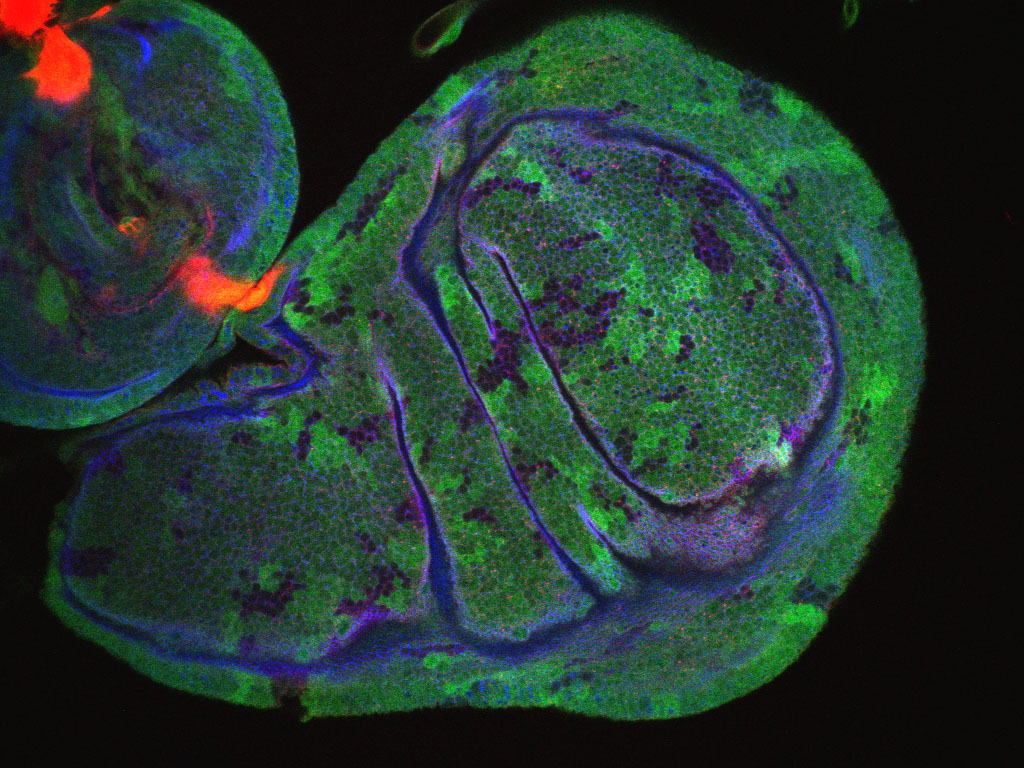
The annual Embryology course at Woods Hole starts again this week. Best of luck to all participants! We thought it was an appropriate time to launch the third voting round to choose a Development cover from images taken by the students in last year’s course.
Meanwhile, the winning image from the first round – a sea urchin chomping down on a piece of seaweed – will appear on the cover of Development vol. 138 issue 13, which will go live online on June 7 (tomorrow!). The image on the left is a sneak preview of the cover. That issue also contains a primer article about sea urchins as model organism, so the cover was a good fit!
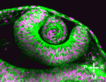
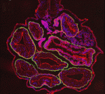
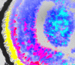
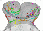
Which of these images will be next to appear on the cover of Development? Please vote in the poll below the images. (Click any image to see a larger version.) You can vote until June 20, 12:00 (noon) GMT
1. Dorsal view of the central nervous system of a Drosophila embryo. Neurons and axons are stained with anti-HRP (red). A distinct subset of neurons express even-skipped (green). Nuclei of the body wall stained with DAPI (blue). This image was taken by Joshua Clanton (Vanderbilt University).
2. Pilidium larvae of the Nermertean, Cerebratulus lacteus. Acetylated tubulin (green), serotonin (red), nuclei (blue, DAPI). This image was taken by Meii Chung (UT Austin).
3. Planaria stained with MF20 (green), phalloidin (red) and DAPI (blue). This image was taken by Valeria Merico (University of Pavia).
4. Drosophila embryo (stage 16) immunostained to detect Tropomyosin (purple) in the developing muscle, the Hox gene Ultrabithorax (green), and Spalt (red). This image was taken by Elise Delagnes and Hannah Rollins (UC Berkeley).
Four and a half years ago I was introduced to the field of clinical ethics while nearing the end of my Doctorate in Molecular Genetics at the University of Toronto. After attending a talk given by Kerry Bowman, a clinical ethicist at one of the University teaching hospitals, I approached him with some additional questions. The ensuing discussion led to a productive working relationship. Initially, I helped him perform an ethics analysis of a complicated genomics project. He then invited me to participate in some of his other professional responsibilities, opening a door into a new profession. This foray into clinical ethics had a lasting effect; in short order I decided to retire my pipette and pursue a career in clinical ethics.
At the beginning of my PhD, my intention was to pursue a career as an academic scientist. Like many graduate students I experienced both euphoric moments when critical experiments worked and frustrating periods when experiments were not informative. While I enjoyed the scientific process, I had a nagging feeling that a scientific career was not the perfect fit for me. Accordingly, I tried to keep an open mind and got involved in extracurricular activities. I participated in student governance, got involved in science outreach and taught with a non- governmental organization overseas. I also regularly attended a life science career development seminar series meant to expose graduate students to careers outside of academic science. I first met Kerry when he talked about clinical ethics at one of these seminars.
What immediately captured my imagination was the interesting breadth of activities performed by clinical ethicists. In Canada, most large hospitals either employ their own clinical ethicist(s) or have access to a regional clinical ethics service. Clinical consultations are the bread and butter of most clinical ethics programs and are usually triggered by an ethical tension in the provision of patient-care. A conflict regarding a treatment decision or plan, complicated end-of-life decision making, whether to withdraw life support, consent and capacity issues, and disagreement about discharge planning are all examples of clinical cases that may benefit from clinical ethics support. On the organizational level, many ethicists are also involved in the development of hospital policy since good policies may help mitigate future ethical tensions. Teaching is another important facet of clinical ethics. Education serves to raise awareness about ethical issues and can increase the capacity of healthcare workers and researchers to deal with ethical issues in their practice. Many ethicists also conduct independent research and serve on hospital research ethics boards (the Canadian equivalent of research ethics committees).
After deciding on a career track, I knew I needed more education. To address my knowledge gap, I enrolled in a Bioethics Masters program at the University of Toronto. I was fortunate enough to get a scholarship and worked hard over the two year degree to immerse myself into the field of bioethics. I took a generalist approach and chose a course-based professional masters program, which allowed me to take more courses compared to the thesis-based program that was also offered. I completed the degree and am currently a fellow in clinical and organizational ethics at the University of Toronto Joint Centre for Bioethics. The fellowship program is analogous to an apprenticeship program in ethics. Each fellow rotates through the ethics programs of four partner healthcare institutions over the course of a year (3 months is spent at each site). This unique and wonderful experience has given me practical ethics experience that complements the theoretical knowledge I learned in graduate school.
How has a PhD in molecular genetics prepared me for a career in ethics? Interestingly, several of my patients and non-ethicist colleagues have asked about my background and only about half immediately appreciate why a background in genetics might be useful in ethics. The other half usually can’t get over the fact that most of my training is not in philosophy. I view my background in science to be of great relevance. I am able to understand the science behind many emerging technologies in medicine, which is critically important in order to discuss the ethical implications of new technology and recommendations on how to proceed. My science training also heavily contributed to the development of my analytic, writing and presentation skills, which nicely compliments my bioethics education.
The combination of science and clinical ethics training is still quite unusual and has afforded me some unique opportunities. For example, I have been embedded in a genomics centre at a large research institute to work on ethical issues in parallel with scientific innovation. I have also been invited to speak at several interesting venues on topics involving genetics and ethics. As my fellowship is drawing to an end, I am seeking out clinical ethics positions that will allow me to perform all the clinical activities I described earlier and also use my genetics knowledge in a research ethics capacity. I hope to continue working closely with scientists and work on the ethics of the new research as it is being developed. Although not yet employed as a clinical ethicist, I was recently asked to give a talk about clinical ethics at the same seminar series that first introduced me to the field. I graciously accepted and am looking forward to describing my exciting field to other life science students.
Project: Apico-basal polarity regulators, Signalling pathways and Tumourigenesis
Available from Sept 2011 – contract initially 1 year with scope for extension
Our lab uses Drosophila as a model of tumour development, and through collaboration to translate this into mammalian systems. We seek a highly motivated, suitably experienced and qualified individual with a PhD (or submitted PhD thesis). Knowledge and experience in Drosophila Cell Biology, Genetics, Molecular Biology and Biochemistry is essential, and expertise in Drosophila imaginal disc biology and tissue culture is desirable. Applicants must have good organisational and communication skills, attention for detail, and the ability to carry out work efficiently and independently.
The Peter MacCallum Cancer Centre is Australia’s foremost Cancer Centre, and has an internationally renowned research division covering Cancer Cell Biology, Genomics and Immunology.
Peter Mac offers its employees the following benefits:
A 2 year post-doc position is available from October 2011 in Delphine DUPREZ’s team, in Paris.
Tendon and ligament injuries are common clinical problems during aging or following accidents. No treatment currently exists to restore injured or defective tendon/ligament to its normal condition. The ultimate aim of this project is to build an in vitro tendon (or ligament) for implantation into patients with defective tendons. The global strategy is to use the knowledge that we have acquired (and that we are currently acquiring) concerning tendon formation during embryonic development using the chick and mouse models (Lejard et al., 2011 J. Biol. Chem286(7), 5855-5867, Wang et al., 2010 Dev Cell, 18, 643-654, for review Tozer and Duprez, 2005, Birth Defects Res C Embryo Today. 75(3), 226-36) in order to establish an artificial tendon.
The project will involve cell culture of adult mesenchymal stem cells and transfection experiments with the appropriate factors in order to trigger cell differentiation towards a tendon phenotype. The project will also require in vivo manipulations in chick embryos, the use of mouse mutant lines and the use of rat tendon injury models.
A background in mesenchymal stem cells and/or cell culture will be an advantage.
Applicants may be of any nationality and should have obtained the equivalent of a PhD. Salary will be at least 2500 Euros per month (depending upon experience). Applications including a CV, a description of previous research experience, and 2 or 3 names of referees should be sent to Delphine DUPREZ to Delphine.duprez@upmc.fr
Each year, three medals to honour extraordinary research achievements in cell and developmental biology are awarded at the joint conference of the British Societies for Cell Biology (BSCB) and Developmental Biology (BSDB). Here on the Node, Eva has recently posted an interview with Carlos Carmona-Fontaine, to whom this year’s Beddington medal was awarded, for his PhD work in Roberto Mayor’s lab at UCL in London. Carlos’ talk on collective migration of neural crest cells was highly entertaining; Eva’s interview gives an impression of the entertainment value – I highly recommend having a look!
The BSCB’s Hooke Medal honours a person who has made an outstanding contribution to UK cell biology within the first 10 years of establishing his or her own lab. 2011’s recipient is Alex Gould (NIMR, London, UK). Together with his team, Alex determined the scheduling mechanism that terminates proliferation of neuroblasts in the Drosophila central nervous system at the end of development. Their second big discovery was uncovering the function of Drosophila hepatocyte-like cells, called oenocytes, which regulate fat metabolism in the fly. In his lecture he mainly presented their most recent research on organ sparing during nutrient restriction: Starvation of fly larvae slows tissue growth, except in the brain. Sparing of the brain is a phenomenon that is also known to occur in mammals. The Gould lab carefully characterised brain sparing in starved fly larvae and identified the molecular mechanism responsible for sparing growth of the central nervous system.
Finally, the BSDB’s Waddington Medal is awarded for outstanding research performance as well as services to the developmental biology community. The awardee is announced only at the meeting; this year it was Chris Wylie (Cincinnati Children’s Hospital Medical Center, USA). His first comment when coming on stage was, “I feel like a dinosaur that’s just been dug out!” Yet, far from resembling a fossil, Chris gave a lucid seminar that described the broad sweep of his research career, which has been largely dedicated to understanding primordial germ cells, the embryonic precursors of gametes. Chris highlighted how the direction of his research has been heavily influenced by the arrival of technological advances over the years. Chris himself has made a major contribution to these advances, being the first to use morpholinos to knock down zygotic genes in the early Xenopus embryo. More recently he has developed an interest in post-natal development of vertebrae and presented some exciting new data on growth and differentiation of intervertebral discs after birth, making a strong case for this kind of research to tackle post-natal disorders.
Chris did not miss the opportunity to give some advice to younger scientists. In his opinion, we should be cautious about believing too strongly in any accepted dogma, since he has seen even the most well established models overturned. He also advises collaboration and, if possible, to find the “perfect partner” – a reference to his long-standing scientific collaboration with his wife, Janet Heasman. Finally, Chris believes we should never follow old scientists’ advice as they grew up in a completely different era – in his case an era when supervisors would allow their PhD students to publish single author papers! Instead, Wylie believes one can benefit from observing the careers of successful senior scientists and copying their methods.
Maurange C, Cheng L, & Gould AP (2008). Temporal transcription factors and their targets schedule the end of neural proliferation in Drosophila. Cell, 133 (5), 891-902 PMID: 18510932
Heasman J, Kofron M, & Wylie C (2000). Beta-catenin signaling activity dissected in the early Xenopus embryo: a novel antisense approach. Developmental biology, 222 (1), 124-34 PMID: 10885751
(This interview originally appeared in Development)
Every two years, the German Society for Developmental Biology (GfE – Gesellschaft für Entwicklungsbiologie) holds a scientific meeting for their members. This year, from 23 to 26 March, their meeting was held in Dresden, jointly with the Japanese Society of Developmental Biologists (JSDB). At this meeting, we sat down with GfE President Elisabeth Knust to learn more about her and about the society’s role in connecting developmental biologists in Germany.
What is your lab working on?
My lab is working on major questions in cell polarity, in particular on the elucidation of the mechanisms that maintain cell polarity, and we are concentrating specifically on polarity in epithelial cells. Some years ago, we identified what is now called the Crumbs complex. We’re now trying to understand how this complex controls cell polarity. For the past few years we have also been working on photoreceptor cells. We know that the Crumbs complex is involved in the function and development of these cells by controlling shape and morphogenesis. Flies that do not have Crumbs in their photoreceptor cells become blind when they’re exposed to constant light, a phenotype reminiscent of a human disease, retinitis pigmentosa 12. Indeed, some of these patients have mutations in one of the homologues of the Crumbs gene, CRB1. Given these different aspects of the function of Crumbs – control of cell polarity, control of cell morphogenesis and prevention of light-dependent degeneration – we are asking what the complex is doing at a cell biological level. I expect that the function is the same but that the readout of each cell is slightly different. However, this is what we have to figure out.
You’re also the current President of the GfE. How long have you been president of the society?
I’ve been president since 2010 and presidency is always a two-year period. The main task of the President is to organise the meeting, which we are currently holding here in Dresden. The society also runs the GfE school, a symposium particularly for young scientists – graduate students, postdocs – to present their work. This school also takes place every other year and is organised by Ulrich Nauber, the treasurer of the society, and one or two additional scientists, who determine the topic.
Is the GfE school just open to members or can anyone attend?
In principle, anyone can attend. For GfE members, at least for member students, participation and accommodation is free. The invited speakers also get free accommodation but they are supposed to pay for their travel themselves. I think that’s a good way to keep this meeting affordable while still getting good scientists to present their work. But a major function of this GfE school is also to provide the opportunity for students and postdocs to present their own work.
How old is the society?
The society was founded in 1975 with the goal of fostering developmental biology in Germany. Initially, it was meant to be the society for all German-speaking countries, including Austria and Switzerland, but the number of members in Austria and Switzerland has gone down with time: currently there are only eleven members from these countries.
When the society was founded in 1975, was that just for West Germany at the time?
Yes, it was only for West Germany, because at that time everything was separated. After the unification of East and West Germany, it was not difficult to merge GfE membership because there was very little developmental biology in the eastern part of Germany. There was one Drosophila group in Halle – the group of Gunter Reuter, whose work on position-effect variegation made major contributions to what is now known as epigenetic regulation of chromatin. Today, only about 12% of the members come from the former east, e.g. from Dresden, Berlin, Halle and Rostock. (more…)
Howard Hughes Medical Institute (HHMI) is launching a documentary film initiative to bring high quality science to TV. Benefiting from HHMI’s direct access to scientific resources and researchers, the $60 million project aims to give the public an accurate overview of the scientific process, while highlighting compelling stories.
Recently, HHMI announced that the film initiative will be led by Michael Rosenblad, a documentary producer and former president of National Geographic Television. In the HHMI news article, he shares his vision for the project:
“Good science films capture the passion of discovery,” said Rosenfeld. “At their best, they give viewers a vicarious sense of what it is like to be a scientist and to be on an adventure. Through film we can help people imagine — in a vivid way – what it would be like to make a discovery themselves.”
Having a top film-maker work with a scientific institute should lead to interesting films, and I can’t wait to watch them.
Although I’m no longer working at the bench, I still think of myself as a scientist. During grad school and much of my post-doc, I assumed that I would follow the “grad student to post-doc to professor track” so that I could continue to be paid to learn for the rest of my life. I’ve come to find out that many alternatives to the traditional academic path, like my current job as a scientific editor at Cell, enable life-long scientific learning.
For as long as I can remember, I’ve always loved learning about the natural world. When I went to college, I thought I wanted to be a medical doctor, but several summers working in labs and one summer studying animal behaviour changed my mind. I was bitten by the basic science research bug. My PhD thesis work focused on mitochondrial morphology and inheritance, but I also pushed myself beyond my cell biology and genetics comfort zone into areas like biophysics, biochemistry, and computational biology. Six years and three first author papers later, the Damon Runyon Cancer Research Foundation awarded me an opportunity of a lifetime. They funded my post-doc fellowship proposal on dissecting the connections between cell proliferation and differentiation in the zebrafish retina.
My post-doc was challenging – it stretched me emotionally, culturally, and intellectually. The transition from yeast cell biology and genetics to zebrafish developmental biology was more difficult than I had anticipated, but my lab mates, husband, and friends provided the support I needed to succeed. Deciding to leave the lab – my projects, my colleagues, and my friends – was one of the most difficult decisions I’ve ever made. When I talked with my advisor about the possibility of working as a scientific editor, he tried his best to be supportive but also tried to convince me to stay on in his lab. Unlike other post-docs who had moved onto non-academic positions, he (and many of my peers and colleagues) told me that I “have what it takes to be an academic researcher”.
Were they wrong? No. I actually agree with them. Intellectually and emotionally, I am suited to academic research. I delight in thinking and discussing biological questions; I enjoy working collaboratively as well as individually; and I am keen to share my knowledge with others by teaching and mentoring. I am, however, not well suited to the uncertainty that comes with tight funding and shrinking university budgets.
Near the end of the third year of my post-doc, with fellowship money running out, I began to worry that my research, while important and interesting to me, wasn’t likely to make into the high-profile journals (this is something that I never really thought about before; I always just wanted to do the best research in an area that interested me). I applied for several research/teaching assistant professorships back in the US, and I received very nice rejection letters. Around this time, I also began to notice that many scientists whom I respected were struggling to secure funding and spending much of their time carrying out administrative duties. Together, these events motivated me to think about what I was really doing in academic science. Was there something besides being a professor that would satisfy my desire to learn and share my enthusiasm for scientific discovery?
Throughout grad school and my post-doc, I participated in scientific outreach events – hosting high school students in the lab during the summer, working with non-scientists to explain our work to the public (you can listen to the result of one of my favorite collaborations, “Fish Eye/Fix Me”), visiting local schools and science fairs, and even starting my own blog called post-doc perspective. After participating in the 2010 Santa Fe science writers workshop, where I met fantastic people interested in learning about the best ways to communicate scientific knowledge and scientific discoveries, I thought I might use my talents to become a science editor/writer. When the job offer from Cell came, it was a no-brainer.
Some people were happy when I told them I would be starting a job as a scientific editor of Cell, a few of them even tried to become my new best friend. Others were horrified, asking how I could dream of leaving science. I explained that I didn’t see it as leaving science at all, simply as participating in a different aspect of the scientific process.
I began my scientific editing career with the hope that I would be able to facilitate the communication of scientific breakthroughs with integrity, honesty, and fairness. I’ve been an editor for less than a year, and in that time I’ve come to appreciate that scientific editing is a very challenging job; it comes with great responsibility, but it is also a lot of fun, especially for someone like me who loves to learn. At this stage of my career, I can say with confidence that this is the right place for me. I love the intellectual challenges that come with being an editor at Cell, and my husband is thankful that I’m no longer frustrated by experiments not working. I enjoy working with authors and reviewers to ensure that the scientific studies we publish are accurate and at the forefront of their respective fields, and I am thrilled to be part of a team of scientists, writers, and illustrators who work hard to communicate scientific discoveries in the best way possible. If you enjoy reading and writing and learning about biology from reading papers and attending journal clubs, scientific editing might be a good fit for you.
For those who are interested, my undergraduate degrees in biology and chemistry (and summer research experiences) are from at Duke University. I earned my PhD in biochemistry, cell and molecular biology from Johns Hopkins Medical School, and I carried out my post-doctoral research at the University College London.
Here are the highlights from the current issue of Development:
How transcriptional silencing goes into reverse
The Polycomb group (PcG) machinery silences terminal differentiation genes in stem cell lineages. Reversal of this epigenetic transcriptional silencing is implicated in the selective activation of these genes during differentiation, but little is known about the mechanism of this process. To find out more, Xin Chen, Margaret Fuller and colleagues have been examining the reversal of silencing in the Drosophila male germline stem cell (GSC) lineage. They report that developmentally regulated sequential events at promoters relieve the silenced state of the GSCs when their offspring commit to spermatocyte differentiation (see p. 2441). These sequential changes include the global downregulation of Polycomb repressive complex 2, the recruitment of hypophosphorylated RNA polymerase II to promoters, the expression and function of testis-specific homologues of TATA-binding protein-associated factors, and the function of the testis-specific meiotic arrest complex. These results provide a paradigm for how epigenetic silencing can be reversed in a gene-selective and stage-specific manner to allow the appropriate expression of terminal differentiation genes.
β-Catenin degraded by Notch in early frog development
In vertebrates, the Wnt/β-catenin pathway is the core of a conserved mechanism that establishes the main body axis during early development. Now, on p. 2567, Andrés Carrasco and colleagues report that Notch restricts dorsal-anterior development in Xenopus by destabilising maternal β-catenin. The blastula chordin– and noggin-expressing centre (BCNE) is a signalling centre in early Xenopus embryos that precedes the Spemann-Mangold’s organiser and that contains brain precursors. BCNE specification depends on the dorsal accumulation of nuclear β-catenin. By injecting early embryos with Notch mRNA and morpholino constructs, the researchers show that Notch antagonises Wnt signalling by degrading β-catenin in the ventral region of the embryo. This degradation process, they report, does not require β-catenin phosphorylation by GSK3, a process that usually marks β-catenin for degradation. The researchers suggest that this interaction between Notch and β-catenin, which has not previously been recognised in vertebrates, restricts the size of the BCNE and controls the size of the brain.
Mon2 takes pole (plasm) position
In Drosophila oocytes, the pole (germ) plasm, a specialised cytoplasm at the oocyte posterior, contains the maternal RNAs and proteins that are essential for germline and abdominal development. Akira Nakamura and co-workers now describe the role that the Golgi-endosomal protein Mon2 plays in pole plasm anchoring (see p. 2523). Pole plasm assembly begins with the transport of oskar (osk) RNA to the oocyte posterior where it is translated. Osk then stimulates endocytosis, which promotes an actin remodelling event that is essential for pole plasm anchoring. The researchers report that Mon2 interacts with Cappuccino and Spire, actin nucleators involved in osk RNA localisation in the oocyte, and promotes the accumulation of the small GTPase Rho1 at the oocyte posterior. In oocytes lacking Mon2, actin remodelling does not occur in response to Osk-induced endocytosis and pole plasm components are not correctly anchored. The researchers propose, therefore, that Mon2 is a scaffold that links Osk-induced vesicles with actin regulators to anchor the pole plasm to the oocyte cortex.
RanBPM – a scaffold for gametogenesis
The recently identified scaffold protein RanBPM belongs to the Ran-binding protein family. Like other scaffold proteins, RanBPM interacts with numerous proteins, but what is its function? Lino Tessarollo and co-workers now report that RanBPM is essential for mouse gametogenesis (see p. 2511). Using gene targeting, the researchers show that adult RanBPM−/− mice of both genders are sterile and have atrophied gonads. They report that in male RanBPM−/− mice the testes develop normally for one week postnatally but that there is then a marked decrease in spermatogonia proliferation. The first wave of spermatogenesis, they report, is characterised by spermatocyte apoptosis towards the end of prophase I. Moreover, experiments in chimeric mice indicate that RanBPM acts in a cell-autonomous way in male germ cells. Finally, they show that fertility in female RanBPM−/− mice is compromised because of germ cell depletion at the end of prophase I. Thus, mammalian RanBPM plays a crucial role in both spermatogenesis and oogenesis.
Gli-ful insights into Hedgehog signalling conservation
Hedgehog (Hh) signalling controls cell fates and cell proliferation in many animals by regulating gene transcription. Some parts of the Hh pathway, including the final transcriptional effectors, are highly conserved. Thus, in flies and mammals, Hh signalling activates full-length Cubitus interruptus (Ci) and Gli family transcription factors, respectively, and prevents Ci/Gli proteolytic processing to repressor forms. But, are the molecules that regulate Ci/Gli protein activities similarly conserved? Steven Marks and Daniel Kalderon address this contentious topic on p. 2533 by investigating the regulation of mammalian Gli proteins in Drosophila cells. They show that the fly kinesin-family protein Costal 2 (Cos2), which directs Ci processing in Drosophila, binds to three regions of the transcriptional activator Gli1, just as for Ci, and that Cos2 silences mammalian Gli1 in Drosophila cells in an Hh-regulated manner. They also show that Gli regulation by protein kinase A is conserved between flies and mammals. Together, these results reveal a greater degree of Hh pathway conservation than was previously recognised.
POU proteins: ancient stem cell regulators
What is the evolutionary origin of pluripotent stem cells? On p. 2429, Uri Frank and colleagues provide new insights into this longstanding question by studying the marine cnidarian Hydractinia echinata. In mammals, pluripotent stem cells are limited to early embryos where they are induced and maintained by the POU domain protein Oct4 and certain other key transcription factors. By contrast, clonal invertebrates such as H. echinata possess pluripotent stem cells throughout their life. Here, Frank and colleagues report that Polynem (Pln), a putative homologue of Oct4, is expressed in the embryonic and adult stem cells of H. echinata and that ectopic expression of Pln in epithelial cells induces stem cell neoplasms and loss of epithelial tissue. Neoplasm cells, they report, downregulate expression of the Pln transgene but express the endogenous Pln gene and other cnidarian stem cell markers. Conversely, Pln downregulation by RNAi leads to differentiation of adult stem cells. Together, these results suggest that POU proteins are conserved regulators of stem cells.
Plus…
Recent evidence suggests that craniofacial muscles are evolutionarily, morphologically and molecularly distinct from those of the trunk. Here, Sambasivan, Kuratani and Tajbakhsh review these studies and discuss the molecular basis of craniofacial muscle development.
See the Review article on p. 2401
Hi there! My name is Melinda, and I’m a postdoctoral researcher at Cambridge University in the UK in the lab of Dr. Clare Baker (http://www.pdn.cam.ac.uk/staff/baker/). I’ve just wrapped up my research trip to work on paddlefish embryos in the southeastern state of Georgia in the United States, generously funding by the Development Travelling Fellowship award!
We are used to experiencing the world with five senses: sight, smell, taste, touch, and hearing. Many of these sensory systems are generated by placodes, which are regions of thickened ectoderm found in the embryonic head that generate a variety of peripheral sense organs, such as the otic and olfactory placodes, which form the inner ear and nasal epithelium, important for hearing and smelling, respectively. Hearing and balance are mediated by the mechanical displacement of tiny ¨hairs¨ on specialized sensory ¨hair cells¨ in our inner ears (also simply called mechanoreceptors). In fish and aquatic amphibians, a series of lateral line placodes generates the lateral line system, which also contains modified mechanoreceptor hair cells, much like those found in the inner ear. These are used to detect changes in the local water environment important for prey or predator detection and schooling behaviors. In addition to the mechanoreceptors, another type of modified hair cell can be found in all major aquatic vertebrate groups: these are the electroreceptors, distributed in fields of “ampullary organs” on either side of the lateral lines of mechanosensory hair cells.
As the name suggests, electroreceptors allow animals that possess them to detect weak electric fields in water. Similar to mechanoreceptors, this is also used to find prey and for orientation. However land vertebrates (including reptiles, birds and mammals), as well as frogs and most modern bony fish (such as teleosts), have lost this ancient ¨sixth sense¨. They are still found in many aquatic vertebrates including jawless fish (lampreys), cartilaginous fish (sharks, rays), primitive bony fish (e.g. sturgeon, paddlefish), and even some amphibians (salamanders). Interestingly, in a few groups of modern bony fish, such as catfish and “electric fish”, electroreceptors have been independently “re-invented”. Although an evolutionarily ancient sense, electroreceptors were only discovered in the 1950s, and very little is known about their development or formation, i.e., how they develop in the embryo, what genes control their development, and what makes the difference between the sensory hair cells that detect changes in electric fields and those that detect water movement.
That’s where the North American paddlefish (Polyodon spathula) can help! This is truly an incredible animal. It has the most electroreceptors of any living vertebrate: between 50,000 and 70,000 “ampullary organs” per adult, many of them located on their rostrum or “paddle”, which is an extension of their cranium that accounts for nearly a third of their total body length (typically 1-2 meters). Although a vulnerable or “threatened” species, conservation and farming efforts have made this primitive fish commercially viable as a source of caviar (No, I’ve never tried it…maybe it’s just me, but I’m not crazy about the idea of eating what I study), thus allowing us to obtain embryos for studying hair cell, and more specifically, electroreceptor development.
Now, contrary to what people initially think about my “field” trips, I don’t even see the adult fish! I go to the lab of collaborator Marcus Davis at Kennesaw State University, which is located on the outskirts of Atlanta, Georgia. The actual process of fertilizing the embryos is done in Missouri at Osage Catfishieries (osagecatfisheries.com) by the Kahrs family, a terrific family owned business that we’ve worked with over the years. Fertilization is external, so mature adults are injected with hormones that, along with weather conditions, dictate whether they are ready to be squeezed (or in the case of males “milked” for sperm). So like buying from Amazon, I get an approximate delivery date and anxiously wait until I get an email saying the box of embryos is on the way. Once there, our game faces come on and, it’s at least 14 days of working all kinds of hours to ensure we maximize the one or two clutches we get per year. That means all experimental manipulations (e.g. injections, electroporations, drug treatments) need to be done in a short amount of time, in addition to the husbandry and collection of fixed specimens for future work. To say it´s intense at times is an understatement.
For me, one of the biggest challenges working at this university is that they are still in “transition” from their previous role as a small two-year college to a large four-year undergraduate college trying to advance scientific research. To give you an idea of what this means, there are no graduate students or postdocs, and I am the first postdoctoral researcher to ever visit this department. While I do get to interact with other faculty and undergraduate students, for the most part, I work alone. While I appreciate the chance to get caught up on all the podcasts I let pile up, it’s a very different environment to what I’m familiar with. Also as a former “commuter” school, it is located just off the major interstate, convenient for drivers, but not close to town. So any excursions require driving, thus making it more difficult to explore the area when you are limited for time.
However, two of my favorite things about going to Georgia (besides working on paddlefish, of course) are southern food and spring thunderstorms. Coming from England, I know drizzling rain. But in Georgia, with little warning, thunder and lightening just roll in. It can be quite a show and then 15 minutes later it’s completely gone and if it’s still daylight, the sun comes back out. Usually, it’s no big deal and quite normal around here. This year was different though. Across much of the southern United States, many states experienced the worst storms and tornados in nearly four decades! Luckily, the area I was visiting was spared much of the destruction: we only had a couple of power outages, but it did make for a few sleepless nights. All in all, not a bad season.
To see a juvenile paddlefish eating, check out this video I took: