The community site for and by
developmental and stem cell biologists

6th International Chick Conference

Posted by , on 6 May 2011

Can I just bring to eveyone’s attention that the 6th International Chick Conference is now to be held at The Roslin Insititute, UK. Sept 17-20, 2011.
This forum often attracts a strong developmental biology contingent and we anticipate the 2011 conference will include many relevant themes (e.g Morphogenesis; Organogenesis; Patterning, Cell Fates and Organizers; Genetic manipulation of chickens;Imaging and Image Analysis). Not only that but this is the first conference to be held at the new and beautiful Roslin Building, in the stunning location of the Pentland hills of Edinburgh.
Please register your interest now at-
http://www.roslin.ed.ac.uk/chick6/

Thanks
Megan

Thumbs up (6 votes)
Loading...

Tags:
Categories: Events, News

Navigate the archive

Use our Advanced Search tool to search and filter posts by date, category, tags and authors.

Healing an injured heart

Posted by , on 5 May 2011

Regenerative medicine and stem cell research go hand-in-hand when it comes to dreaming up future strategies for treating disease and injury in humans.  Today’s image is from a recent Development paper discussing how damaged heart tissue regenerates in zebrafish, and serves as a great model for devising strategies to help human heart attack patients.

When a person suffers a heart attack, white blood cells move into the injured area of the heart and create scar tissue.  This scar tissue is important to maintain the structural integrity of the heart, but causes long-term changes in the heart’s architecture that may lead to heart failure.   A recent paper in Development looks at this process in zebrafish, and describes how the zebrafish heart can undergo regeneration after injury to cardiac tissue.  In this paper, researchers used cryocauterization to cause localized injury to the heart that appears similar to that seen in humans after a heart attack.  Cryocauterization caused myocardial cell apoptosis within the injured area, followed by formation of scar tissue, followed by complete regeneration.  This regeneration included key cardiac tissue types, including epicardium, myocardium, endocardium and coronary vasculature.  This amazing regeneration ability of the zebrafish heart may provide a framework for how this process may be engineered for human patients after suffering heart attacks.

The images above show zebrafish heart tissue after injury (dpi = days post-injury), with bottom images showing higher magnification views of the boxed regions.  The injured area (IA) lacks tropomyosin staining (red).  Shortly after injury (A), the presence of Mlck (myosin light chain kinase, green) at the border of the injury indicates the presence of activated platelets, which promote scar formation.  After a few days, the Mlck-positive cells in the injured area (B,C) indicates the presence of smooth muscle scar tissue.  Many days after injury (D), the lack of Mlck suggests that the scar tissue has been replaced with new, healthy tissue.

For a more general description of this image, see my imaging blog within EuroStemCell, the European stem cell portal.

ResearchBlogging.orgGonzalez-Rosa, J., Martin, V., Peralta, M., Torres, M., & Mercader, N. (2011). Extensive scar formation and regression during heart regeneration after cryoinjury in zebrafish Development, 138 (9), 1663-1674 DOI: 10.1242/dev.060897

Thumbs up (5 votes)
Loading...

Categories: Images, Research

Map of Life: A guide to convergent evolution

Posted by , on 4 May 2011

The Map of Life is a recently published guide to convergent evolution produced by the University of Cambridge that has been touring science festivals and events across the world. It contains hundreds of article about structures and adaptations that have evolved independently in unrelated organisms such as camera eyes in jellyfish and snails to gliding in lizards and mammals.

This project is co-ordinated by the Professor of Evolutionary Palaeobiology, Simon Conway Morris in the Department of Earth Sciences. Professor Conway Morris and his team have spent several years on the project and the depth of the Map of Life really reflects this. The articles are all interlinked to each other, making it less of a list of convergent adaptations and more of a well linked database. I found myself wandering from camera eye evolution (they evolved 7 times!) to cognition in birds!

Map of  Life Homepage

All the information presented here comes from peer reviewed journals and other scientific literature. Although this seems primarily aimed at students and academics it is written in a way that also makes it accessible to members of the public (with some basic understanding of science).

The Map of Life does a great job in showing off the beauty (and laziness!) of evolution and how it arrives at the same or similar adaptations independently. It also tells us that evolution can be predictable when faced with similar environmental or selective pressures and interestingly, could also give us some clues about how life could evolve on other planets or moons.

The main message that the Map of Life presents is that evolution is true. Whilst there are may proponents of evolution, what really separates this message from others is that it is funded by the John Templeton Foundation, an organisation that aims to align science and religion. Whilst this organisation has been at the centre of controversy recently, it is quite refreshing to see them displaying the marvels of evolution with a resource that is so accessible and absorbing.

Links:

Map of Life – http://www.mapoflife.org
Map of Life on Facebook – http://www.facebook.com/mapoflife
Press release from Cambridge University – http://www.admin.cam.ac.uk/news/dp/2011030302

Thumbs up (5 votes)
Loading...

Categories: Discussion, Resources

March of Dimes Prize Announced

Posted by , on 4 May 2011

The March of Dimes Prize in Developmental Biology was jointly awarded this April to David Page, Director of the Whitehead Institute, and Patricia Ann Jacobs, professor of human genetics at Southampton University Medical School and co-director of research at the Wessex Regional Genetics Laboratory. Both Page and Jacobs specialize in research on human sex chromosomes and the development of sex disorders.

David Page

Page has been studying the human Y chromosome for nearly three decades. He and his colleagues have changed the scientific community’s perception of the Y, revealing the mechanism by which it maintains its genetic diversity through recombination at palindromic regions on the chromosome. He has also investigated the developmental effects that arise when this process doesn’t occur properly, which can lead to a loss of sperm production, sex reversal, and Turner’s syndrome.

Jacobs’ research has also played a fundamental role in establishing current understanding of human sex chromosomes. She is best known for a 1959 paper that first described Klinefelter syndrome, in which males carry an extra X chromosome.

The March of Dimes Prize has been awarded annually since 1996 to scientists whose research has advanced the understanding and treatment of birth defects. Jacobs and Page jointly received the award, worth $250,000, on May 2nd, at the annual meeting of the Pediatric Academic Societies in Denver, Colorado.

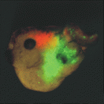
(Image from Whitehead Institute for Biomedical Research)

Thumbs up (3 votes)
Loading...

Tags: , ,
Categories: News, Research

Second cover image winner: mouse pharyngeal arch

Posted by , on 4 May 2011

Congratulations to Hozana Andrade Castillo of the University of Sao Paulo in Brazil, whose image of a mouse pharyngeal arch jumped from third place to first place in the last few days of voting. Her image will appear on a cover of Development in the next few months.


Developing pharyngeal arch region of mouse embryo showing immunofluorescence detection of neurofilaments (green), DiI labeling of trigeminal ganglion (red), and nuclei detected with DAPI (blue).

Second place in this round went to the cilia-stained squid image taken by Davalyn Powell of the University of Colorado Denver, followed by the flower-like image of cell division in the slipper limpet (by Anna Franz – University of Oxford) and an ascidian muscle (by Christine Carag Krieger – University of Pennsylvania).

We had over a thousand people vote in this round as many people forwarded the link to their friends. Thanks to everyone for participating and voting!

The next round of images will be up on June 6th. Around that same time, the winning image from the first round will appear on the cover of Development, and that week also marks the start of the 2011 Woods Hole Embryology Course. We’re looking forward to the images from this year!

Thumbs up (1 votes)
Loading...

Categories: Images

An After Thought to Evolution: Exceptional ways of Controlling Gene “Expression”

Posted by , on 3 May 2011

More and more, the central dogma is becoming well, dogged, for being a dogma at all. As humans, we have 3 billion nucleotides. Only 1% of it makes up our protein coding genes, which led to the development of the central dogma: DNA is transcribed to RNA and translated into proteins. During undergrad, we’re taught that transcription factor proteins bind to sequences in the DNA to either enhance or shut off transcription, thereby discontinuing protein production. Gene regulation seems to follow certain pathways and rules itself.

Then, 20 years ago RNAi was discovered, a whole new field where RNA can also regulate protein production. Generally, small RNAs repress protein coding genes by either causing their mRNAs to decay or inhibiting their translation into protein. They can be naturally produced for the sake of controlling the native genes, following a similar central dogmatic pathway: DNA is transcribed to a precursor RNA, which is converted into teeny RNA strands. These strands are selected by Argonaute proteins (AGOs) and used to target specific protein coding genes to halt their expression.

The progression from DNA to RNA to “something”, is like the foundation of genetics. And gene regulation is about how to control that progression. Evolution develops these elegant pathways to produce “something”, and then it evolved ways to control the pathway.

Well, not always.

Sometimes you can get “unintelligent design.”

Which is what a student joked, at a journal club meeting I’d attended today. The meeting started off as a presentation of a recently published article that assigned a role to an AGO protein in Arabidopsis. It ended up as a digression on evolution and how we perceive it, among other things.

The protein of interest was AGO10, which is somehow involved in miRNA/small RNA directed repression of target genes. But how it exactly it does this, was hazy to us, the observers. By inference, previous studies on its loss of function mutant, ago10/zwille/pinhead had deformed shoot apical meristems. After embryogenesis, the meristems at the junctions of leaves are where new ones form. In the mutants, the meristem is the size of pinhead, smaller than normal (Pictured in F, on the left, versus wild type in C, by Tucker et al., 2008). The protein expression of some transcription factors & signalling peptides changed in the ago10 mutants as well. But what does AGO10 specifically do?

(Picture: A, B = AGO10 expression in the yellow YFP areas of the embryo, at heart stage & torpedo stage. CLV, a stem-cell specific signalling peptide. its expression is weak is in the embryo ago10, as pictured in D, E. (it’s not strong in WT either though) CLV needs AGO10 to be maintained past embryogenesis, in the meristem Tucker et al. 2008).

One group discovered that AGO10 doesn’t really help in repression. It actually indirectly causes the activation of target genes. AGO10 is a decoy for the main AGO1, that performs 90% of miRNA directed repression in Arabidopsis plants. However, it’s a decoy for one miRNA alone (so it seems). It binds and sequesters miR166/165, a well studied miRNA that is deeply conserved in mosses and higher plants. By sequestering it, miR166/65 is prevented from binding to AGO1 and regulating its targets (HD ZIP proteins). Effectively it’s as if miR166/65, the silencer, has been silenced itself. By a massive protein no less.

Oddly, AGO10 is most abundant in one tissue type, the developing embryo. So it only catches up all the miR166/65 population in that one tissue. If it doesn’t, as suggested by ago10, then the embryo develops into mutant plant. 

But why would plants bother developing this? Couldn’t nature have just evolved so that miR166/65 isn’t transcribed in the embryo? So that some transcription factor or miRNA shuts it off. Like, why produce something and then evolve a whole protein to muffle its activity? It’s like..evolution forgot to turn off the tap or shut the door, and created a massive sink or club bouncer instead. Energy wise, it seems a waste though. Evidently there probably is an unknown purpose.

The authors of the paper suggest that miR166 is required in specific cells, but not all cells of the developing embryo. So miR166 is allowed to be expressed but AGO10 prevents it from being spread elsewhere..assuming that miRNAs can travel…

(A-C: WT, CLV expression is maintained throughout embryogenesis and meristem development. Without AGO10 in D-F, its expression disappears).

ResearchBlogging.orgIt’s funny. We have these commonly accepted notions about gene regulation engrained in us from years of training. With the result that when we’re faced with something altogether new, we’re not sure what to make of it. We’re not even sure if we can accept it, but there it is. It’s like realizing black swans with ghoulish red eyes exist in Australia.

Sources/References:

Lab discussion with students at the Research School of Biology at ANU, led by PIs Tony Millar and Iain Searle.

Zhu H, Hu F, Wang R, Zhou X, Sze SH, Liou LW, Barefoot A, Dickman M, & Zhang X (2011). Arabidopsis Argonaute10 Specifically Sequesters miR166/165 to Regulate Shoot Apical Meristem Development. Cell, 145 (2), 242-56 PMID: 21496644

Pictures:
Tucker MR, Hinze A, Tucker EJ, Takada S, Jürgens G, & Laux T (2008). Vascular signalling mediated by ZWILLE potentiates WUSCHEL function during shoot meristem stem cell development in the Arabidopsis embryo. Development (Cambridge, England), 135 (17), 2839-43 PMID: 18653559

Flickr CC by Kyknoord

Thumbs up (5 votes)
Loading...

Categories: Research

Science – The Bigger Picture

Posted by , on 1 May 2011

This is a retelling of the student and post-doc workshop from the second day of the BSDB/BSCB joint spring meeting that took place in Canterbury at the University of Kent. The session emphasised the need for accurate science and scientific involvement in public communication. It ended up a bit longer than I’d intended, but this is something I’m really enthusiastic about and felt it needed to be shared in detail. I hope you find it helpful.

Panellists:

Dr Peter Wilmshurst – A consultant cardiologist, known for his refusal to falsify or withhold data in pharmaceutical studies. He was being sued for libel and slander by NMT medical until the company entered liquidation in April.

Rose Wu – A representative for the charity Sense about Science which works tirelessly despite limited funding to improve the public image of science, aids accurate reporting of scientific issues in the media and campaigns for further government support for research.

Dr Jenny Rohn – A UCL post-doc by day. Also known for her punditry, she runs the popular science communication website lablit.com has been interviewed numerous times for tv and radio. She has published numerous stories and editorials and two fictional novels Experimental Heart and The Honest Look, communicating science through the engaging and emotional personal lives of scientists. She was also central to the campaign to save UK science funding.

(more…)

Thumbs up (4 votes)
Loading...

Tags:
Categories: Discussion, Funding, News, Outreach, Research

Rethinking X-chromosome Inactivation

Posted by , on 30 April 2011

I’ve been asked to present the back-story behind our recently published manuscript in Development “Transcription precedes loss of Xist coating and depletion of H3K27me3 during X-chromosome reprogramming in the mouse inner cell mass.”

Mammalian dosage compensation occurs by silencing one X-chromosome in female cells, termed X-chromosome Inactivation (XCI).  Balancing X-linked gene transcription is critical for female development, and in most cells, the absence of XCI is lethal.  Interestingly, female mouse germ cells and inner cell masses (ICMs) appear to be an exception, capable of handling imbalances in X-linked gene dosage and exhibiting activation of both X-chromosomes.  These uncommitted cells have the unique ability to reset the epigenetic profile of inactive X-chromosomes.  As a graduate student, I was drawn to investigate the mechanism of X-chromosome reactivation because I wanted to understand how epigenetic reprogramming occurs naturally in the embryo.

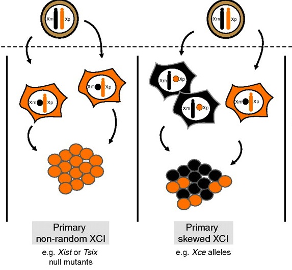
Most of what is known about the molecular events that initiate XCI has been extrapolated from observations of XCI in mouse cells.  In the mouse embryo, the choice of which X-chromosome to be silenced is made stochastically; maternal and paternal X-chromosomes have an equal chance of being silenced and the end result is a random pattern of XCI.   In contrast, extraembryonic tissues exhibit an imprinted form of XCI, and the paternal X-chromosome appears to be predetermined for silencing.  It has been the prevailing opinion in the field that a master controller, the Xist non-coding RNA, regulates XCI.  Xist RNA has been linked to almost every step in the XCI mechanism, including choice, initiation, and maintenance; however, its exact role is still unknown.  What is clear is that Xist transcripts coat the X-chromosome in cis, recruiting epigenetic modifying proteins that are responsible for forming inactive-X heterochromatin.

It had previously been shown that in all cells of the female preimplantation mouse embryo, Xist transcripts coat and recruit the repressive histone modification H3K27me3 to the Xp.  However, X-linked gene expression at single cell resolution had not been directly analyzed, relative to cell fate in the embryo.  In our manuscript, we definitively show that all cells, irrespective of lineage, establish imprinted XCI by the early blastocyst stage.   This sets up a situation in the ICM; epiblast cells must undergo a reactivation/inactivation cycle.  During ICM maturation, epiblast cells exhibit progressive upregulation of Nanog expression, coincident with their transition to ground-state pluripotency.  Curiously, Xist is a target of OCT4, SOX2, and NANOG.  Based on the concept that Xist is the master regulator of XCI, it had been predicted that progressive upregulation of NANOG in the ICM leads to Xist repression, resulting in loss of Xist coating and triggering reactivation of Xi genes.

We used a knockout of Grb2 to upregulate Nanog expression in the ICM.  As might be predicted, increased NANOG dosage led to Xist repression, indicated by loss of Xist coating.  However, X-linked gene silencing is maintained in cells that lack Xist coating, perhaps suggesting that induced epigenetic reprogramming is incomplete in these cells.  These provocative results point to other factors, in addition to Xist, that are needed initiate Xi gene silencing.  Recent results from Kalantry et al. Nature 2009, Namekawa et al. Mol. Cell Biol. 2010, and Okamoto et al. Nature 2011 support these conclusions and the need for the field to reconsider mechanisms regulating XCI.
(more…)

Thumbs up (17 votes)
Loading...

Tags: , ,
Categories: Research

Stem cell patent case could have far-reaching impact

Posted by , on 28 April 2011

Last month, the advocate-general of the European Court of Justice gave his opinion on a long-running legal debate about a patent filed several years ago in Germany. If the Court follows his recommendation, patenting of applications using embryonic stem cells will be prohibited on moral grounds.

13 leaders of major stem cell projects in Europe responded to the advocate-general’s statement with an open letter published in Nature this week. They express serious concerns about the impact of a patenting ban on European research.

http://www.eurostemcell.org/stem-cell-patentsEuroStemCell, Europe’s stem cell hub, has collected comments and information about this case on its Stem cell patents topic page. Visit the site to read the open letter and find out more about the case.

Post a comment on the site, or add your signature to the letter if you wish: http://www.eurostemcell.org/stem-cell-patents

Thumbs up (2 votes)
Loading...

Categories: Discussion, News, Research

Got the Blues? How Plants Respond to Blue Light

Posted by , on 28 April 2011

Physiologically speaking of course…

As humans we can see a limited assortment of electromagnetic wavelengths, known as the visible spectrum of light, a.k.a. colours. (Other wavelengths we cannot see include UV rays, X rays or infrared). Why is it that we can differentiate light colours? Its likely to do with adaptation to the environment. Plants and animals have evolved different physiological responses to various wavelengths of light.

As mammals we have biological clocks, or circadian rhythms that respond to light. Dark or blue light set off signalling mechanisms, which ultimately regulate the melatonin levels in our system. Melatonin is a hormone which can induce sleep, if at sufficiently high levels in our system. (Hence your doctor prescribing you melatonin if you have trouble sleeping at night. Suffice to say countless grad students are probably on this stuff right now). (Photo: Festival Colours by jmtimages, Flickr CC)

Plants also have a light-sensing system, which can respond to different times of day and seasonal changes. This can determine  fruit maturation and changing colour in the leaves etc. For plants, blue light has a revitalizing (instead of drowsy) effect. If you’ve ever left your plants without light for too long, they go yellow. Irradiate them with a little blue, and the green will return within a couple of days. (Photo: Autumn Colours in Australia’s National Capital, personal collection).

Interestingly, plants and animals have the same type of photoreceptors to blue light, called Cryptochromes (CRYs), even though they elicit a different response. (Photoreceptors = light absorbing molecules or pigments.)

CRYs initiate the light response, analogous to a molecular domino effect. The pigments bind specialized proteins that in turn, regulate transcription factors that can switch on the light response genes involved in development. Termed as photomorphogenesis, light dependent changes in development can include initiation of flowering, or extension of roots in germinating seeds.

Currently, characterizing the molecular interactions in the plant blue-light/CRY1 response seems to be a hot topic. Two highly similar articles were just published in Genes & Development, which was drawn to my attn by Eva (cheers). They were produced by two different research crews, using virtually the same methods to similar results. It’s not by total coincidence either. Characterizing molecular interactions in plants involve the same gold standard biochemical and genetic assays (i.e. transgenic plants, yeast hybrid systems, loss-of-function mutants etc.)

In summary: the blue-light/CRY1 pathway doesn’t directly switch genes on. instead, it switches off the default dark condition mechanism. In the dark, SPA1 and COP1 proteins negatively regulate the light signalling pathway. Active COP1 normally marks transcription factor HY5 for degradation. SPA1 binds COP1 to enhance this activity. If HY5 were allowed to accumulate, it promotes the expression of multiple genes involved in photomorphogenesis, such as flowering, or germination.

What the groups found: Blue-light triggers CRY1 to bind SPA1. This in turn inhibits SPA1 binding to COP1. In 2001, another group found that CRY1 can also interact directly with COP1 to inhibit its activity in a 2nd way. Result: no COP1 activity, HY5 accumulates. Photomorphogenesis is observed. (CRY1 could not bind SPA1 in the dark, or under red light).

(photo: blue light, by psychiks, Flickr CC. does this make you feel sleepy?)

Some discordance over the CRY1-SPA1-COP1 interaction: One group claims that CRY1 causes SPA1 to dissociate from COP1. (exact mechanism unclear, allosteric binding = conformational change?) the other group suggests that CRY1 impedes COP1 and SPA1 binding, possibly by sandwiching itself between them. Refer to sandwich model in Liu et al. 2011 Figure 4D, or kit-kat dissociation model in Lian et al. 2011 Figure 5G. Devil’s in the details: both groups developed their models based on two biochem assays ~ yeast two/three hybrid and co-immunoprecipitation. However, they only tell you if things bind or not. Interestingly, blue-light and CRY1 were required to prevent SPA1 and COP1 binding. Under blue light, CRY1 could bind both COP1 and SPA1, just not sure how.

The interaction hasn’t been characterized in animals, although they do contain the CRYs. Mammals even have COP1, which can also mark other transcription factors for degradation. Inexplicably, breast cancers were found to have over-expressed levels of COP1. Not sure if CRY also inactivates mammalian COP1, although, one group did stick the mammalian COP1 into Arabidopsis, and found it reacts to blue-light. Interestingly, photoreceptors and COP1 were initially found in Arabidopsis. (score 1 for the plant model of genetics).

ResearchBlogging.org Plants:

Liu B, Zuo Z, Liu H, Liu X, & Lin C (2011). Arabidopsis cryptochrome 1 interacts with SPA1 to suppress COP1 activity in response to blue light. Genes & development PMID: 21511871

Lian HL, He SB, Zhang YC, Zhu DM, Zhang JY, Jia KP, Sun SX, Li L, & Yang HQ (2011). Blue-light-dependent interaction of cryptochrome 1 with SPA1 defines a dynamic signaling mechanism. Genes & development PMID: 21511872

Both research groups hail from China and the US. They’re also based in some of the top universities in the world (Peking Uni, UCLA etc.), which makes for some hefty competition, among neighbours no less.

Human Photoreceptors and COP1:
Yi C, & Deng XW (2005). COP1 – from plant photomorphogenesis to mammalian tumorigenesis. Trends in cell biology, 15 (11), 618-25 PMID: 16198569

Yi C, Wang H, Wei N, & Deng XW (2002). An initial biochemical and cell biological characterization of the mammalian homologue of a central plant developmental switch, COP1. BMC cell biology, 3 PMID: 12466024

Cashmore AR (2003). Cryptochromes: enabling plants and animals to determine circadian time. Cell, 114 (5), 537-43 PMID: 13678578

Thumbs up (2 votes)
Loading...

Categories: Research