The community site for and by
developmental and stem cell biologists

SDB meeting in Albuquerque

Posted by , on 5 August 2010

Today is the first day of the SDB meeting in Albuquerque. The program looks amazing, and I’m looking forward to many of the talks. The meeting is held jointly with the Japanese Society for Developmental Biologists, so many of the attendees came all the way from Japan.

Development and the Node are here in Albuquerque as well. You can find us at the Company of Biologists booth as well as in the audience at the talks.

We hope to summarize the meeting on the Node afterward, but could use some help, because try as we might, we can’t be everywhere at once! So if you’re at the SDB meeting, and would like to help us out by summarizing part of the event for the readers of the Node, please get in touch. You can leave a comment here, send an email, or track me down in person. We ask, at this particular meeting, that you get in touch before writing, because we’ve agreed with the conference organizers to write a report the “old fashioned way” on the Node – meaning that speakers’ permission will be asked before posting the report. If you want to write something, please do let us know and we’ll contact the speakers for you.

Here are some reports from previous society meetings on the Node:
– BSDB/BSCB meeting part I, part II, part III
SFBD/JSDB meeting
ISSCR meeting
See also our general tips for blogging from meetings.

I’m looking forward to an interesting meeting, and hope to see some of you here!

Thumbs up (No Ratings Yet)
Loading...

Tags: ,
Categories: Events

Navigate the archive

Use our Advanced Search tool to search and filter posts by date, category, tags and authors.

Would you like a zebrafish with your tea?

Posted by , on 4 August 2010

Model organisms never looked so delicious… The baking blog Not So Humble Pie features science cookies made by the author or by her fans. Many of the cookies are biology-themed, and I think the model organisms are my favourites.


These zebrafish have edible glitter on them to give them that familiar fishy shine


wild-type Drosophila cookies

Of the fly-cookies, the baker-blogger writes “I found it hard to balance a realistic look with something you’re supposed to want to eat.”

But the cookies are not just animal-shaped. Creative tricks with different kinds of icing can make square cookies look like agarose gels and turn round cookies into a tasty plate of bacteria. Yum!

For more biology cookies, visit the science section of Not So Humble Pie. Her own cookies all come with the recipe so you can try making your own at home.

Thumbs up (4 votes)
Loading...

Tags: , ,
Categories: Images

Of whales and lice

Posted by , on 3 August 2010

Imagine an ordinary louse. You might be thinking of the small insects that infest the human scalp and cause intense itching. What you may not know: lice actually exist on great variety of birds and mammals. Even whales have ‘lice,’ which are not closely related to human head lice and are giant in comparison (can be over 25 mm long!), but these lice do have a similar lifestyle to the common head lice. Whale lice are amphipod crustaceans that exist as ectoparasites in the folds of the whale’s skin and feed on dead skin and debris. Below is a photo of an adult whale louse, almost 2 cm long.

Adult Whale Louse

As a student in the MBL’s Embryology class, I had the unique opportunity to get a close look at embryonic whale lice. Whale lice brood developing embryos in a brood chamber called a marsupium. Thus, they have no free-swimming stage and spend their entire life on the whale. Whale lice (Cyamus ovalis) collected from a beached whale were procured from Jon Seger and Heather McGirk of University of Utah for the course. In class, we dissected out embryos from the marsupium of two gravid females. Using techniques and materials provided by the course, we looked at expression of developmental patterning genes by floursecence immunostaining. See confocal stack below with DAPI (nuclei) in teal, phoso-histone H3 (mitotic cells) in magenta, and HRP (neurons) in orange.

Stained Whale Louse Embryo
Whale Louse Embryo

Thumbs up (7 votes)
Loading...

Categories: Research

The Intestinal Crypt

Posted by , on 2 August 2010

It’s not often that the introductory part of a research talk is beautiful as well as informative, but Hans Clevers achieves both by using this video about the intestinal crypt in his presentations. (Click either screenshot to see the video)

The video shows how stem cells at the base of the intestinal crypt produce the epithelial cells of the intestines, and how the cells are pushed up toward the tip of the villi. Once at the top, the cells die, but are immediately replaced by the next cells. It’s a “clonal conveyor belt”, constantly moving new cells up.

When Clevers’ lab identified crypt base columnar cells as the stem cells responsible for the generation of the rapidly renewing intestinal epithelium, they simultaneously identified the gene Lgr5 as marker for intestinal stem cells. Until their discovery, other groups had used the method of detection the retention of labeled DNA, which suggested other crypt cells as stem cells.

The last part of the video shows how they used the unique expression pattern of LGR5 to create a knock-in mutant in which the tumour suppressor APC is no longer expressed in stem cells, and show that this is sufficient to trigger adenoma formation.

Clevers had the animation made through a company called Digizyme, which specializes in multimedia approaches to present scientific information. It’s a very welcome addition to the home-made powerpoint slides that you usually see in talks, and I appreciate the effort made to pack this information on intestinal stem cells in an, if you’ll pardon the pun, easy to digest format.

(more…)

Thumbs up (10 votes)
Loading...

Tags: ,
Categories: Research, Video

Node update

Posted by , on 29 July 2010

A few bits of news and reminders about the Node, just to keep you all up to date:

-We’ll be interviewing Jorge Cham, creator of the grad student comic Piled Higher and Deeper (PHD), this weekend. Do you have any questions you’d like to ask him? I asked for input via our Twitter account, and have one question so far (plus my own). I’m sure many of you are familiar with the comic, and this is your chance to ask away, so leave a comment with your question!

– Ah yes, the Twitter account. All the bits of news we find that are too short for a proper post end up there. You can follow us if you have a Twitter account yourself, or just look at the page to see what’s there and stay even more up to date than you already are.

– For example, we spread the word on Twitter that EMBO is looking for official bloggers for their annual meeting in September. They ask that you are already registered for the meeting and contribute to an existing blog, but that can be a group blog, like the Node. So if you don’t have your own blog, but would like a chance to blog for EMBO, make sure to register for the Node before you apply. If you’re selected as official blogger, they provide you with direct access to the speakers for interviews, and you receive free registration at next year’s meeting. EMBO’s application deadline for bloggers is August 15. Good luck!

– Finally, you may have noticed comments on the Node disappearing and then reappearing. This is the result of a too sensitive comment reporting system. We’re working to get this solved by the end of August, and will keep you updated about any changes.

Thumbs up (No Ratings Yet)
Loading...

Categories: News

Embryological Discovery in Woods Hole : Vertebrates

Posted by , on 27 July 2010

We are in our last week of the Embryology Course at Wood’s Hole now, and currently working on annelid and squid embryos. Things are still going at a frenetic pace but I’d like to take this chance to talk a little more about some of the work we did using vertebrates.

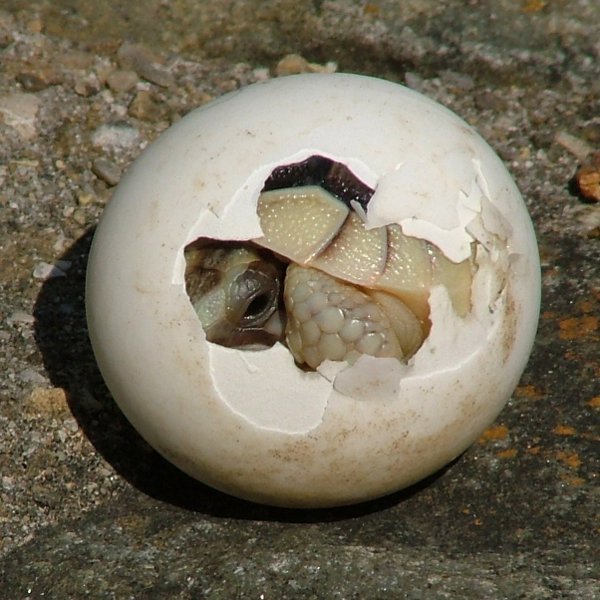
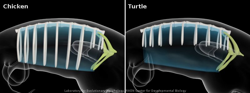
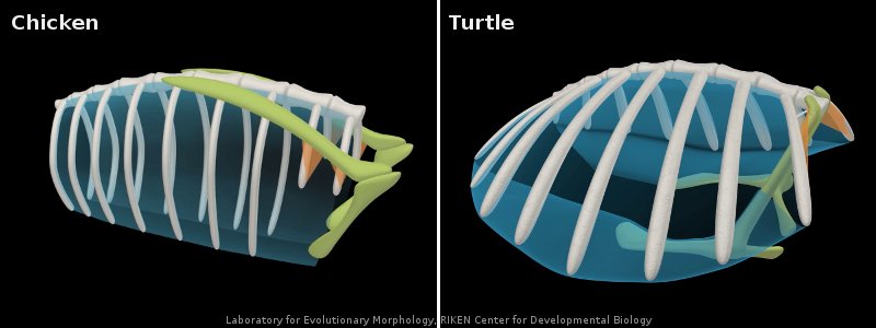
The course covers many vertebrate model organisms such as mice, chick, frog and zebrafish but we were also able to carry out some work on other organisms like sticklebacks, bats and even turtles! This was mostly thanks to Richard Behringer who brought an amazing array of different embryos for us to experiment on. On the left you can see a beautiful lizard, which I stained with alcian blue to show cartilage and alizarin red to show bone. This was particularly interesting for me as I am working on the development of the sternum for my Ph.D. project, so it was great to be able to see the morphological diversity in the sternum between species.

To complement the study of this wide range of species, we also revisited some classical experiments carried out in chick and frog to illustrate a number of key developmental principles. One example was the Spemann-Mangold organizer graft in Xenopus laevis, which involves transplanting the dorsal marginal zone of an early gastrula stage embryo into the ventral region of an intact embryo at the same developmental stage. This tissue has the capacity to organize surrounding tissues to form a dorsal axis, so this experiment should generate a tadpole with a secondary axis. Although this required a lot of patience and very steady hands, a few people in the class did successfully grow some two-headed tadpoles!

I wasn’t one of the lucky ones to grow a monster tadpole, but I did get some interesting results for a project I carried out with others in the class working on the establishment of the dorso-ventral axis. We used lithium chloride treatment to dorsalize embryos, and UV irradiation to ablate the dorsal axis. The resulting tadpoles were very messed up, and it was interesting to see which features were lost and which were radialized after each treatment.

We also tried out some transplantation and bead insertion experiments in chick embryos, and used whole embryo and organ culture in mouse. Now the course is drawing to a close and we are trying to frantically gather together and organize all of the data we have on so many of different microscopes. Hopefully I can make some sense of it all and I’ll write back with an overview of the entire course once I’m back in London.

Sorrel

Thumbs up (5 votes)
Loading...

Tags: , ,
Categories: Images, Research

In Development this week (Volume 137, Issue 16)

Posted by , on 27 July 2010

Here are the highlights from this week’s issue of Development:

Lymphatic networks follow arterial lead

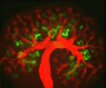
The vertebrate lymphatic system consists of lymphatic vessels, which collect fluid from the tissues and return it to the blood, and lymph nodes, which are involved in immune defence. Lymphatic vessels, like blood vessels, form a complex vascular network, but what guides the development of this network? According to Stefan Schulte-Merker and co-workers, arteries provide crucial guidance cues to the lymphatic endothelial cells (LECs) that form the lymphatic network in the zebrafish trunk (see p. 2653). Using transgenic zebrafish lines that allow the direct visualization of arteries, veins and lymphatic vessels in single embryos, the researchers show first that intersegmental lymphatic vessels (ISLVs) in the zebrafish trunk align with arterial intersegmental vessels (aISVs) but not with venous intersegmental vessels. Then, using time-lapse confocal imaging, they show that LECs migrate exclusively along aISVs and that LEC migration is blocked in zebrafish mutants that lack aISVs. Together, these data reveal a crucial role for arteries in LEC guidance; future research will unravel the mechanism underlying this guidance.

Flowering plant fertilization model over turned

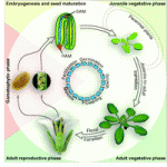
Fertilization in flowering plants involves two sperm cells and two female gametes – the egg cell and the central cell, progenitors of the embryo and endosperm, respectively. A previous study suggested that Arabidopsis plants carrying loss-of-function mutations in cyclin dependent kinase A1 (CDKA:1) make a single sperm cell that preferentially fertilizes the egg cell to produce an embryo that triggers central cell division. Now, Frédéric Berger and colleagues overturn these widely accepted results by showing that a significant proportion of cdka;1 pollen actually delivers two sperm cells (see p. 2683). Delivery of a single cdka;1 sperm cell to a wild-type ovule can fertilize either female gamete, they report. However, when two cdka;1 sperm cells are delivered, one fertilizes the egg cell and the other activates central cell division. Fusion of the gamete nuclei fails in the central cell, however, which prevents paternal genome incorporation and causes seed abortion. Thus, this new analysis of the cdka;1 phenotype reveals an essential role for the paternal genome during early seed development.

Med(12)iating Wnt signalling in mouse development

Mediator, a conserved multiprotein complex that connects DNA-bound transcription factors to the RNA polymerase II machinery, is part of the intricate mechanism that regulates eukaryotic transcription. The Med12 mediator subunit is required for gene-specific functions during zebrafish development, but are its developmental functions conserved in mammals? On p. 2723, Heinrich Schrewe and colleagues address this question by examining embryos generated from mouse embryonic stem (ES) cells in which Med12 has been targeted. Embryos generated from Med12 hypomorphic ES cells fail to develop beyond embryonic day 10, the researchers report, and have severe defects in neural tube closure, axis elongation, somitogenesis and heart formation. The Wnt/planar cell polarity pathway and canonical Wnt/β-catenin signalling are both disrupted in the Med12 hypomorphic embryos, they note. Furthermore, embryos generated from Med12 null ES cells fail to establish the anterior visceral endoderm, activate brachyury expression or complete gastrulation. Together, these results indicate that Med12 is necessary for gene-specific functions and for correct Wnt/β-catenin and Wnt/PCP signalling during early mouse development.

Mechanics of morphogenesis revealed

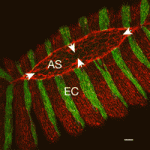
The morphogenetic movements that shape embryos depend on the forces generated by embryo’s cells and on the resistance of its tissues to these forces. Microtubules and F-actin are largely responsible for both these cellular properties but the contribution of these structural elements to morphogenesis is unclear. Now, Lance Davidson and colleagues unexpectedly report that nocodazole-induced depolymerization of microtubules stiffens the converging and extending dorsal tissues in Xenopus embryos (see p. 2785). The researchers attribute this result to the release of Xlfc – a guanine exchange factor that binds to microtubules and that regulates actomyosin contractility by activating Rho family GTPases. Consistent with this idea, drugs that reduce actomyosin contractility rescue nocodazole-induced embryonic stiffening and partly rescue the morphogenetic defects of stiffened embryos. Other experiments that combine drug treatments and Xlfc activation and knockdown indicate that microtubules have no direct role in maintaining bulk tissue stiffness in Xenopus embryos. The researchers conclude, therefore, that microtubules indirectly regulate the mechanical properties of embryonic tissues through RhoGTPase pathways.

Bicoid gradient: precision through hunchback

Morphogenetic gradients determine cell identity during development through the concentration dependent activation of target genes, but how the precision of the response to morphogens is determined is unclear. On p. 2798, Nathalie Dostatni and co-workers provide new insights into this developmental puzzle by examining the transcriptional response to Bicoid in Drosophila embryos. The Bicoid gradient is established in Drosophila embryos after eight nuclear divisions (cycle 9) and target protein expression is specified by cycle 14 with a precision that corresponds to a 10% Bicoid concentration difference. To understand how this precision is achieved, the researchers analyze nascent transcripts of the Bicoid target gene hunchback. They report that hunchback is already transcribed from both alleles in most anterior nuclei in cycle 11 interphasic embryos. This synchronous expression is specified within a 10% difference of Bicoid, a precision that is compatible with the fast mobility of Bicoid in the nucleus measured using fluorescent correlation spectroscopy. Finally and importantly, genetic experiments reveal that maternal Hunchback contributes to the early synchrony of the Bicoid response.

Thumbs up (No Ratings Yet)
Loading...

Tags: , , , , , , , , , , , ,
Categories: Research

The Maggot Meeting-2010

Posted by , on 26 July 2010

Greetings!
It’s been an overcast, windy and gloomy Monsoon here in Bangalore, but the past few weeks at NCBS have been bright and exciting! The “Maggot Meeting – 2010”: Neural Circuits and Behaviour is currently on till the 27th of July and I look forward to the seminars and interactive discussions with eminent scientists. Early last week the Center hosted the EMBO Global Exchange Lecture Course on ‘Systems neuroscience of the Drosophila larva: genetic and circuit bases of behaviour’. This series of lectures and demonstrations were devoted to the basics of neuroscience, development and behaviour. I was invited to participate as a Teaching Assistant to demonstrate live cell calcium imaging in Drosophila melanogaster primary neuronal culture. I am very pleased to have been given this opportunity to share my training and knowledge on new methods and techniques with other fellow participants. It has truly been a wonderful experience!

Thumbs up (1 votes)
Loading...

Categories: Events

Too many postdocs and PhD students?

Posted by , on 23 July 2010

There was a nice piece on the Naturejobs site this week, written by postdoc Katherine Sixt. She describes how she started to realize that not every postdoc will eventually become a professor. There simply aren’t enough positions available, so postdocs should look at other careers. But as a postdoc, and even as a PhD student, you are being trained as if you were aiming for a job similar to the one your supervisor has, and anything else is considered strange and different. Katherine writes: “I feel as though I have to sneak off to careers seminars where scientists describe their non-traditional paths. Thoughts of alternative career choices are still dirty secrets for some.”

Over the past few years, I’ve read many similar articles. Some from the position of the postdoc looking for work, others more reflective and distant – considering ways in which to deal with the simple truth that there are far more postdocs than there are academic positions for them to fill.

What do you think: Should there be fewer postdoc and PhD positions? Or different kinds of trainee positions, where some include training for scientific careers outside of the lab? Have a read through the following articles and blog posts to see what others have to say about it:

In Which I Dream of Revolution – Jenny Rohn
Quote: “Yesterday morning I woke up and realized that the entire logistical edifice underpinning the scientific profession is flawed. What’s more, I didn’t just see the problem; I had a glimpse of its solution.”

Do We Produce too Many Biomedical Trainees? – Jeff Sharom [Link to PDF]
Hypothesis Journal, 6(1), 17-29 (2008)
This is a review article that looks at evidence for and against the idea that there are too many trainees.
Quote: “Paradoxically, while research aims to recruit rational individuals, research may not be a rational career choice”

Are we training too many scientists? – Bijal Trivedi (in The Scientist, 2006)
Quote: “With rising numbers of newly minted life science PhDs, fewer tenure track positions open, and bulging ranks of increasingly frustrated postdocs, many want to know why the number of PhDs and the focus of their education is out of balance with job prospects and career expectations.”

Are there too many PhDs? – Jason Hoyt (on the Mendeley blog)
Quote: “Only then, do students realize the road that lies ahead is dotted with pit stops leading, not to Nobel glory, but a journeyman career with salaries well below that of their friends who went into business, law, or medicine.”

Thumbs up (1 votes)
Loading...

Categories: Careers, Discussion

Science on Screen

Posted by , on 20 July 2010

If you’re annoyed by the unconvincing lab work on shows like CSI, and would like to show the world what real scientists are like, why not make a film yourself? You’d be surprised to find out how many films by or with scientists there are, both fiction and non-fiction. This month alone, two organizations involved with science films have either screened or collected a number of submissions, and I also recently found out that one of my favourite documentaries (about the daily life and careers of science graduate students) is available to view online.

Last week, Planet SciCast screened the winners of a competition in which schools and scientists had made fun and informative films about science. All submissions are available to watch on their website, and the awards ceremony was held at the Royal Institution in London last Friday. If you liked the plasticine embryo model previously featured on the Node, you might also enjoy this stop motion clay video about mitosis at Planet SciCast.

While the SciCast films are predominantly factual and made by students, the Imagine Science Film Festival in New York is currently collecting a wide range of both amateur and professional, fictional and factual, films for their annual festival in October. Their late deadline is on August 15, so if you happen to have a film lying around, you can still submit it. Last year my own tiny five-minute film about Lab Waste screened in the ISFF “quirky shorts” program, and I was impressed with all the other (far more professional!) films that were shown that night. ISFF lasts more than a week, and screens full length films as well as shorts.

ISFF promotes interaction between scientists and film makers, and many of their submissions are made by film professionals, but involve scientists as main characters or writers or consultants. My favourite film that I saw at ISFF last year was a French film called MEPE, which also won one of the festival’s awards.


MEPE, a French detective film about biology… It’s entirely in French and this version is unfortunately without subtitles, but even if you don’t speak the language you can appreciate the style and quality, and maybe even some of the humour.

My own little movie (more a slideshow…), which screened on the same night as MEPE at ISFF last year, is quite literally a work of garbage, and a completely different kind of film, so you can see the variety at ISFF – even within one evening of programming.

Lab Waste from Eva Amsen on Vimeo.

If film festivals, artsy French dark comedy, school projects, or pictures of garbage are not your thing, then you might still be interested in watching the full-length documentary Naturally Obsessed about three graduate students in Larry Shapiro’s lab at Columbia University in New York.  No matter what you’re working on and no matter where you are in your career – whether you’ve just started graduate school or finished long ago – you have to see this film. The three students are completely different, and you’ll undoubtedly identify with at least one of them and understand their frustrations and/or celebrations. It’s available to watch online.

All these films involved working scientists either as subjects, producers, advisers, or judges, so there are lots of opportunities to get involved and do your part to represent science on screen.

Thumbs up (1 votes)
Loading...

Tags: , ,
Categories: Careers, Video