The asymmetry in cell morphology and the asymmetric distribution of intracellular organelles, proteins, nucleic acids and other components are the hallmarks of cellular polarity, possessed by most, if not all, cells. Polarity plays important roles in cell differentiation and function, and its dysregulation is intimately related to developmental defects, tumor formation and cancer metastasis. The commonality of cell polarization events is that some polarity-regulating proteins (such as the Par3/Par6/aPKC complex, or the Frizzled/Dishevelled/Diego complex) are specifically recruited to the designated subcellular area and form local aggregates, attaching to the inner surface of the plasma membrane. These protein aggregates are dynamic and assemble and disassemble quickly in response to cellular signals [1-3]. How these protein complexes can achieve polarized concentration on the premise of open contact with the cytoplasm while maintaining a high degree of dynamics has been an open question.
In recent years, the “liquid-liquid phase separation” (LLPS) of biomacromolecules has emerged as an important mechanism underlying the formation of a variety of membraneless structures within cells [4,5]. The aggregates of polarity proteins share some similar characteristics with these membraneless structures, such as high condensation and dynamic equilibrium with proteins in cytoplasm.
We showed previously that during the asymmetric divisions of Drosophila larval neural stem cells (or neuroblasts), cell fate determinants including Numb and Pon undergo LLPS. Multivalent interactions lead to the polarized enrichment of the Numb/Pon complex on the basal cortex of dividing neuroblasts, which regulates the differentiation of the ganglion mother cell (GMC), the differentiating daughter of the neuroblast, upon cell cycle exit [6].
On May 8, 2020, we published a research work titled “Par complex cluster formation mediated by phase separation” online in Nature Communications [7]. The paper provides evidence showing that the apical Par protein complex is also assembled via a LLPS mechanism, thereby regulating the establishment of the neuroblast apical-basal polarity and consequently the distinct cell fate of neuroblast daughters.
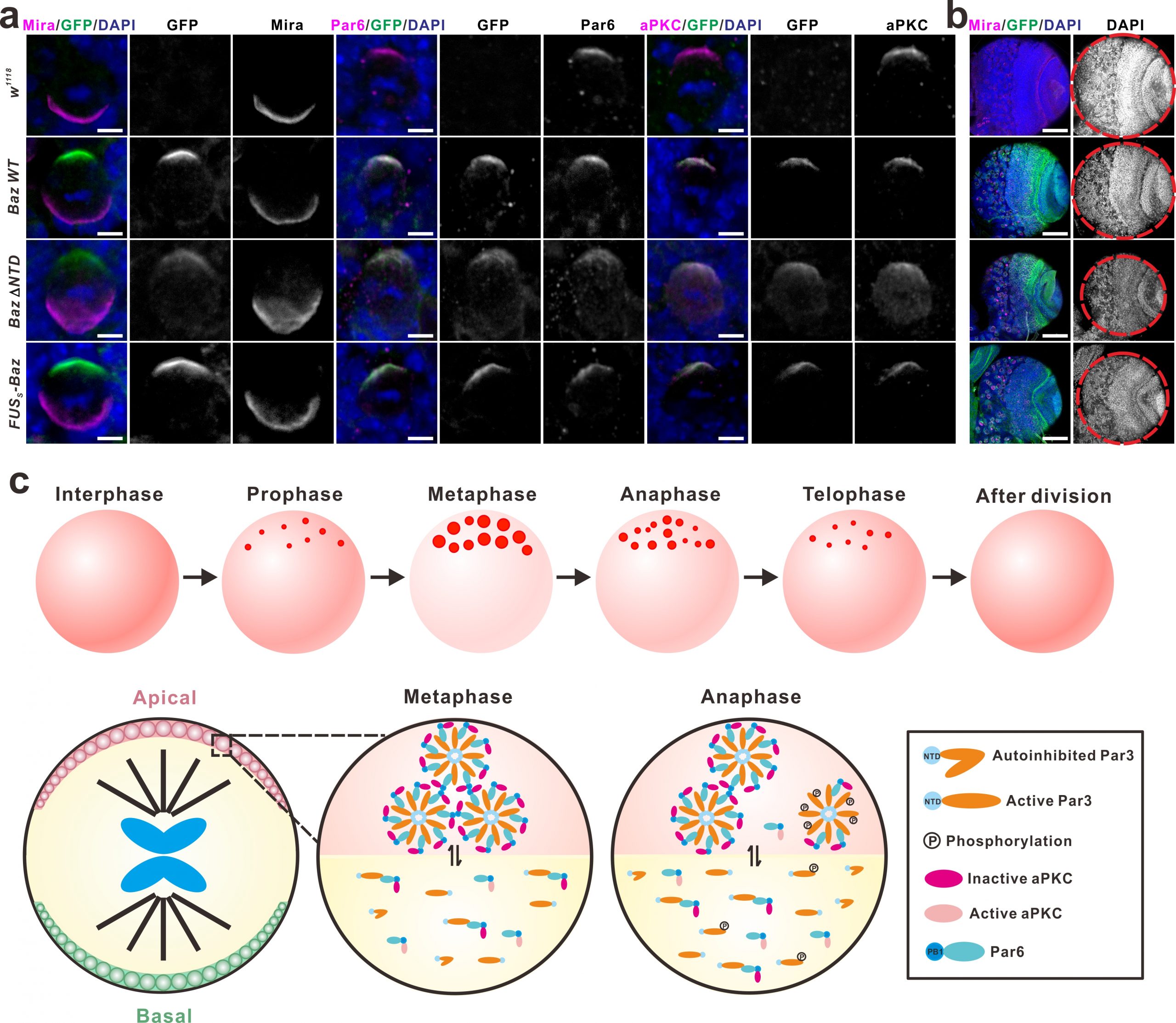
One of the best-studied polarity complexes, the highly conserved Par complex is composed of Par3 (Bazooka in Drosophila), Par6 and aPKC, which can interact with each other to form a polarity core and further recruit other polarity proteins. During the interphase of neuroblasts, these polarity proteins are distributed throughout the cytoplasm. Upon mitotic entry, Par3, Par6 and aPKC gradually accumulate underneath the apical cortex and form a multiprotein-containing apical complex, which directs the basal enrichment of those cell fate determinants including the Mira/Pros/Brat/pros mRNA complex and the Pon/Numb complex, via aPKC-mediated phosphorylation events. Upon the completion of mitotic cycle, the Par3/Par6/aPKC complex remains in the neuroblast daughter and the complex is dissembled, whilst those cell fate determinants are preferentially segregated into the GMC daughter to promote its differentiation. In this latest work, we showed upon mitotic entry Par3 (or Baz) forms apically-localized liquid droplets, which is mediated by its N-terminal domain (NTD) harboring oligomerization activity. These liquid droplets subsequently fuse and assemble into a highly concentrated cluster around the apical pole, forming the condensates underneath the apical cortex. Par6 is recruited to the Par3 condensate through the specific binding of its C-terminal motif to PDZ3 domain of Par3. Interestingly, the incorporation of Par6 into Par3 condensates greatly promotes the LLPS of Par3 via multivalent interactions.
As the only known kinase in the complex, aPKC can also be recruited and enriched in the Par3/Par6 condensates, but aPKC in the droplets is surprisingly in an inactive state. Interestingly, activated aPKC can phosphorylate Par3 and promote dissolve of the Par3/Par6 aggregates. Based on these observations, we speculate that the formation of the Par condensed droplets may provide an effective platform to recruit and concentrate the limited aPKC from the cytoplasm to the apical sub-membrane area, where aPKC can be activated by cell cycle regulatory factors (such as Cdc42) upon mitotic entry, and subsequently phosphorylate Par3, which eventually leads to the dissembly of the Par protein aggregates. Meanwhile, the activated aPKC also mediates the basal segregation of the fate determinants through its kinase activity. Interfering with the formation of Par3/Par6 LLPS will disrupt the establishment of the apical-basal polarity during the asymmetric divisions of Drosophila neuroblasts, leading to the defects in the development of neuronal lineages (Figure 1). This study, together with our previous work, shows that the LLPS, mediated by the multivalent interactions among these polarity proteins, plays important roles during the establishment of Drosophila neuroblast polarity.
Although LLPS provides a new perspective for the formation of a large number of membraneless structures in cells and the selective enrichment and separation of cellular components in various physiological processes, whether it regulates these biological processes under physiological conditions has always been the focus of debate. One main reason is that LLPS is largely dependent on the protein concentration, and the concentration of the target protein used in cell overexpression systems and in vitro experiments is often higher than that under the physiological conditions.
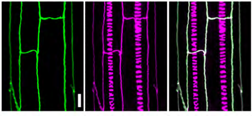
To test this, we established GFP knock-in Drosophila strains expressing GFP-tagged Baz under its endogenous regulatory elements, and explored the role of LLPS (of Baz) in the establishment of neuroblast polarity at an endogenous setting. In sharp contrast to the overexpression assay results showing that Baz ΔNTD (deleting the N-terminal domain, thus compromising LLPS ability) lost its apical condensation in both WT and baz mutant backgrounds, the knock-in mutant Baz ΔNTD surprisingly exhibited obvious apical condensation (crescent formation). However, it also exhibited significant cytoplasmic diffusion, which likely explains the observed smaller brain phenotype. More importantly, replacing the Baz NTD domain with a low complexity domain (LCD)-containing fragment of FUS, which has no secondary structure but can phase separate, also significantly rescued brain development abnormalities caused by NTD deletion (Figure 1). These results provide strong support for the hypothesis that the polarized distribution and establishment of the Par complex is mediated by LLPS, and also points out the importance of protein concentration when assessing the LLPS for protein of interest. It further suggests that the results obtained form in vitro or over-expression study need to be interpreted with caution.
In summary, our study reveals that the LLPS mediated by the multivalent interactions among the polarity proteins Par3, Par6 and aPKC promotes their condensation beneath the apical membrane, and the rapid response of the phase-separated condensates to cellular signals also ensures a high dynamics in the assembly and disassembly of cellular polarity. Since most polarity complexes contain multidomain proteins able to form condensed puncta, and these puncta are in dynamic equilibrium with proteins in cytoplasm, the LLPS proposed in this study may be a general mechanism for the establishment of cellular polarity.
Figure 1. Phase separation of Par complex regulates the polarity establishment of neuroblast. Figure 9 in Liu, et al. Nature Communications 2020.
Fudan University doctoral student Liu Ziheng, master student Gu Aihong, and National University of Singapore Dr. Yang Ying are co-first authors. Fudan University PI Wen Wenyu and Singapore National University Professor Cai Yu are co-corresponding authors.
In early June, as Black Lives Matter protests were gathering momentum across the globe, a group of publishers were brought together (virtually of course) by the Royal Society of Chemistry (RSC) to discuss equality, diversity and inclusion (EDI) in academic publishing activities. While the timing of this workshop was completely coincidental, the experiences shared by Black researchers through, for example, #BlackintheIvory on Twitter lent a sense of urgency to our meeting. Many publishers, The Company of Biologists included, had already begun to think about disparities and biases in publishing, particularly in terms of gender imbalance (as discussed in our recent editorial), but we all recognise that there is much more we need to do.
What came out of that workshop was a Joint Statement committing to action – to better understand our research communities, better reflect that diversity, and to set and share policies to improve EDI practices across multiple publishers. In the video below, I spoke to our Science Communications Officer, Annabel, about the joint statement and the Company’s plans for the future.
We are very much at the beginning of our EDI journey, but we’re looking forward to working with other publishers to come up with concrete actions we can take. And we’re excited about the opportunities that the Node Network has to help diversify our conferences, departmental seminars and other activities – particularly in the virtual world, geography is no boundary to invitations to speak! If you haven’t already signed up for, or looked at, the Node Network, we encourage you to do so.
And, as always, we’re keen to hear from the community about what we should be doing from an EDI perspective, so please do get in touch!
The Department of Biology at the University of Mississippi is seeking a qualified individual to run a new optical microscopy core with several imaging systems including a new Leica LSCM.
The successful candidate will be expected to 1) manage the use of the core imaging systems, 2) train new users on the use of the microscopes and in image analysis, 3), assist new users obtain the best possible images and analyze these images, 4) develop new imaging protocols, 5) consult with users on sample preparation, and 6) help maintain the microscopes and imaging systems.
Applicants are expected to have expertise with confocal microscopy and advanced optical microscopy techniques. Applicants should also be familiar with techniques for image analysis, and experience with imaging glycans is a plus. This position includes opportunities for continued professional development. Women and individuals from underrepresented groups are strongly encouraged to apply.
To apply, please visit our Online Employment Service at https://careers.olemiss.edu/. Applications should include: (1) cover letter outlining interest in and suitability for the position, (2) a curriculum vitae, (3) names and contact information for at least three references.
About the employer
Founded in 1848, the University of Mississippi (UM), affectionately known to alumni, students and friends as Ole Miss, is Mississippi’s flagship university. Included in the elite group of R-1: Doctoral Universities – Highest Research Activity by the Carnegie Classification, it has a long history of producing leaders in public service, academics and business. The University of Mississippi, consistently named by The Chronicle of Higher Education as a “Great College to Work For,” is located in Oxford, MS, which is ranked one of the “Top 10 Best College Towns.” With more than 24,000 students, UM is the state’s largest university and is ranked among the nation’s fastest-growing institutions. The University of Mississippi, which has aggressively implemented many health and wellness initiatives for its more than 2,900 employees, has consistently been named one of Mississippi’s Healthiest Workplaces.
Touted as the “Cultural Mecca of the South”, creativity abounds in Oxford as musicians, artists and writers alike find inspiration in Oxford’s rich history, small town charm and creative community. Oxford is a one-hour drive south of Memphis, TN and is known as the home of Nobel Prize winning author William Faulkner. Over the years Oxford has also been known for offering exceptional culinary experiences and as the home of the University of Mississippi and the Ole Miss Rebels, there is always something here to immerse yourself in. Oxford has also been featured as a literary and arts destination in such publications as The New York Times, Southern Living, Condé Nast Traveler, and GQ. Among other cultural activities, annual events include the Oxford Film Festival, a thriving local music scene, and the Ford Center Performing Arts Series. Oxford is a vibrant university town, filled with unique shops and galleries, eclectic restaurants and clubs, historic landmarks, and comfortable inns.
This Editorial by Aidan Maartens, Katherine Brown and James Briscoe was published yesterday in Development. Don’t forget that we’re celebrating 10 years with a free online networking event on July 29 – more details here.
In 2009, we asked our authors and readers for feedback on how the journal was doing and for suggestions of new ways we could serve and support the community. Many responses concerned our rather limited online presence. Our website, for instance, was deemed outdated, and so in January 2010 we relaunched it with a new design and improved navigation. But there were also calls to think about doing something different: could we create an online space for the global developmental biology community to interact, exchange ideas, advertise positions and events, and so on? Nothing like it existed at the time, and the idea seemed timely. As a community journal, Development was also well-placed to host such a site, and so we decided to go ahead with the project, with new Editor in Chief Olivier Pourquié at the helm. Funding from our publisher, The Company of Biologists, allowed us to develop and market the site, and to hire a dedicated website Community Manager, Eva Amsen, to run it. And so, in June 2010, the Node (thenode.biologists.com) went live. Named both after a connection point in a network and an eminent embryonic organiser, the Node would be a ‘community-based, one-stop shop for developmental biologists’ (Amsen et al., 2010).
In the decade since launch, although the Node has undergone a couple of redesigns and added some new features, the format and functionality has stayed the same: a blog (with newest posts appearing at the top of the homepage), a jobs board and an events calendar. It’s perhaps still under-appreciated that anyone can contribute to the Node: all you have to do is register for an account and start posting. Any moderation by the Community Manager is ‘post-publication’, although of course we’re happy to provide feedback on drafts. Readers can also comment on and rate posts. The Node is intended to be your site – a place where anyone in the community can post on any topic of relevance.
Blog posts (of which there have been 2488 so far) have covered everything from meeting reports to research highlights, image competitions to topical discussions, career stories to society calls. They have been written by authors from across the world, from undergraduates to professors, singly or in groups. Posts can be as simple as a call for information, or as extensive as a five and a half thousand word historical portrait (by Máté Varga, thenode.biologists.com/doctor-delayed-publications-remarkable-life-george-streisinger/careers). We have also run regular series of posts: ‘Forgotten Classics’, on unjustly neglected papers in developmental biology; ‘A day in the life of an X lab’, showcasing the diversity of developmental model organisms (Xenopus inaugurated the series; onychophorans are the most recent addition); ‘The people behind the papers’, our interview series that puts faces to the names on the author list (and that, in 2018, migrated to print in Development); and an alternative careers series, on all the other things scientists can do with their PhDs. Our jobs board (1172 jobs posted so far) is consistently well-read, and we often hear from people who got their ideal job, or their ideal student or postdoc, thanks to it. Our events calendar (1654 events posted so far) provides a promotional platform for organisers of meetings big and small. Finally, in 2015 we added a dedicated resources page, which contains useful links covering advocacy and outreach, education, audiovisuals and research methods. The page was greatly improved in 2017 thanks to an intern, Sarah Morson, who worked with the British Society for Developmental Biology’s Communication Officer, Andreas Prokop.
Although Community Managers have changed – Eva was followed by Catarina Vicente in 2013, and Catarina by Aidan Maartens in 2016 – the role continues to be vital, as we have discussed previously (Vicente et al., 2017). As well as maintaining the site and commissioning new content, the Community Manager can act as a new blogger’s first reader or editor – we know it can be daunting to put writing online. Our experience of running the Node has also helped guide the design and implementation of The Company of Biologists’ two newer community sites, preLights (preprint highlighting by early career researchers, prelights.biologists.com) and FocalPlane (Journal of Cell Science’s site devoted to imaging, focalplane.biologists.com). Like the Node, these sites embody The Company of Biologists’ motto of ‘supporting biologists, inspiring biology’.
Another way the Node serves the developmental biology community is via social media: our Twitter account (twitter.com/the_Node) is fast approaching 15,000 followers and has become a hub for developmental biology in a much more dynamic and responsive way than the blog format provides. We hope the feed serves as a resource in itself – follow us and you’ll find the latest research, discussions, beautiful images and movies, historical perspectives and job offers, and the odd groan-inducing pun.
More recently, in January 2020 we launched the Node Network (thenode.biologists.com/network/), a global database of developmental and stem cell biologists. The Network aims to make it easier for you to find people for professional purposes (reviewers, panellists or speakers, for example), and importantly can also be used with diversity in mind (members can voluntarily add information about gender, ethnicity and disability status). We strongly believe in the benefits of diversity and inclusion in science (Briscoe and Brown, 2020), and hope that the Network will help diversify conferences, reviewer pools and panels. The Network currently has 717 members from 40 countries, with new PIs being the most represented career stage. Please consider using the Network if you are struggling to find the right scientist, and do consider becoming a member if you want to increase your visibility.
Ten years in, and the numbers are good. The Node has been viewed over two million times, and we now regularly receive more than 30,000 page views per month. We are helped by a continual stream of new content: a blog post, job or event is typically uploaded every day. But as well as reflecting on the journey so far, we want to use this anniversary to think about where we’re going, and how we can continue to serve and stay relevant to the community in a changing scientific and communication environment. In April we conducted a community survey (11 years after the one that spurred the Node into being), and noises were generally positive from the respondents – most of our features were considered useful, and most of our content enjoyable to read. We also asked what kind of things readers would like to see more of and were given some great ideas to work on, like more interactive content (‘ask me anything’ posts or webinars), ‘how to’ and technical posts, historical features and pieces on the scientific career ecosystem. We are currently developing these ideas and, as ever, will need authors: if you are interested in trying out scientific writing in an informal context about any of the above (or indeed anything relevant to the community), just get in touch or register for an account.
Ten years ago, the Node was an experiment, and it really was not clear whether it would still be around a decade later. Today, the Node is a vibrant hub for researchers worldwide, with an ever-increasing readership and a host of new ideas for the future. Who knows where we’ll be in 2030?
We are seeking a highly-motivated candidate, who will support an interdisciplinary project on cell dynamics and tissue morphogenesis.
The main tasks of the engineer will be the preparation of cell aggregates from mouse embryonic stem cells (“embryonic organoids”), and the development of cell lines expressing fluorescent reporter proteins to monitor gene expression dynamics through imaging. Specifically, the job requires expert knowledge in cell biology (cell cultures, immuno-staining), imaging (e.g. confocal microscopy) and molecular biology (cloning of large DNA fragments, routine and advanced PCR technology). Expertise in stem- cell biology is desirable.
The working language in the laboratory is English. Candidates are expected to be fluent in English.
A letter of motivation, a CV and the names of two referees should be sent to Pierre-François Lenne
FocalPlane is a new microscopy community site hosted by Journal of Cell Science (JCS), Development’s sister journal, and like the Node funded by our not-for-profit publisher, The Company of Biologists. Launched yesterday, it encompasses all fields in the biological sciences where they meet microscopy.
Here the FocalPlane team introduce the site:
From conversations the JCS editorial team had with the microscopy community, it was clear there was the need for a trusted, curated and centralised online meeting place to connect people, products, resources and information relating to microscopy. The idea for FocalPlane was born.
The ability to tackle ever-more-refined biological questions is improving as microscopy and image analysis become increasingly more complex and sophisticated. However, this has made it more and more difficult for non-experts to access user-friendly resources or tools tailored to their questions. Thus, there is a need for a platform for both microscope/software developers and researchers to exchange ideas and information to help the field develop and progress.
To encourage these interactions, the website includes primers on new techniques and interviews with the people developing them, technique validation and short video tutorials. We are also featuring case studies in which experts in microscopy and image analysis describe a problem that they have had, and how they went about solving it.
This is your site, so please use it! Read, post, comment, connect and feed back to us about what you like, what you don’t like, what’s missing, or what you’d like to see more of. We want to hear from you.
FocalPlane is supported by a distinguished Scientific Advisory Board, and will be run by Community Manager Dr Christos Kyprianou – you can read his welcome message here. Also check out Sharon Ahmad’s FocalPlane origin story. Like the Node, FocalPlane is a community site, so we encourage you to get involved – considering how fundamental microscopy is to developmental biology, we’re sure you’ll find it a useful resource.
In this episode, supported by the Medical Research Council, we discover how researchers are letting the light shine in, literally, by bringing discoveries about the underlying genetic faults that cause eye diseases all the way through to game-changing clinical trials of gene therapy designed to save sight.
Our stay-at-home roving reporter Georgia Mills has been speaking with sight loss charity campaigner and fundraiser Ken Reid about his experiences of living with the genetic eye condition Retinitis Pigmentosa (RP).
She also chats to researchers Chloe Stanton and Roly Megaw from the MRC Human Genetics Unit in the Institute of Genetics and Molecular Medicine at the University of Edinburgh, who are researching the genes and mechanisms underpinning the disease, and Robin Ali at King’s College London who is running clinical trials of gene therapy for inherited eye disorders.
If you enjoy the show, please do rate and review on Apple podcasts and help to spread the word on social media. And you can always send feedback and suggestions for future episodes and guests to podcast@geneticsunzipped.com Follow us on Twitter – @geneticsunzip
This post highlights the approach and findings of a new research article available in preprint on BioRxiv. This feature was written by members of the Solana lab, authors of that paper.
Single cell techniques are revolutionising biology, but at the moment they are largely limited to traditional model organisms and require access to specialised equipment in traditional laboratory settings. We are excited to introduce ACME, a novel technique which promises to democratise access to the world of single cell analysis, simplifying the extraction and processing of samples. We think this will be a ground-breaking innovation for cell biology, developmental biology and evo devo research.
ACME (when not written on an anvil in a Warner Bros cartoon), stands for ACetic acid-MEthanol dissociation. This principle stems from a 19th century approach for cell dissociation for microscopy. For instance, see its application in Hydra and in planarians. We show that, with modification, this same principle works for single cell transcriptomics, profiling the cellular atlases of two planarian species using SPLiT-seq.
What are the benefits of ACME dissociation?
An ongoing problem in single cell sequencing analysis is the time taken between sampling and fixing cells. During this time, cells are alive and under stress, which causes changes to their transcription and cellular identity. Nuclei based methods can get around this problem to an extent, but yield much lower RNA content and have their own technical requirements.
ACME cells are fixed from the time of dissociation and can be cryopreserved one or several times at several points throughout the process. This can be right after dissociation, for instance, in the field or when doing multi step protocols. In the lab, this can also be after FACS enrichment. This will allow exchange of samples between labs, preservation of fixed patient material, and freezing large sample sets for parallel analysis (avoiding some batch effects). RNA from ACME dissociated cells has high integrity, provided that RNAse-free conditions are met. Even after 5 cycles of freezing and thawing we recover RNAs with acceptable integrity – but one or two freezing steps should be enough for most applications.
ACME disassociation also preserves cell morphologies – this could be a game-changer by allowing a variety of integrated approaches to studying cellular evolution and homologies, which are not possible with most current cell fixation methods for scRNAseq.
In short, ACME greatly streamlines the preparation of single cells for scRNAseq, while allowing approaches to answer questions beyond the scope of current methods.
How is ACME dissociation performed?
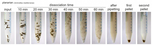
ACME solution is made from common lab reagents, including (unsurprisingly) acetic acid and methanol. We show that this method efficaciously dissociates planarians in ~1h. Cells are then resuspended in PBS with BSA for molecular biology. We have successfully used ACME dissociation with animals including zebrafish, fruit flies, spiders, annelids, snails and sea anemones. Sometimes the harder outer layers of animals or embryos must be removed, but this is usually a simple process, and normally can be adapted from standard protocols.
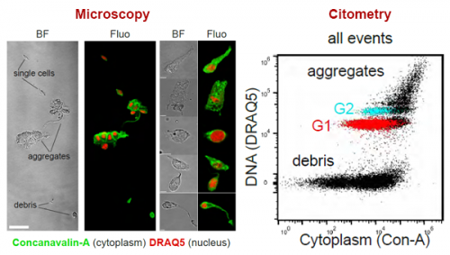
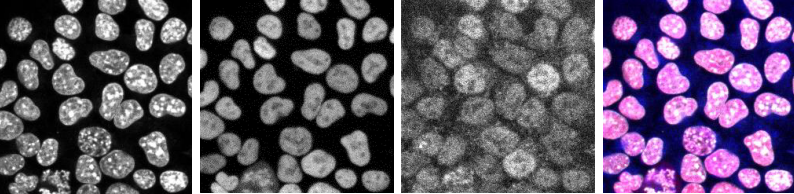
We demonstrate a simple staining process for flow cytometry and FACS. This is based on a nuclear stain (DRAQ5) and a cytoplasmic stain (Concanavalin-A). This allows us to distinguish ACME dissociated single cells from aggregates and debris.
Test-driving ACME
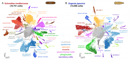
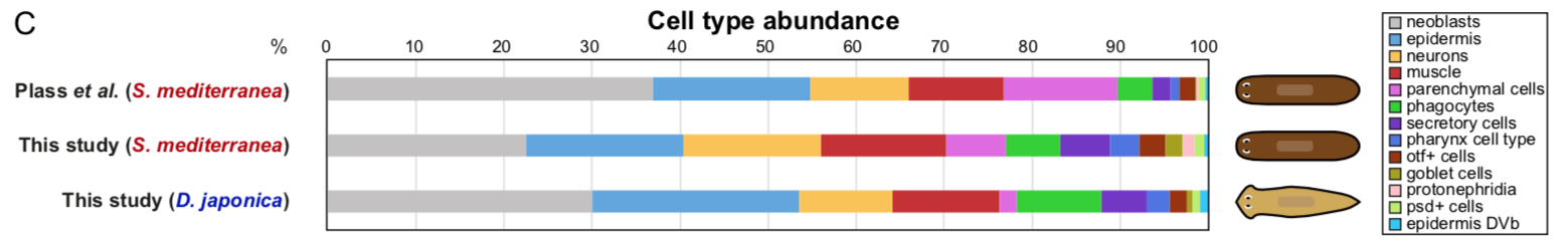
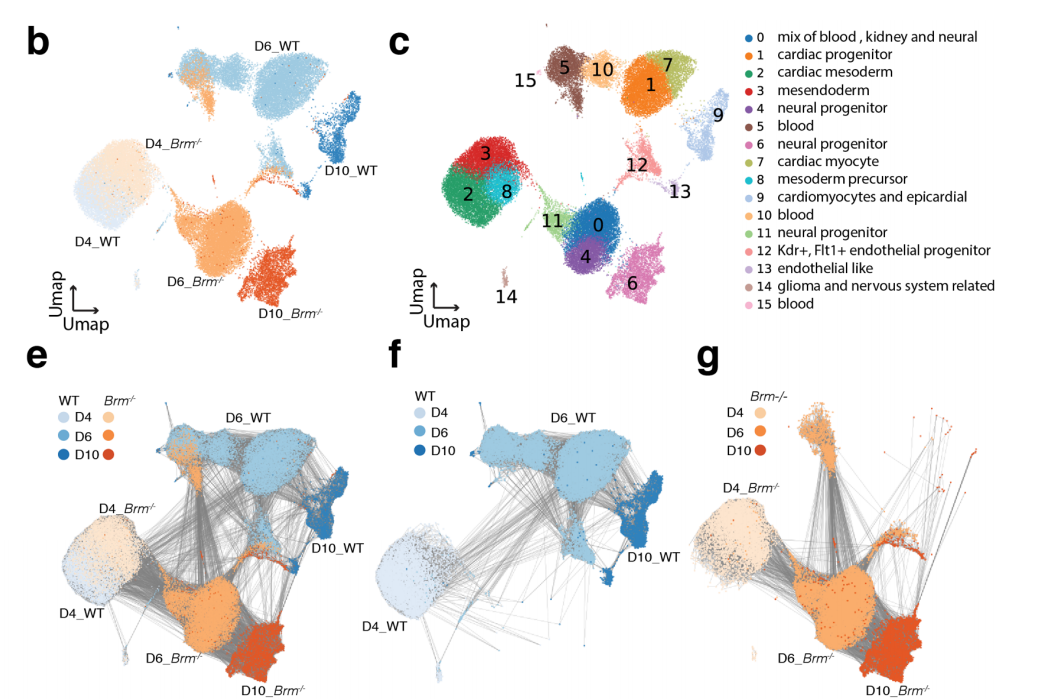
To test that ACME dissociated cells can be used for combinatorial barcoding scRNAseq we performed SPLiT-seq in a species mixing experiment, including two cryopreservation steps. We managed to sequence 34K cells from the two species, with excellent species separation. Our models were the planarian Schmidtea mediterranea (which has been subject to scRNAseq before, allowing us to benchmark our results) and a second planarian species, Dugesia japonica (which hasn’t been sequenced, allowing us to prove that ACME copes with novel species just as well!).
The cellular identity of clusters of Schmidtea mediterranea cells (gif)
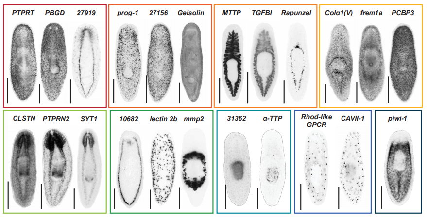
In the planarian Schmidtea mediterranea we find markers of all previously described cell types, showing that ACME dissociation does not artificially deplete any known kind of cell. As a bonus, we find some new clusters, such as the nanos+ germ cell progenitors, which were known to exist, but had not been noted as distinct clusters in planarian scRNA seq data before. The early fixation provided by ACME potentially allowed these cells to be fixed quickly, while it is possible they lost their distinct expression profile in previous experiments.
We also describe for the first time the cell type atlas of a second planarian species, Dugesia japonica. This shows many initial similarities with Schmidtea mediterranea – the most abundant cell type groups in both species are epidermal, neural and muscular cells, present at comparable proportions. The depth of sampling, however, will allow us to compare even rarer cell type abundance and gene expression, allowing species vs species comparison at excellent resolution, even off a single cell sequencing run.
Future possibilities, and please try it out!
We plan to use ACME to explore cell type evolution in a variety of non-model organisms, However, it could also be of broad use in more traditional model organisms, particularly when fixing multiple samples for simultaneous analysis. While we use SPLiTseq, there is no conceptual reason why it would not also work for droplet-based techniques, and could be adapted to solve many problems in current single cell sequencing protocols. Further optimisation will of course continue to take place, and we will be happy to offer advice to anyone willing to try it in their own odd organism.
In short, ACME is a versatile and powerful cell dissociation method for single-cell transcriptomics, providing early fixation and easy storage of material. This will greatly enhance investigations of cell type diversity and dynamics in multiple different organisms – particularly in challenging conditions.
This work was lead by Helena García-Castro and Jordi Solana, with Nathan Kenny, Patricia Álvarez-Campos and Vincent Mason from the Solana Lab, Oxford Brookes, and collaborators from across the UK and Europe (Anna Schonauer, Vicky Sleight, Jakke Neiro, Aziz Aboobaker, Jon Permanyer, Marta Iglesias, Manuel Irimia and Arnau Sebé-Pedrós). Our thanks to the Node for letting us share it with you here.
This is the first paper from Jordi’s new lab at Oxford Brookes – more coming soon. And we will be soon recruiting a bioinformatician. Please contact Jordi Solana if this sounds like you!
References:
Baguñà, J. and Romero, R., 1981. Quantitative analysis of cell types during growth, degrowth and regeneration in the planarians Dugesia mediterranea and Dugesia tigrina. Hydrobiologia, 84(1), pp.181-194. https://link.springer.com/article/10.1007/BF00026179
García-Castro, H., Kenny, N.J., Álvarez-Campos, P., Mason, V., Schönauer, A., Sleight, V.A., Neiro, J., Aboobaker, A., Permanyer, J., Iglesias, M. and Irimia, M., 2020. ACME dissociation: a versatile cell fixation-dissociation method for single-cell transcriptomics. bioRxiv. https://www.biorxiv.org/content/10.1101/2020.05.26.117234v
Plass, M., Solana, J., Wolf, F.A., Ayoub, S., Misios, A., Glažar, P., Obermayer, B., Theis, F.J., Kocks, C. and Rajewsky, N., 2018. Cell type atlas and lineage tree of a whole complex animal by single-cell transcriptomics. Science, 360(6391). https://science.sciencemag.org/content/360/6391/eaaq1723
Rosenberg, A.B., Roco, C.M., Muscat, R.A., Kuchina, A., Sample, P., Yao, Z., Graybuck, L.T., Peeler, D.J., Mukherjee, S., Chen, W. and Pun, S.H., 2018. Single-cell profiling of the developing mouse brain and spinal cord with split-pool barcoding. Science, 360(6385), pp.176-182. https://science.sciencemag.org/content/360/6385/176
Phox2a defines a developmental origin of the anterolateral system in mice and humans
R. Brian Roome, Farin B. Bourojeni, Bishakha Mona, Shima Rastegar-Pouyani, Raphael Blain, Annie Dumouchel, Charleen Salesse, W. Scott Thompson, Megan Brookbank, Yorick Gitton, Lino Tessarollo, Martyn Goulding, Jane E. Johnson, Marie Kmita, Alain Chédotal, Artur Kania
AP-2γ is Required for Maintenance of Pluripotent Mammary Stem Cells
Vivian W. Gu, Edward Cho, Dakota T. Thompson, Victoria C. Cassady, Nicholas Borcherding, Kelsey E. Koch, Vincent T. Wu, Allison W. Lorenzen, Mikhail V. Kulak, Trevor Williams, Weizhou Zhang, Ronald J. Weigel
Induction of Muscle Regenerative Multipotent Stem Cells from Human Adipocytes by PDGF-AB and 5-Azacytidine
Avani Yeola, Shruthi Subramanian, Rema A. Oliver, Christine A. Lucas, Julie A. I. Thoms, Feng Yan, Jake Olivier, Diego Chacon, Melinda L. Tursky, Tzongtyng Hung, Carl Power, Philip Hardy, David D. Ma, Joshua McCarroll, Maria Kavallaris, Luke B. Hesson, Dominik Beck, David J. Curtis, Jason W.H. Wong, Edna C. Hardeman, William R. Walsh, Ralph Mobbs, Vashe Chandrakanthan, John E. Pimanda
Salamander-like tail regeneration in the West African lungfish
Kellen Matos Verissimo, Louise Neiva Perez, Aline Cutrim Dragalzew, Gayani Senevirathne, Sylvain Darnet, Wainna Renata Barroso Mendes, Ciro Ariel dos Santos Neves, Erika Monteiro dos Santos, Cassia Nazare de Sousa Moraes, Ahmed Elewa, Neil Shubin, Nadia Belinda Froebisch, Josane de Freitas Sousa, Igor Schneider
Patient-specific functional genomics and disease modeling suggest a role for LRP2 in hypoplastic left heart syndrome
Jeanne L. Theis, Georg Vogler, Maria A. Missinato, Xing Li, Almudena Martinez-Fernandez, Tanja Nielsen, Stanley M. Walls, Anais Kervadec, Xin-Xin I Zeng, James N. Kezos, Katja Birker, Jared M. Evans, Megan M. O’Byrne, Zachary C. Fogarty, André Terzic, Paul Grossfeld, Karen Ocorr, Timothy J. Nelson, Timothy M. Olson, Alexandre R. Colas, Rolf Bodmer
Modulation of root growth by nutrient-defined fine-tuning of polar auxin transport
Krisztina Otvos, Marco Marconi, Andrea Vega, Jose O’Brien, Alexander Johnson, Rashed Abualia, Livio Antonielli, Juan Carlos Montesinos, Yuzhou Zhang, Shu-Tang Tan, Candela Cuesta, Christina Artner, Eleonore Bouguyon, Alain Gojon, Jiri Friml, Rodrigo A Gutiérrez, Krzysztof Wabnik, Eva Benková
Xyloglucan remodelling defines differential tissue expansion in plants
Silvia Melina Velasquez, Xiaoyuan Guo, Marçal Gallemi, Bibek Aryal, Peter Venhuizen, Elke Barbez, Kai Dünser, Martin Darino, Aleč Pěnčik, Ondřej Novák, Maria Kalyna, Grégory Mouille, Eva Benkova, Rishikesh Bhalerao, Jozef Mravec, Jürgen Kleine-Vehn
The gene cortex controls scale colour identity in Heliconius
Luca Livraghi, Joseph J. Hanly, Ling Sheng Loh, Anna Ren, Ian A. Warren, Carolina Concha, Charlotte Wright, Jonah M. Walker, Jessica Foley, Henry Arenas-Castro, Lucas Rene Brenes, Arnaud Martin, W. Owen McMillan, Chris D. Jiggins
PP2A:B56 Regulates Meiotic Chromosome Segregation in C. elegans Oocytes
Laura Bel Borja, Flavie Soubigou, Samuel J.P. Taylor, Conchita Fraguas Bringas, Jacqueline Budrewicz, Pablo Lara-Gonzalez, Christopher G. Sorensen-Turpin, Joshua N. Bembenek, Dhanya K. Cheerambathur, Federico Pelisch
20 years of African Neuroscience: Waking a sleeping giant
MB Maina, U Ahmad, HA Ibrahim, SK Hamidu, FE Nasr, AT Salihu, AI Abushouk, M Abdurrazak, MA Awadelkareem, A Amin, A Imam, ID Akinrinade, AH Yakubu, IA Azeez, GM Yunusa, AA Adamu, HB Ibrahim, AM Bukar, AU Yaro, LL Prieto-Godino, T Baden
The Center of Regenerative Medicine at Washington University in St. Louis invites applications for the Rita Levi-Montalcini Postdoctoral Fellows in Regenerative Medicine program. These fellowships honor Rita Levi-Montalcini, whose Nobel-winning discovery of Nerve Growth Factor was among the first regenerative biology research to be conducted at WUSTL.
The Center of Regenerative Medicine (CRM) seeks individuals of outstanding talent with a doctoral degree to provide them with the opportunity to pursue research within a CRM lab. As Rita Levi-Montalcini was herself an international scholar working at WUSTL, we strongly encourage international applicants to apply. Additional information about the Center of Regenerative Medicine can be found at: https://regenerativemedicine.wustl.edu/. Full program details can be found at: https://regenerativemedicine.wustl.edu/about/rlm_fellowship/rlm_programdetails/. We anticipate awarding one fellowship in 2020.
Qualifications
RLM Fellowships are intended for exceptional scientists of great promise who have recently been awarded, or who are about to be awarded, the doctoral degree. Fellows are required to work in the lab of a CRM faculty member on a project that directly focuses on regenerative medicine. Current employees, fellows, and students of Washington University in St. Louis are not eligible. Applicants currently on H-1B visas are not eligible.
Terms of Appointment
RLM Fellowships will be granted for a period of two years.
A Ph.D./D.Sc./M.D. must be awarded and proof furnished to the CRM before the start of the Fellowship.
The RLM Fellowship provides annual compensation of $55,000, as well as fringes and health insurance, research funds, and relocation and travel funds.
Review of applications will begin on August 30, 2020.
Please provide a full current CV, two letters of reference, a brief description of scientific accomplishments and long-term goals, and indicate a potential CRM faculty host (https://regenerativemedicine.wustl.edu/people-page/).
Washington University is an Equal Opportunity Employer. All qualified applicants will receive consideration for employment without regard to race, origin, religion, age, sex, sexual orientation, gender identity or expression, national origin, genetic information, disability, or protected veteran status.
 (1 votes)
 (No Ratings Yet)
 FocalPlane
 In this episode, supported by the Medical Research Council, we discover how researchers are letting the light shine in, literally, by bringing discoveries about the underlying genetic faults that cause eye diseases all the way through to game-changing clinical trials of gene therapy designed to save sight.