An exciting opportunity has arisen to join the Childhood Leukaemia Research Group led by Professor Anindita Roy at the University of Oxford. The group’s research explores how childhood leukaemia develops before birth, with a focus on developmental haematopoiesis and treatment resistant infant leukaemia. This post has funding for a total of five years from Wellcome Trust.
In the latest episode we’re getting our hands dirty by delving into the poop-ome – the trillions of bacteria that live inside our guts and make up what’s known as the microbiome. Rather than simply being a bunch of bugs, the microbiome is now believed to play a role in virtually every aspect of health and disease. But what are they up to? How do we even know what species are in there? And can you blame your stinky farts on your gut bacteria?
If you enjoy the show, please do rate and review and spread the word. And you can always send feedback and suggestions for future episodes and guests to podcast@geneticsunzipped.com
Joining an experienced and successful team, including Editor-in-Chief Michael Way, this is an exciting opportunity for a talented scientific editor to make a significant contribution to an important journal in the field of cell biology. Journal of Cell Science publishes outstanding primary research articles, reviews and topical comment, and continues to expand its services to authors and readers.
Applicants should have a PhD, and ideally postdoctoral experience, in cell biology or a relevant scientific field, and a broad knowledge of cell biology. The successful candidate will have strong communication, networking and interpersonal skills. We are also looking for enthusiasm, motivation, commitment, and a broad interest in science and the scientific community. Editorial experience is preferable but not essential.
Core responsibilities include:
Commissioning, handling peer review and developmental editing of review-type content
Maintaining and developing the ‘Cell Scientists to Watch’ interviews section of the journal
Writing content for the Research Highlights section of the journal
Representation of the journal at local and international conferences and within the wider scientific community
Creative involvement in the journal’s development and marketing activities
Additional responsibilities may be available for the right candidate. The Features and Reviews Editor will work alongside both an experienced Executive Editor and Senior Editor. This is a permanent, full-time position, and is based in The Company of Biologists’ attractive modern offices on the outskirts of Cambridge, UK.
The Company of Biologists exists to support biologists and inspire advances in biology. At the heart of what we do are our five specialist journals – Development, Journal of Cell Science, Journal of Experimental Biology, Disease Models & Mechanisms and Biology Open. We take great pride in the quality of the work we publish. We believe that the profits from publishing the hard work of biologists should support scientific discovery and help develop future scientists. Our grants help support societies, meetings and individuals. Our workshops and meetings give the opportunity to network and collaborate.
Applicants should be eligible to work in the UK and are requested to send to recruitment@biologists.com: a CV; a 400-word summary of a recent ground-breaking development in cell biology; and a cover letter explaining their interest in the post. Initial application deadline is 6 November 2019, but we will consider applications on a rolling basis so encourage candidates to apply as soon as possible.
Journal of Cell Science, published by The Company of Biologists, is seeking enthusiastic and motivated applicants for the role of Features & Reviews Editor.
Joining an experienced and successful team, including Editor-in-Chief Michael Way, this is an exciting opportunity for a talented scientific editor to make a significant contribution to an important journal in the field of cell biology. Journal of Cell Science publishes outstanding primary research articles, reviews and topical comment, and continues to expand its services to authors and readers.
Applicants should have a PhD, and ideally postdoctoral experience, in cell biology or a relevant scientific field, and a broad knowledge of cell biology. The successful candidate will have strong communication, networking and interpersonal skills. We are also looking for enthusiasm, motivation, commitment, and a broad interest in science and the scientific community. Editorial experience is preferable but not essential.
Core responsibilities include:
• Commissioning, handling peer review and developmental editing of review-type content
• Maintaining and developing the ‘Cell Scientists to Watch’ interviews section of the journal
• Writing content for the Research Highlights section of the journal
• Representation of the journal at local and international conferences and within the wider
scientific community
• Creative involvement in the journal’s development and marketing activities
Additional responsibilities may be available for the right candidate. The Features and Reviews Editor will work alongside both an experienced Executive Editor and Senior Editor. This is a permanent, full-time position, and is based in The Company of Biologists’ attractive modern offices on the outskirts of Cambridge, UK.
The Company of Biologists exists to support biologists and inspire advances in biology. At the heart of what we do are our five specialist journals – Development, Journal of Cell Science, Journal of
Experimental Biology, Disease Models & Mechanisms and Biology Open. We take great pride in the quality of the work we publish. We believe that the profits from publishing the hard work of
biologists should support scientific discovery and help develop future scientists. Our grants help support societies, meetings and individuals. Our workshops and meetings give the opportunity to
network and collaborate.
Applicants should be eligible to work in the UK and are requested to send to recruitment@biologists.com: a CV; a 400-word summary of a recent ground-breaking development in cell biology; and
a cover letter explaining their interest in the post. Initial application deadline is 6 November 2019, but we will consider applications on a rolling basis so encourage candidates to apply as soon as
possible.
The Company of Biologists is looking to recruit an experienced Scientific Copy Editor to work across our portfolio of five life-science journals.
The role entails copyediting articles to a high standard, compiling author corrections, overseeing the journal production process, and liaising with authors, academic editors, external production suppliers and in-house staff to ensure that articles are published in a timely and professional manner.
Candidates should have a degree (ideally a PhD) in a relevant scientific area. Previous copyediting experience is essential. Additional requirements include excellent literacy skills, high attention to detail, a diplomatic communication style, good interpersonal and IT skills, a flexible approach and the ability to work to tight deadlines.
The position gives an experienced copy editor the opportunity to work on our highly successful life-science journals and offers an attractive salary and benefits. The position will be based in The Company of Biologists’ attractive modern offices on the outskirts of Cambridge, UK.
The Company of Biologists (biologists.com) exists to support biologists and inspire advances in biology. At the heart of what we do are our five specialist journals – Development, Journal of Cell
Science, Journal of Experimental Biology, Disease Models & Mechanisms and Biology Open – two of them fully open access. All are edited by expert researchers in the field, and all articles are
subjected to rigorous peer review. We take great pride in the experience of our editorial team and the quality of the work we publish. We believe that the profits from publishing the hard work of
biologists should support scientific discovery and help develop future scientists. Our grants help support societies, meetings and individuals. Our workshops and meetings give the opportunity to
network and collaborate.
Applicants should send a CV to recruitment@biologists.com, along with a covering letter that summarises their relevant experience, why they are enthusiastic about the role, and their current salary.
All applications must be received by 6 November 2019.
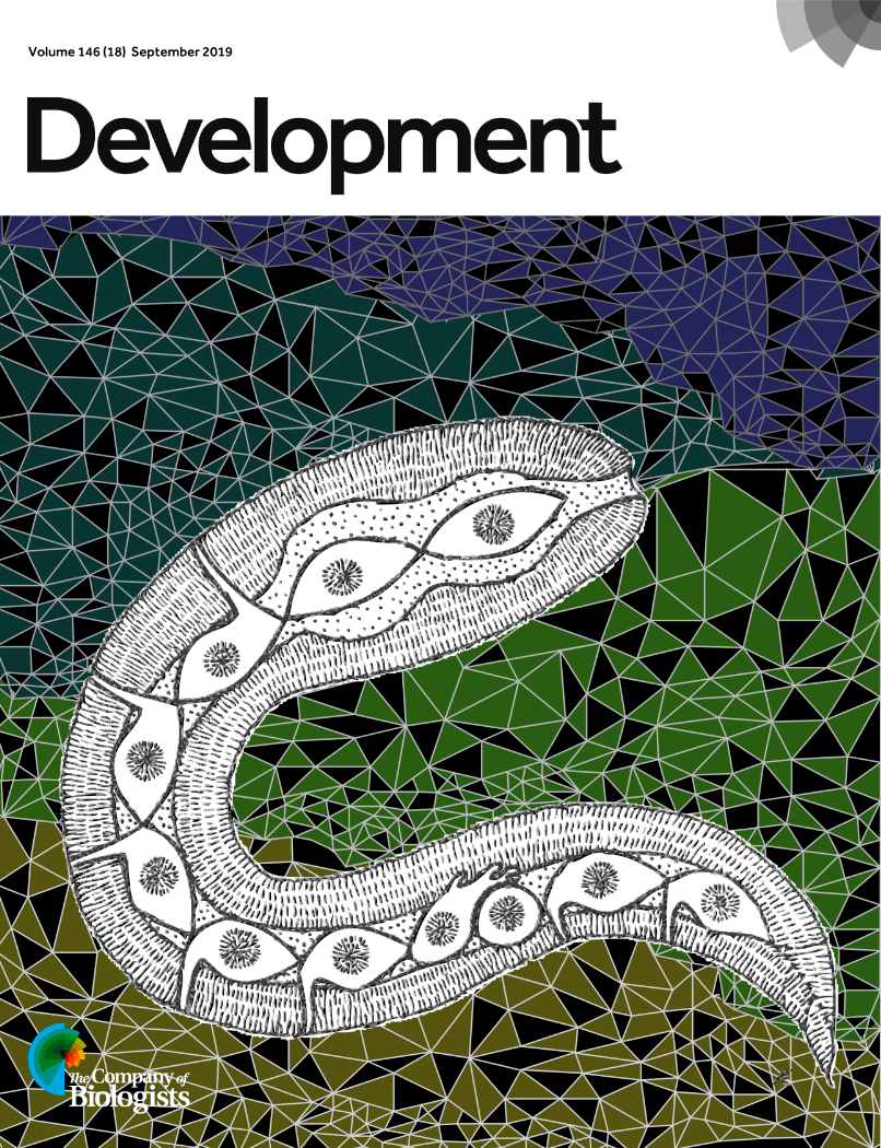
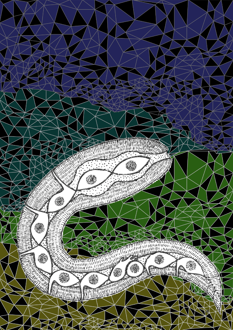
Development covers showcase the beauty of developmental biology. Embryos, tissues and cells are rendered in striking colour palettes and artistic arrangements. We mainly receive confocal image submissions but sometimes also EM and standard light microscopy. And sometimes, art – like our most recent cover, a schematic overview of C. elegans created by Annabel Ebbing, PhD student in Hendrik Korswagen’s labat the Hubrecht Institute in The Netherlands and first author of a new paper on neuroblast migration. We caught up with Annabel to find out how her beautiful cover came about and how she thinks about art and science.
Annabel’s cover
Can you tell us about your PhD project and your new Development paper?
The project started a while back (in 2013), when I was still a Master’s intern student at the Korswagen lab. My direct supervisor was Teije (the shared first author on this paper) who helped me with the imaging and epistasis experiments to elucidate the role of the Fat-like cadherins CDH-4 and CDH-3 in Q neuroblast migration. In the meanwhile, he did similar experiments for UNC-40, DPY-19, and MIG-21. Finally, when I started my PhD I set out to combine the two projects. This was during the emergence of CRISPR as a standardized tool for C. elegans, which really helped the project get to the state where it is now. We managed to study the effects of tissue specific protein depletions and endogenous protein localization, which together really resolved some of the outstanding questions we had concerning the interplay between proteins and a possible molecular mechanism. All in all, there are several proteins involved in Wnt independent Q neuroblast migration; apart from their own roles they have overlapping functions as well, making it a complex and highly regulated mechanism controlling protrusion formation and directionality.
Have you always been interested in art as well as science?
Both art and science have always been fascinating subjects to me. Actually, I wanted to pursue a more artistic education at a certain point, but I decided to study biology in the end to feed my general curiosity. Pursuing a career in science did not make me leave the artistic side altogether though. I especially love thinking about scientific projects in a more abstract way. For me the process of visualization helps in my understanding of a certain subject.
Where did you get the idea for submitting cover art rather than something more standard?
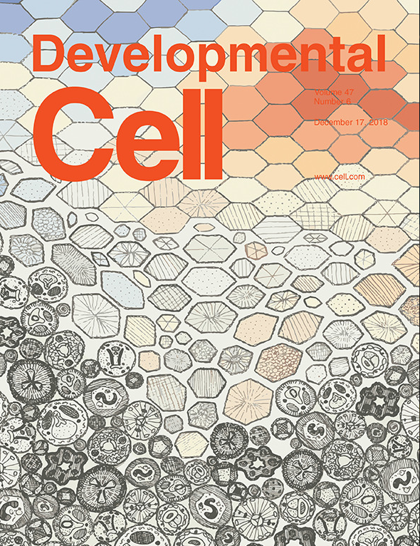
Well, last year we published a paper in Development Cell, for which I made the cover. So when we were asked to submit a cover proposal for Development, I decided to take a similar approach. I love drawing and painting and honestly thought the immunofluorescence images for this project were rather dull (sometimes you see these amazing structures of cells, organs, and even organisms and our imaging was just rather 2D). Let’s say, the choice to draw was quickly made.
Annabel’s Developmental Cell cover from last year
Can you tell us a little about the process of designing the piece?
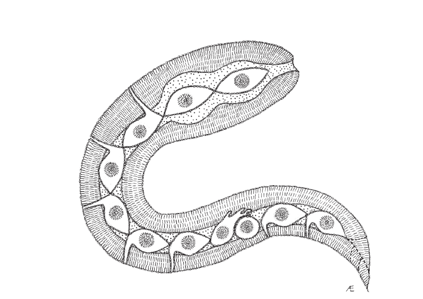
I started the piece by trying to make an abstract overview of a worm, including a migrating Q neuroblast, using simple lines and dots. Later on, I imported the file in illustrator and made the background. Most covers of Development (if not all) have a dark/black background, so I decided to use dark colors, which made a nice contrast with the black and white worm.
The abstract worm drawing at the heart of Annabel’s piece
How did Aboriginal art influence the work?
Aboriginal art is an artform of storytelling; using simple lines and dots the Aborigines manage to tell entire stories and legends. Since the mechanism underlying the process of Wnt independent Q neuroblast migration is rather complex, I thought there would be no better way to illustrate it than in such a narrative-rich manner.
Annabel’s final piece (minus the Development augmentations)
What’s next for you after this paper?
I am submitting my thesis as we speak! So I guess the main focus for the coming months is the defense. Art wise that means I am illustrating a cover for every chapter, which is a lot of fun! Moreover, together with colleagues we are preparing another manuscript at the moment, which will hopefully be accepted sometime this winter.
Some more worm art from Annabel
Some previous artistic Development covers
This picture pays homage to M. C. Escher’s tessellation studies and Alan Turing’s reaction-diffusion mechanism. Both Turing and Escher were interested in pattern generation and the changes of organic forms, two phenomena central to evolutionary developmental biology, as addressed here in the context of periodic patterning in the turtle shell. From Moustakas-Verho, et al. https://dev.biologists.org/content/141/15/3033?iss=15
Taking the iconic example of the Nautilus pompilius shell from Darcy Thompson’s On Growth and Form, the rules of logarithmic spiral growth were abstracted as the foundation to develop a computational model. By experimenting with its parameters, an array of new shapes was created, highlighting the important role that computational modelling has in advancing our understanding of complex physical form in the field of developmental biology. Image created by Jennifer Ma (Zandstra lab, University of Toronto, Canada) and Matthew Spremulli (Living Architecture Systems Group and University of Toronto, Canada). To find out more, visit http://thenode.biologists.com/behind-the-cover/interview/.
Recently, two iconic developmental biology models entered into the single cell genomics era: chick and zebrafish. In this image, line art was traced using real embryo images for reference and filled with individual dots to represent the reduction of the whole embryo to its smallest structural, functional and biological unit: the cell. This cover was chosen by Special Issue guest editors Allon Klein and Barbara Treutlein from entries to Development’s cover competition. By Martin Estermann (Monash University, Australia).
Applications open for the Graduate School of Life Science
The Graduate School Life Science Munich (LSM) offers an international doctoral programme to highly motivated and academically qualified next generation researchers at one of Europe’s top Universities. LSM members are internationally recognized for their innovative research approaches and technologies, they are aiming to answer essential questions relevant to basic and applied biological and biochemical research. Within their own research group or in collaboration with a specialized research group on campus, LSM doctorates are given the opportunity to learn and command a variety of techniques. Furthermore, the graduate programme holds various workshops and seminars that strengthen and prepare doctorates for a successful career as scientists.
With over 40 research groups from the Faculty of Biology and the Faculty of Chemistry and Pharmacy of Ludwig Maximilian University (LMU) München, the LSM is located in the Biocenter. Its prominent location within the HighTechCampus in Martinsried south of Munich, contributes to the enormous possibilities for support, interdisciplinarity and constant scientific input from the surrounding laboratories. Available research projects cover areas from Biochemistry, Bioinformatics, Cell and Developmental Biology, Epigenetics, Genetics, Human Biology and Bioimaging, Pharmacology, and Plant Biology.
All courses, lectures and seminars at the LSM are held in English. Thus, selected candidates have to be fluent in both written and spoken English.
Successful students of the graduate school will be awarded a doctoral degree (Dr. rer. nat.) by the LMU after 3 to 4 years. Candidates need to prove a strong qualitative background as well as interest and ability to conduct independent research.
The doctoral programme is open for students who hold either a master´s or diploma degree, as well as to exceptional candidates with a four years bachelor degree (with written thesis).
LSM calls for doctoral applications on a yearly basis, currently open from the 14th of October until the 29th of November 2019. Applicants are selected in a multi-step process through our online portal, thus ensuring openness and fairness throughout the application procedure. Every complete submission is evaluated by the LSM coordinator. Applications will be independently reviewed by several faculty members of the LSM Graduate School. Based on academic qualification, research experience, motivation, scientific background and the letters of recommendation, candidates will be selected to participate in the LSM Interview week. After thorough evaluation through the LSM committee board members, successful candidates will be invited to join the LSM Graduate School.
We are looking for a highly motivated and talented PhD candidate to investigate the role of enteric glial cells in the assembly and maintenance of the intrinsic neural circuits that regulate gastrointestinal function and homeostasis.
During the course of this PhD research project, which will take place at Maastricht University (The Netherlands), novel genetic and neurophysiological tools will be combined with cutting-edge live cell microscopy techniques to study the role of enteric glial cells in enteric neuron connectivity. You will make use of in vivo models to investigate how enteric glial cells adapt to safeguard gastrointestinal function and homeostasis. You will run a research project in an international multidisciplinary lab and collaborate with (inter)national research groups to complement the know-how within our research group with state-of-the-art external expertise. This PhD position is part of a NWO-funded Vidi project, entitled “Glia of the bowel, landscapers in the second brain” that was recently awarded to Dr. Werend Boesmans.
Additional information about the vacancy can be obtained from: dr. Werend Boesmans (w.boesmans[at]maastrichtuniversity.nl). Interested candidates should send a letter of motivation, a detailed CV and the contact details of two referents.
The Buckley lab at the department of Physiology, Development and
Neuroscience (PDN), University of Cambridge is recruiting a Research
Assistant or Research Associate to help further develop their
optogenetic technology. The lab uses optogenetic and live confocal
imaging approaches within the whole zebrafish neural tube and mammalian
embryo stem cell (mESC) culture to manipulate the polarity of single
cells. In combination with CRISPR-mediated functional knock down
experiments, we are directly testing the role of cell polarity in
building epithelial integrity during organ development and breaking in
it during normal developmental processes such as EMT and during abnormal
disease states.
The post is for 1 year in the first instance from early 2020 and funding
is available for potential contract extension.
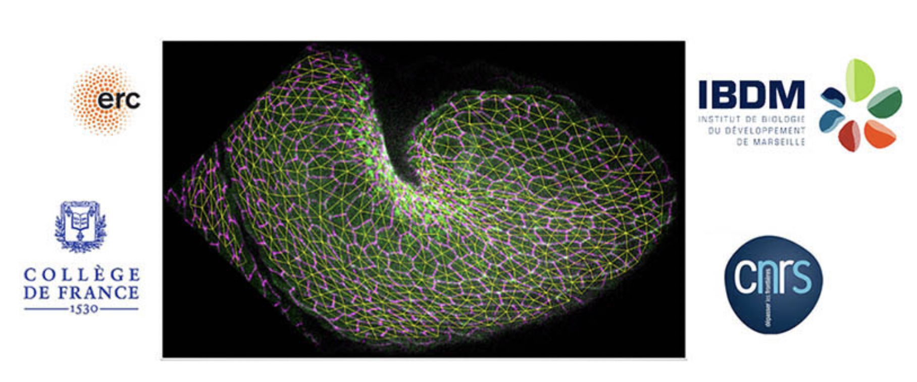
An Engineer position in Molecular Biology is available starting January 2020 in the group of Thomas Lecuit at the Developmental Biology Institute of Marseille (IBDM, CNRS UMR7288), Marseille, France. Funding is provided by an ERC grant. The appointment will be made for 1 year, with a possible extension to up to 4 years.
We are seeking a highly-motivated candidate with strong expertise in Molecular Biology. Experience in CRISPR/CAS9 editing and/or Drosophila genetics is valued. The working language in the laboratory is English, so the candidate should have a good practice in English.
The Engineer will perform standard molecular biology techniques and CRISPR/Cas9 gene editing in vivo. We are seeking a highly motivated and flexible individual to join our team.
Essential Skills
Advanced knowledge of molecular biology and associated practical techniques. Experimental dexterity and attention to detail; quality driven.
Excellent organizational and planning skills.
A high level of interpersonal and communications skills.
Ability to follow instructions accurately and efficiently, correctly interpret scientific data and work under supervision.
 (No Ratings Yet)


 (8 votes)