At the end of each month, I pick the same month from a random year from the past 15 years of the Node, and take a look at what people were talking about back then.
Since this is the first time I’m travelling via the Time Machine, I’ve picked the first ever February that the Node experienced, back in 2011. Let’s step into the machine, and turn the dial to February 2011…
If you scroll down to the comments section, you might find a few familiar names discussing the definitions of stem cells vs progenitors. 14 years on, what are people’s views on this?
Here, Sarah Gibb told the story of how she took the leap and applied for a job at the Glasgow Science Centre after her PhD. Since that blog post, we have featured many interesting career stories on the Node. Browse through the collection.
How amazing is this? Dresses that represent different human embryonic developmental stages. Head over to the website about the exhibit to find out more!
Emma Kemp from EuroStemCell was very active on the Node from 2011-2012. In 2020, EuroStemCell was superseded by EuroGCT, but people can still find useful resources around stem cell research on their website.
Did you know that The Company of Biologists’ journals – Development, Journal of Cell Science, Journal of Experimental Biology and Disease Models & Mechanisms – offer Travelling Fellowships to early-career researchers (graduate students and postdocs) so that they can make collaborative visits to other research laboratories across the world? The first cohort of Travelling Fellowships was launched for Development in 1990, which was later extended to Journal of Cell Science (JCS) in 1992 and Journal of Experimental Biology (JEB) in 1996. Disease Models & Mechanisms (DMM), being a relatively younger journal (you can read about DMM’s origin in this Editorial), offered Travelling Fellowships to ECRs since it was launched in 2008.
As we celebrate the Company’s 100th birthday in 2025 (you can find out more about us in this recent Editorial), we would love to hear stories from you – our community – about how the Company has helped you in your scientific journey. If you ever received a Travelling Fellowship that enabled you to travel to other research labs, particularly in the period from 1990 to early 2000s, do get in touch and let us know about your experience and how this has impacted your career.
You can tell us your story by sending your own digital ‘message in a bottle’ in honour of the founder of the Company, George Parker Bidder III.
You can also share your story by replying to this post or to any of our social media posts with #biologists100 hashtag on Bluesky and X.
The Company of Biologists is looking for one or more interns, through the BBSRC DTP/PIPS or equivalent schemes, to work on our community sites – the Node, preLights and FocalPlane. This is a great opportunity to gain experience in the rapidly growing online science communication environment, to develop writing skills, and to learn about academic publishing.
Together, the Company’s three community sites provide platforms for the research community to share news, discuss issues relevant to the field, and read about the latest research and events.
The Node is a field-specific site for developmental biologists, FocalPlane aims to bring together the microscopy and biology communities, and preLights is focussed on highlighting the preprint literature and exploring the journey from preprint to publication. The intern will be involved in the day-to-day running of one or more of the sites and will be mentored by the relevant Community Manager(s). The internship will be based in our office in Cambridge, though hybrid working opportunities may be available.
Core responsibilities of the position include:
Creating and commissioning content, including writing posts and soliciting content from the academic community, societies and other organisations
Providing user support
Helping to run the community site social media accounts.
The successful intern will have:
Relevant scientific expertise (ideally in the developmental or cell biology field, though we are open to applications from individuals working in other areas of biology)
Strong writing and communication skills
Keen interest in science communication, ideally including experience in blogging and/or social media. Familiarity with WordPress would be a bonus.
We are looking for an intern to start in early summer 2025 and encourage interested candidates to submit their application as soon as possible.
To apply, please send a CV and cover letter, stating why you are interested in this opportunity, to recruitment@biologists.com and Katherine Brown (Development’s Executive Editor) at katherine.brown@biologists.com. Please also direct informal enquiries to the same addresses.
Join us at the EMBO Workshop “EvoDevoTempo – Developmental Timing Across Species: From Mechanisms to Evolutionary Insights”.
Topics will include theoretical modelling, metabolism, and evolutionary biology, thus bringing a uniquely interdisciplinary perspective to the meeting.
Sessions:
Developmental timing in vivo
in vivo & in vitro models for timing
Metabolic rates and timing
in vitro & in silico models for timing
Cell dynamics and timing
Timing in homeostasis
New molecular mechanisms of timing
Where: Paris Brain Institute, France
When: May 6-9 2025
Abstract submission and registration deadline: 1 April 2025
Brief description of the project: The marine environment is under increasing thermal stress due to climate change. This is a major issue for development in poikilothermic animals that have thermal limits for correct development at the egg stage. Degradation in eggs quality has been a recent major issue in additionally for agriculture and human health. Prof Sato’s research group has been investigating the molecular basis of developmental robustness under thermal stress (https://www-p.sci.ocha.ac.jp/bio-atsuko-en/) and how environmental stress could have altered developmental pathways in the evolutionary process with a special focus on maternal factors. The goal of this project is to predict development and body patterning from initial maternal input by mathematical modelling. We also aim to identify heritable phenotypes by maternal environment, but not by the genome, using surrogate fish technology.
The student will be engaged in computer programing to create a mathematical model and molecular work involving collecting tissue and RNA samples from fish and/or tunicates. Molecular work will involve collecting tissue and RNA samples and the student will have opportunity to learn RNA extraction, PCR, immunohistochemistry, HR and imaging analysis, under the guidance of Dr. Sato. In the computational work, the student will learn programming and simulation, but this requires experience and basic skills in programming using python and R.
Applicant eligibility: A female student who has Master’s degree or going to obtain Master’s degree in a relevant field of study, either in mathematical or computer science or biological sciences with a strong computational or bioinformatics component by the starting date of PhD. We are looking for motivated student interested in animal biology, developmental biology, programming with problem solving abilities as well as resilience to solve daily problems. Research experience in molecular biology and basic programming would be advantageous. This position is open to applications from all nationalities.
Application deadline: 1 June 2025 midnight JST, open until filled.
The studentship will start 01 October 2025 the earliest. Full time only.
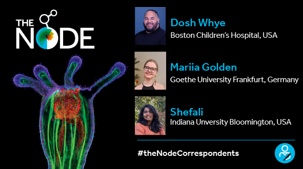
Let’s welcome the three new Node correspondents – Dosh Whye, Mariia Golden and Shefali! We look forward to working with them to bring you a wide range of content and perspectives for the Node. Stay tuned for their posts over the coming year.
Dosh Whye serves as the assistant director of the Human Neuron Core, where he leads the cell development and differentiation efforts to build complex 3D neural organoid models using induced pluripotent stem cells (iPSCs) from pediatric patients with rare genetic neurodevelopmental disorders. Dosh has close to 20 years in the stem cell field, and he continues to be fascinated by the power of pluripotent cells and the technological advancements of stem cell applications in the fields of science & medicine. He plans to serve as a correspondent for several international stem cell research conferences, and he’s also inspired to write a Q&A blog series profiling many brilliant scientists in the stem cell field.
Mariia Golden is a third-year PhD student moving from Goethe University Frankfurt to Marburg University, Germany. She investigates dynamical morphogenetic events in insect development. She is passionate about live imaging and working with non-model organisms. As a Node correspondent, Mariia wants to promote diversity among the organisms we choose for our research questions through a series of interviews with established scientists who are enthusiastic about the topic. She also would like to highlight the topic of motherhood and scientific career, because she truly believes that the status quo should change in order to put a stop to the “brain drain” through female scientists.
Shefali is a fifth-year PhD student in the Tennessen Lab at Indiana University, Bloomington. She is broadly interested in inter-organ metabolic signaling, and her current research focuses on how glucose metabolism coordinates the brain-body growth signaling axis during Drosophila melanogaster brain development. Shefali also serves as the elected graduate student representative on the FlyBoard, where she has been working towards creating more mentoring opportunities for early career scientists. Beyond work, she enjoys learning various Latin-style dances and singing classic Hindi songs (which is her mother tongue). As a Node correspondent, she is eager to combine her scientific interests with her commitment towards building a strong and empowered scientific community. She looks forward to writing not only about the emerging field of inter-organ metabolic signaling, but also about unconventional scientific journeys, community resources, importance of mentoring, and bring in her perspective as a trainee to support incoming graduate students.
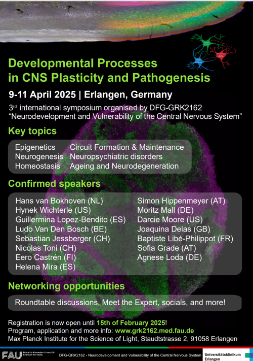
The Research Training Group: Neurodevelopment and Vulnerability of the Central Nervous System (GRK2162) is excited to host its 3rd International Symposium: Developmental Processes in CNS Plasticity and Pathogenesis. The program will bring together junior scientists with experts in neurodevelopmental biology, stem cell biology and translational neurobiologists. Next to a great set of lectures, the program offers ample opportunities for scientific networking and presentation of your scientific work including round tables / meet the expert sessions, interactive poster sessions, flash talks and short talk opportunities.
Researchers in developmental neuroscience, neurobiology, and related fields—including PhD students, postdocs, and principal investigators—are encouraged to participate!
Date: April 9-11, 2025 Location: Max-Planck-Institute for the Science of Light, Erlangen, Germany
You can register here until 15th February 2025 or until all spots are taken. There is no registration fee but the number of participants is limited by the space we are using. EXTENDED DEADLINE: 28th February 2025
Join us for stimulating discussions and networking opportunities in the vibrant research environment of FAU Erlangen-Nürnberg!
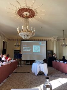
By Claudio Hernández-López and Aditya Singh Rajput
This is one of three reports about the “Physics of the Early Embryonic Divisions” Workshop, organised by The Company of Biologists. Read the other two reports for different perspectives on the science that was discussed:
After a taxi ride from Heathrow to Buxted Park, the first day of the conference started on a high note as everyone was treated to a hearty lunch with a view of the sprawling gardens. Later, having found our way through the many corridors of the hotel, we had a few words from the organizers of the conference, Lendert Gelens and Julia Kamenz, and also from Laura Hankins on behalf of The Company of Biologists. All the speakers introduced themselves and pitched their research interests and out-of-the-lab hobbies. And so, the stage was set for the first session of the conference: actin and its biochemical regulation
Actin I: Swirling biochemical waves in natural and artificial cortices
Andrew Goryachev, professor at the University of Edinburgh, introduced everyone to Rho-Actin (RhoA) waves in the cortex of Starfish oocytes. A critical part of the cell cycle is cytokinesis, i.e. the ability of the cell to constrict its membrane and form two separate cells. These waves are a result of cortical excitability before this process. Andrew showed us their mesmerizing spatio-temporal evolution in fluorescence microscopy movies, displaying correlated activity in regions with sizes of about 10 microns [1]. Biochemical reaction-diffusion systems are able to produce spatio-temporal patterns and spread waves. However, depending on the molecular interactions between the chemical species, the properties of these waves can change. Two competing models have been proposed in the literature to explain these waves, differing in the presence or absence of an explicit chemical species that inhibits RhoA. Current experimental data measures either the total RhoA concentration or active RhoA concentration in space and time, and these two models cannot be distinguished from the observed dynamics of one concentration alone. Andrew presented new experimental data from simultaneous space-time recordings of total and active RhoA. These measurements indicate that changes in active RhoA precede changes in total RhoA, in agreement with a model including an explicit RhoA inhibitor.
A major push in the field is also to study and recapitulate the intrinsic propensity of the actomyosin cortex to form patterns. In this direction, the lab of Jennifer Landino, an assistant professor at Dartmouth College, has been working on developing reconstituted cortices to study the emergence of spatio-temporal patterns by an interplay of F-actin and Rho. These Supported Lipid Bilayers (SLBs) [2] display waves and localized oscillations that have previously been observed in frog and starfish oocytes. This provides strong evidence of the self-organized nature of these patterns, with the mutual feedback between F-actin and Rho rendering the cortex excitable. The observed dynamics in SLBs present crucial differences compared to living cells, in particular, lacking periodic traveling waves. Changes in material properties after Xenopus extract addition allowed more than a single wavefront to propagate, thus bringing attention to the importance of the composition and design of these reconstituted systems.
Actin II: Geometry, flows, and deformations in early development
The second part of the session on actin started with Claudio Hernández-López, who recently finished his PhD at ENS Paris and is currently a postdoc in AMOLF Amsterdam, discussing the early development of Drosophila Melanogaster. The pre-gastrulation embryo, i.e. from cell cycles 1-14, is a syncytium: all the nuclei share a common cytoplasm. Before gastrulation, the nuclei migrate towards the cortex, forming their cellular membranes during cell cycle 14. The synchrony of the cell cycle along the 500-micron embryo depends on the nuclear density in space, hence, it is important that the nuclei achieve a uniform distribution in the embryo before cellularization. Previous experiments have shown that the expansion of the nuclear cloud along the anterior-posterior axis is mediated by cytoplasmic flows driven by cortical actomyosin contractions [3]. Claudio presented a new modeling framework comprising two fluids: an active gel (actomyosin), and a passive cytosol [4]. A mechanochemical coupling between the position of the nuclei and localized activation of myosin-II at the cortex reproduced previous experimental measurements on the flows and nuclear positioning. Remarkably, this self-organized positioning is robust, meaning that no matter the location of the initial nucleus after fertilization, the final nuclear distribution will remain uniform.
Aditya Singh Rajput, a PhD student at ICTS-TIFR, Bengaluru, continued the session by discussing asymmetric ingression in embryos. This phenomenon seems to be highly conserved amongst multiple phyla, with comb jellies being one of the major examples. The discussion began by looking at ingression in the nematode C. elegans and highlighting the emergence of myosin inhomogeneities that lead to this asymmetry. He then discussed his work on understanding this from the lens of active matter physics and treating the actomyosin cortex as an active fluid surface. Due to an emergence of differing timescales of ingression and cortical flow, the cytokinesis can be symmetric or asymmetric depending on the cortical contractility. The predictions from this theory also seem to hold true when compared to the dynamics of the first cleavage in the C. elegans embryo.
Hervé Turlier, a research group leader at the Collège de France, discussed his group’s work to build physical and computational models for the early development of multicellular organisms. The group has been developing general computational tools to simulate [5], as well as reconstruct [6] cell and tissue surfaces, which could help understand, for example, cleavage patterns in early embryos. One of the foremost open problems in the field of developmental mechanobiology is the experimental measurement of forces in tissues as they undergo growth and morphogenesis. To this end, Dr. Turlier’s group has been working on analysing fully three-dimensional networks of cellular contacts to infer cellular stresses. This method, called foambryo, relies on mesh reconstruction to accurately capture the cellular geometry and then infer the relative tensions from the junctional lengths. These physical and computational tools not only provide a unique window into the mechanical behavior of early embryos but are also demonstrated to be generalizable and scalable to different species and various stages of development.
Information processing across scales
Switching gears, the session on information was kicked off by Rob Phillips with a discussion on language, words, and the information therein. By comparing the information content in examples from daily life- works of literature, encyclopedias, and entire libraries – with the information content of genomes, Rob brought to light the vast unknown regions of genome sequences that remain understudied till now. With his lab, he has been developing novel tools [7] to understand the transcriptional crosstalk between genes in a high-throughput manner to develop a genomic Rosetta stone that helps to bridge our understanding of the genotype-phenotype map. He talked about how this work attempts to infer the underlying gene regulatory networks by studying thermodynamic interactions and binding affinities and combining this with tools from information theory. In addition to the problem of deciphering the genotype-phenotype map itself, Rob highlighted how the many-to-one nature of this map comes with added complexities of different genes having varying degrees of impact on their associated phenotypes, which is also another aspect this toolbox can help bring to light in a more quantitative manner.
Sophie de Buyl, professor at the Vrije Universiteit Brussel, closed this session by talking about the development of the Ascidian embryo. These filter feeders present two characteristics that make them particularly attractive for experimental studies in developmental biology. First, their cleavage pattern is invariant. Second, there is no cell migration, death, or embryo growth during their development. Previous theoretical work performed in her lab focused on the differentiation of neural tissue in the very early stages of development of this species. This process depends on the activation of ERK, which is regulated by the concentration of external signaling cues. In particular, the ERK activator is localized at the basal side of the outermost cells, and the ERK repressor is localized at the cell-cell junctions. Hence, the geometry of the cells is a relevant variable to study in relation to cell differentiation [8]. One overarching question in developmental biology is how living systems integrate signals from their environment to decide cell fate robustly, and the tools of information theory allow us to cast this problem in a tractable mathematical form. As a key result of this new study, Sophie showed that maximizing the information transmission from the external input to cell expression yields a predicted cell geometry that closely matches experimentally measured values. Furthermore, this maximal information transmission supports reliable differentiation between four different possible cell fates [9].
Finishing thoughts
The early embryonic cell divisions arise from an interplay between biochemical and mechanical cues, spanning multiple temporal and spatial scales. Given that regulatory pathways are generally less active at that stage, we feel that a comprehensive dialogue between theory and experiments on early development can be fertile ground for a broader understanding of not only cell division but also the emergence of complex behaviors in metazoan cells. Going beyond, perhaps a more biophysical understanding of the embryonic divisions can tell us something about the evolution of the diverse regulatory networks that we observe in extant organisms, potentially learning more about cell division in the first multicellular organisms.
Landino, Jennifer, Marcin Leda, Ani Michaud, Zachary T. Swider, Mariah Prom, Christine M. Field, William M. Bement, Anthony G. Vecchiarelli, Andrew B. Goryachev, and Ann L. Miller. “Rho and F-actin self-organize within an artificial cell cortex.” Current Biology 31, no. 24 (2021): 5613-5621.
Text written by Olga Afonso, Helena Cantwell, and Shuzo Kato
This is one of three reports about the “Physics of the Early Embryonic Divisions” Workshop, organised by The Company of Biologists. Read the other two reports for different perspectives on the science that was discussed:
Microtubules: bridging meiosis to mitosis and cellular differentiation
In a workshop dedicated to early embryonic divisions, we could not miss a session focused on microtubules. The session started with a talk from Helena Cantwell (University of California, Berkeley) who combined two model systems, Ciona robusta and Xenopus laevis to study the molecular mechanisms that mediate the meiotic to mitotic spindle transition. Helena found that Casein Kinase II is as a key regulator of spindle morphology that acts through the Ran-GTP pathway of spindle assembly [1]. Further work on how the Ran-GTP gradient itself changes during the meiotic to mitotic spindle transition would ultimately close the loop. Simone Reber, from the Max-Planck Institute for Infection Biology, showed recent work from the lab, focused on the changes of spindle morphology during cell differentiation. They used optical diffraction tomography to show that differentiated cells have a more dilute cytoplasm resulting in a shift of microtubule mass from the mitotic spindle bulk to centrosomes and astral microtubules [2]. Remaining questions relate to how the cytoplasm is diluted during differentiation and what the biological function of increased astral microtubules is. The session concluded with a presentation from Nicolas Minc, Institute Jaques Monod, Paris. Nicolas Minc uses sea urchin embryos to study how the position of the metaphase spindle is maintained in a large cell, where microtubules are far away from the cell’s boundary. By using a combination of optical tweezers and computational analysis of cytoplasmic flows, Nicolas showed that spindle positioning is maintained by the viscoelastic properties of the cytoplasm [3,4,5]. Overall, this session covered the role of microtubules in every step of embryonic development: from the very first meiosis to mitosis transition to what happens to the spindle morphology when cells start to differentiate.
Opening session with the organizers Lendert Gelens and Julia Kamenz.
Energetics of early development
Cellular processes in embryonic development are powered by energy metabolism. Although the biochemical pathways of metabolism have been characterised in past centuries, how embryos organise limited energy sources for their accurate development is poorly understood [6,7]. Specifically, what are the energetic costs of specific processes such as cytoskeletal assembly and cell cycle regulation? Also, how do intracellular energy fluxes constrain these processes? And, what kind of quantitative tools do we need to measure or infer energy fluxes in development? Qiong Yang (the University of Michigan) addressed these questions by quantifying how energy sources limit spatiotemporal control of the cell cycle in embryos using Xenopus laevis cytoplasmic extracts. Shuzo Kato (TU Dresden) discussed the role of energy fluxes in regulating mitotic spindle organisation using cell-free and embryo systems. Jonathan Rodenfels (the Max Planck Institute of Molecular Cell Biology and Genetics) shared insights into the spatial control of energy metabolism during development, and the possible role energetics play in evolution. Assessing these challenges and questions will advance our understanding of the energetic basis of embryonic development.
Cell fate decisions in early embryos
The focus of the workshop then shifted to cellular differentiation with a group of talks describing work tackling the question of what drives cell fate decisions in early embryos using a range of different systems and approaches. Silvia Santos (The Francis Crick Institute) discussed her lab’s work exploring the interplay between cell cycle signature and cell fate in embryonic stem cells (ESCs) and ESC-based organoid models [8]. Amber Rock (Harvard University) then presented her work using acoel worms [9] to explore the minimum components required for, and contribution of positional information to, viable embryonic development. Jordi Garcia-Ojalvo (Universitat Pompeu Fabra) finished the session with a discussion of his group’s work using physical approaches, in combination with experimental data, to uncover circuits driving cell fate specification in embryogenesis [10]. This session brought together a range of perspectives from experimental embryology and cell biology to modelling based on physical principles and sparked interesting discussions around cell autonomy, identity and decision making in the context of embryogenesis.
Early embryonic cell division is a complex phenomenon involving both physical and biological processes. Overall, the workshop was truly a unique opportunity to sit at the same table established group leaders in the field of early embryonic divisions and young researchers in a relaxed and informal setting that fostered open discussions on longstanding and emerging problems, as well as collaborations across traditional disciplines. Moreover, the meeting gathered experimentalists and theoreticians, a much-needed synergy to tackle complex challenges in the field. As an emerging property of such collaborative atmosphere, we built a robust network among all participants – a network that will undoubtedly strengthen the “early embryo” research community. Exciting times lie ahead, as we uncover the principles of early embryonic development.
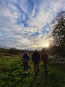
The group going for an afternoon walk in the fields near Buxted Park.
References
[1] Cantwell H, Nguyen H, Kettenbach A, Heald R. Spindle morphology changes between meiosis and mitosis driven by CK2 regulation of the Ran pathway. Biorxiv (2024) DOI: 10.1101/2024.07.25.605073.
[2] Kletter T, Muñoz O, Reusch S, Biswas A, Halavatyi A, Neumann B, Kuropka B, Zaburdaev V, Reber S. Cell State-Specific Cytoplasmic Material Properties Control Spindle Architecture and Scaling. Biorxiv (2024) DOI: 10.1101/2024.07.22.604615
[3] Nommick A, Xie J, Minc N. Manipulation of Spindle Position Using Magnetic Tweezers in Sea Urchin Embyos. Methods Mol Biol. (2025) 2872:87-100.
[4] Tanimoto H, Sallé J, Dodin L, Minc N. Physical Forces Determining the Persistency and Centering Precision of Microtubule Asters. Nat Phys. (2018) 14(8):848-854.
[5] Najafi J, Dmitrieff S, Minc N. Size- and position-dependent cytoplasm viscoelasticity through hydrodynamic interactions with the cell surface. PNAS (2023) 120(9)e2216839120.
[6] Ghosh S, Körte A, Serafini G, Yadav V, Rodenfels J, Developmental energetics: Energy expenditure, budgets and metabolism during animal embryogenesis. Semin. Cell Dev. Biol. (2023) 138, 83–93.
[7] Yang X, Heinemann M, Howard J, Foster P, Physical bioenergetics: Energy fluxes, budgets, and constraints in cells. PNAS (2021) 118(26)e2026786118.
[8] Padgett J, Santos S. From clocks to dominoes: lessons on cell cycle remodelling from embryonic stem cells. FEBS letters (2020) 594(13), 2031-2045.
[9] Srivastava M. Studying development, regeneration, stem cells, and more in the acoel Hofstenia miamia. Curr. Top. Dev. Biol. (2022) 147:153–172.
[10] Saiz N, Mora-Bitria L, Rahman S, George H, Herder J, Garcia-Ojalvo J, Hadjantonakis, AK. Growth Factor-Mediated Coupling between Lineage Size and Cell Fate Choice Underlies Robustness of Mammalian Development. eLife (2020) 9, e56079.
Written by Irene Li, Magdalena Schindler and Isaac Wong
This is one of three reports about the “Physics of the Early Embryonic Divisions” Workshop, organised by The Company of Biologists. Read the other two reports for different perspectives on the science that was discussed:
We recently had the chance to be amongst 30 developmental biologists and theoretical biophysicists who tackled the complex dynamics of early embryonic cell divisions in a workshop organized by The Company of Biologists. This scientifically stimulating event took place at Buxted Park in the picturesque Sussex countryside of England. The small size of the workshop fostered conversations and encouraged sharing of unpublished work, creating an ideal environment for both established and early-career researchers to engage in scientific discourse that wouldn’t have been possible at larger-scale meetings.
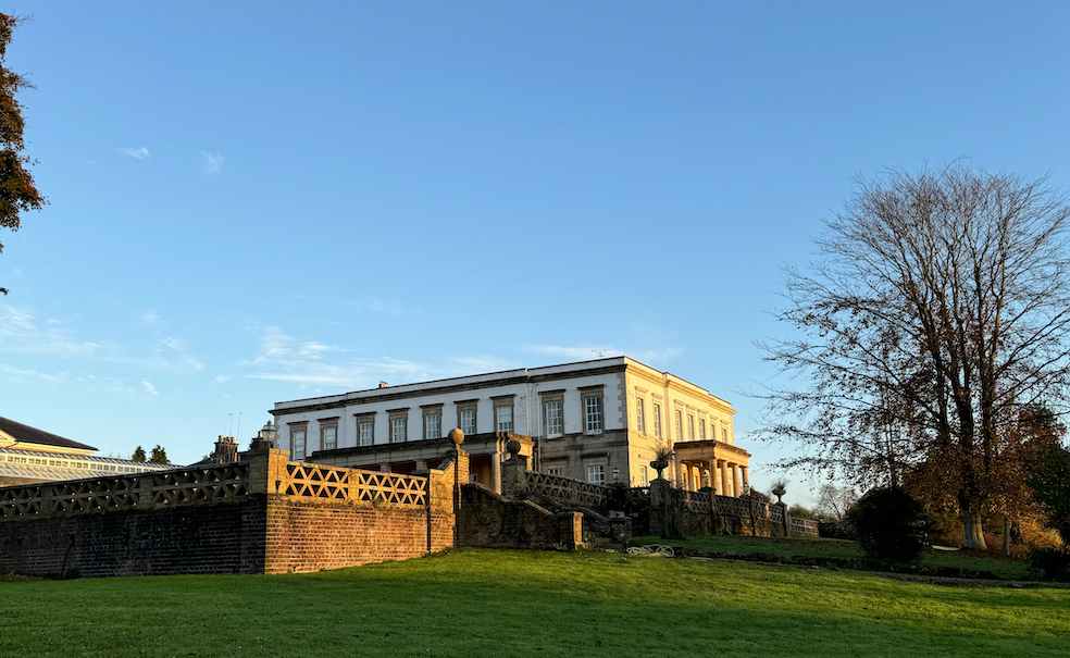
The historic Buxted Park Hotel, gracefully overlooking the picturesque countryside
Thanks to the hard work of the organizers Lendert Gelens and Julia Kamenz, scientific conversations flowed seamlessly from morning presentations into afternoon walks around the countryside and evening minglings at the bar. The program’s design struck an excellent balance between structured sessions and informal discussions, while the venue’s inviting spaces encouraged participants to delve deep into scientific exchanges. The atmosphere of openness was further enhanced by the break-out sessions in the evening, where the participants brainstormed on the bigger picture for early embryonic studies. These interactions throughout the workshop laid excellent groundwork for new friendships and exciting future ventures. We summarised here some, to us, outstanding sessions:
Session on cell cycle control
Recent studies are painting a vivid picture of just how finely tuned and adaptable the cell cycle really is. Stefano di Talia opened the session with a fascinating talk on the relationship between nearest-neighbour network topology and spindle packing. He showed that embryonic cells start deciding their future nuclear identities surprisingly early, hinting that fate is determined sooner than we might expect [1]. Next, Julia Kamenz introduced a new sensor that gives us a clearer view of how the cell cycle progresses. Yuting Irene Li’s research uncovers a kind of “tug-of-war” between two mechanisms that keep the meta-synchrony of cell cycles in early embryonic divisions: pre-set gradients of cell cycle lengths and short-range interactions [2]. Finally, Lendert Gelens highlighted how even temperature can influence these delicate processes, showing that environmental factors can shift the gears of cell division [3]. Taken together, these insights reveal just how dynamic and responsive the cell cycle can be.
Session on the cytoskeleton and cytoplasmic flow
Despite many years of research, there are still countless open questions on the interplay of the cell cycle, the cytoskeleton and the cytoplasm. How these components interact within the embryo to achieve proper tissue-scale development is even more elusive. Olga Afonso addressed this problem by analysing how scaling of cytoplasmic flows can happen despite the massive cell size changes throughout the reductive cell cleavages of the embryo. The session concluded with Isaac Wong’s presentation, which examined how variations in the cytoplasmic concentration of centrosomal proteins influence centrosome growth. His findings offered new insights into the mechanisms that enable early embryos to maintain centrosome size homeostasis.
Session on multicellular dynamics
In the last session, the participants explored how one egg cell can give rise to complex multicellular dynamics. Nathan Goehring opened the discussion with exciting research on cell polarity and its propagation during early embryonic divisions in C. elegans [4]. Next, Sebastian Streichan addressed how cytoskeletal anisotropy arises in the first place. Magdalena Schindler then asked the question of how changes in cell cycle synchrony can impact the tissue material state of a developing embryo. She suggested that an optimum level of variability in cell cycle synchronicity driven by cell lineage is key in controlling tissue fluidization, which is essential for developmental progression. Diana Pinheiro’s research then explored how cell fate and macroscopic patterns may be impacted by material properties in Warmflash patterns. Lastly, Nicoletta Petridou’s work showed how cell scale dynamics dictate emergent tissue mechanical properties and how using optogenetics to control those properties revealed unexpected changes in cell signalling. The talks altogether gave a great insight into the impact of cell divisions and other cellular dynamics across scales.
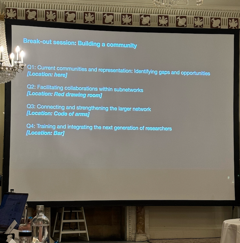
Breakout sessions
The workshop included 3 breakout sessions, during which the participants were split into subgroups to discuss big-picture questions. The three workshops respectively addressed community building within and across fields, important scientific questions we need answered and the tools we might use to do so. Given the interdisciplinarity of the participants, many different problems and hopes for technologies came up. As young researchers, we really appreciated this opportunity to exchange with leaders in our field and the engagement in this higher-level thinking. Altogether, we may have not been able to agree on one common goal in these sessions, but we are excited to see where our field will be going in the future and how we can shape it.
A sample slide displaying the key questions discussed during one breakout session, focused on building a strong and collaborative scientific community
[1] Xu Y, Chao A, Rinaldin M, Kickuth A, Brugués J, Di Talia S. The cell cycle oscillator and spindle length set the speed of chromosome separation in Drosophila embryos. Curr Biol. 2025 Feb 3;35(3):655-664.e3.
[2] Mishra N, Li YI, Hannezo E, Heisenberg CP. Geometry-driven asymmetric cell divisions pattern cell cycles and zygotic genome activation in the zebrafish embryo. bioRxiv [Preprint]
[3] Rombouts J, Tavella F, Vandervelde A, Phong C, Ferrell JE Jr, Yang Q, Gelens L. Mechanistic origins of temperature scaling in the early embryonic cell cycle. bioRxiv [Preprint]
[4] Rodrigues NT, Bland T, Ng K, Hirani N, Goehring NW. Quantitative perturbation-phenotype maps reveal nonlinear responses underlying robustness of PAR-dependent asymmetric cell division. PLoS biology. 2024 Dec 9;22(12):e3002437.
 (No Ratings Yet)