Want to organise one of The Company of Biolgists’ Workshops? We’re now accepting topic proposals for Workshops in 2027.
As the scientific organiser, your involvement will be focused on the science. We will undertake all of the logistical arrangements, liaise with the venue, organise speaker travel, assist with the programme construction and fund the meeting.
We are continuing our efforts to diversify our Workshops programme to support a wider selection of research communities around the world. We will be reserving one of our Workshops for an application from a Global South country.
We are delighted to announce sustainable travel incentives for those travelling to our Biologists @ 100 conference. Sustainable rail incentives up to £150 will be available for international train journeys and we are offering a prize of £700 in our sustainable travel blog competition. See more details here.
Arteries are an essential part of any tissue. For the tissue or organ to survive, it needs to be perfused with blood carrying nutrients and oxygen, which gets distributed through capillaries and keep cells alive and healthy. We use artery development as a model to ask questions about cellular responses, interactions, heterogeneity and plasticity. Specifically, we use a special kind of artery¾the collaterals (Figure 1), to ask important questions about biological processes.
Collateral arteries are special because these connect two arterial trees. So, if/when there is a clog in one of the artery branches, the collaterals reroute the blood flow and continue perfusing the underlying tissue without interruption. This ensures healthy and functional organs.
Figure 1: A collateral artery segment connecting two artery trees. Image was taken from an adult mouse brain (pial layer). Red, artery endothelial cells; Green, proliferating nuclei. Arrows point to the direction of blood flow. Image credit: Dr. Suraj Kumar
To date, we have not identified a molecular marker for collaterals, which, prevents us from distinguishing collaterals from conventional arteries on a tissue section or in the cellular clusters obtained via analyses of single cell sequencing data. The only identifying characteristic of collateralsis the fact that they connect two artery trees. So, we use imaging of whole organs (in our case, mouse hearts) to identify the coronary arteries and subsequently, the coronary collateral arteries which connect them together. Along with imaging whole hearts with cellular resolution, we heavily use mouse genetics and gene expression datasets to build hypotheses and test them both in silico and in vivo.
Like all scientific studies, this work went through a number of roadblocks which inspired our team to take up the challenges in a systematic manner and tackle them one at a time. It was not straight forward. In the process, we made some inspiring discoveries, developed some new tools and opened avenues for several questions in the field of vascular biology. Here is our story.
The inspiration: Same but Different
While it seems like the properties of cells which compose vessels within an organism are same, scientific evidence points to the contrary. We and others (Arolkar et al., 2023; McDonald et al., 2018) have time and again shown that, the building block of arteries─artery endothelial cells─can be quite different. Research groups around the world have used a variety of experimental models to highlight the differences in their origin, function, plasticity and regenerative abilities. The molecular heterogeneity of endothelial cells within a given vessel segment is getting more and more attention (Augustin and Koh, 2017; Trimm and Red-Horse, 2022).
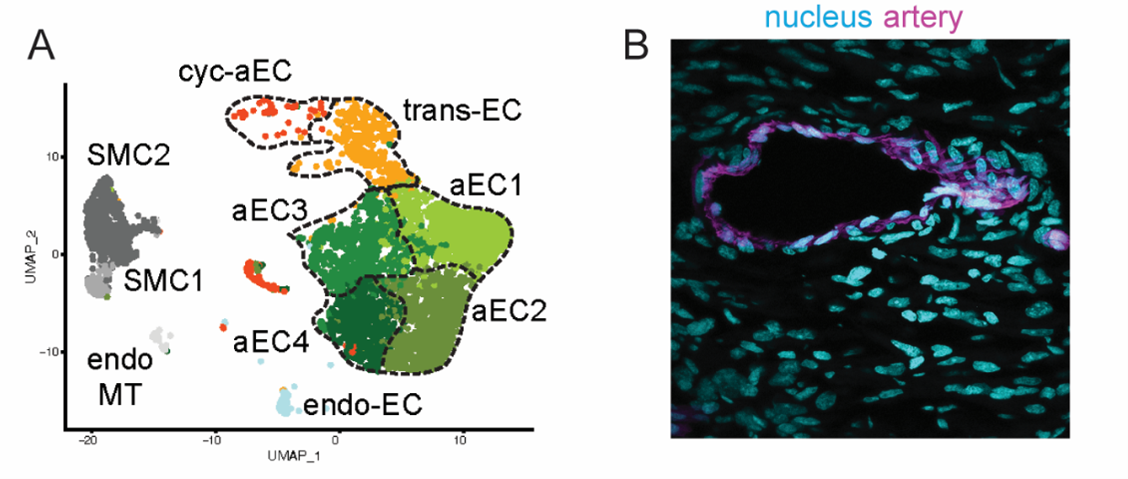
When we take a section of any vascularized tissue, and immunostain for arterial markers, like Cx40 or SMA, we cannot distinguish one cell from the other. We also know that these cells are different in their gene expression profiles (Arolkar et al., 2023). So, while the artery cells all look the same, they can be very different from each other (Figure 2). Cellular heterogeneity could be determined by their origin, which, consequently regulates the plasticity. This is a hypothesis, which we would like to test in as many ways as possible.
Figure 2: Heterogeneity of artery endothelial cells. (A) UMAP from single cell RNA sequencing data showing heterogeneity of artery cells. aEC1, aEC2, aEC3 and aEC4 are the mature artery cell clusters. Figure adapted from Arolkar et al. 2023 (B) Tissue section of neonatal heart showing artery (in magenta). Image credit: Bhavnesh Bishnoi
In our prior work we have done in silico and in vivo analyses of molecular properties of cardiac artery endothelial cells. In these studies, we have shown that only a small subset of artery cells is plastic enough to give rise to new cells, and that this plasticity is coupled to age¾the younger the hearts, the more plasticity (Arolkar et al., 2023; Das et al., 2019). That being said, we were always curious to test if this molecular heterogeneity within arteries is also observed in other organs.
The question and where to look
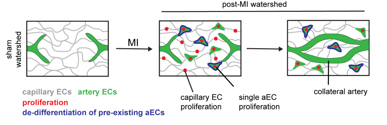
In mouse hearts, induction of myocardial infarction leads to dissociation of individual artery endothelial cells from pre-existing arteries. Only a subset of these dissociated single cells undergo proliferation, which results in massive expansion of this population. Eventually these cells come together and coalesce to build a collateral artery in the heart (Figure 3). We still do not understand what makes a small population (~8.4%) of artery endothelial cells more plastic than its neighbors. What is the relevance of such functional heterogeneity? Can these “plastic” cells, with proliferative properties, be distinguished molecularly from other artery cells? We continue to ask these questions in our lab.
Figure 3: Schematic diagram of the process of artery reassembly involved in the formation of collateral artery in neonatal heart. Upon myocardial infarction (MI), artery endothelial cells migrate, dedifferentiate, proliferate and coalesce to form collateral arteries (Adapted from Arolkar et al. 2023 with modifications)
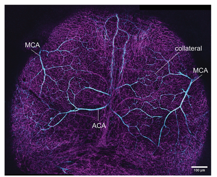
We are also curious about the occurrence of this unique biological process elsewhere in the mouse. So, we asked, if other critical (ischemia-prone) organs such as the brain, would demonstrate similar molecular heterogeneity within a given artery cell population. Luckily for us, apart from circle of Willis, which is embedded deep, the brain’s superficial layer, the pial layer, also has an extensive network of collaterals. This collateral network is formed between two major arterial trees, the middle cerebral artery and the anterior cerebral artery (Figure 4).
Figure 4: An embryonic brain vasculature. Arteries are shown in cyan and magenta shows all vessels. Collateral arteries are located between middle cerebral artery (MCA) and anterior cerebral artery (ACA) in each hemisphere of brain. Image credit: Swarnadip Ghosh
At the time, the brain was a new model to work on. The team did a thorough research on the anatomy and structure of mouse brain, especially how the arteries run through different segments of the brain, the time-lines, and the drivers. We read about different structures in the mouse brain and analyzed the gene expression patterns of various anatomic structures. We were surprised how little was known about the brain vasculature. Though stroke is one of the leading causes of deaths world-wide and have been known to us since ages, our approaches in its prevention or treatment is extremely limited (Bam et al., 2022; Grossman and Broderick, 2013). With all this in mind, we set off to ask our question: How do collateral arteries develop in a mouse brain? We chose to study pial collaterals to look for answers.
The hiccups: Mind the brain
While we were excited to start this new venture, we encountered the first bump very early on. A big challenge was to deduce a timeline. From earlier studies, we understood that pial collaterals in mouse brains were present at the time of birth, meaning, these structures developed during embryogenesis. We knew that tracking a developmental process could easily become very tricky. For starters, multiple overlapping cellular events during development could complicate our assessment of the timeline for the origin of pial arteries (middle and anterior cerebral arteries) and pial collateral arteries. Lack of a single molecular marker for collaterals would not let us distinguish arteries from collateral arteries.
The second challenge was more technical. The brain tissue was nothing like the heart¾it was squishy and lost its integrity post-processing. Preserving the integrity of the tissue and the embedded vasculature was a non-trivial task. We tried a variety of approaches, from isolating the pial layer (as a sheet) to sectioning and reconstructing different slices of brain tissue. But nothing worked.
The detours: Bypassing the blocks
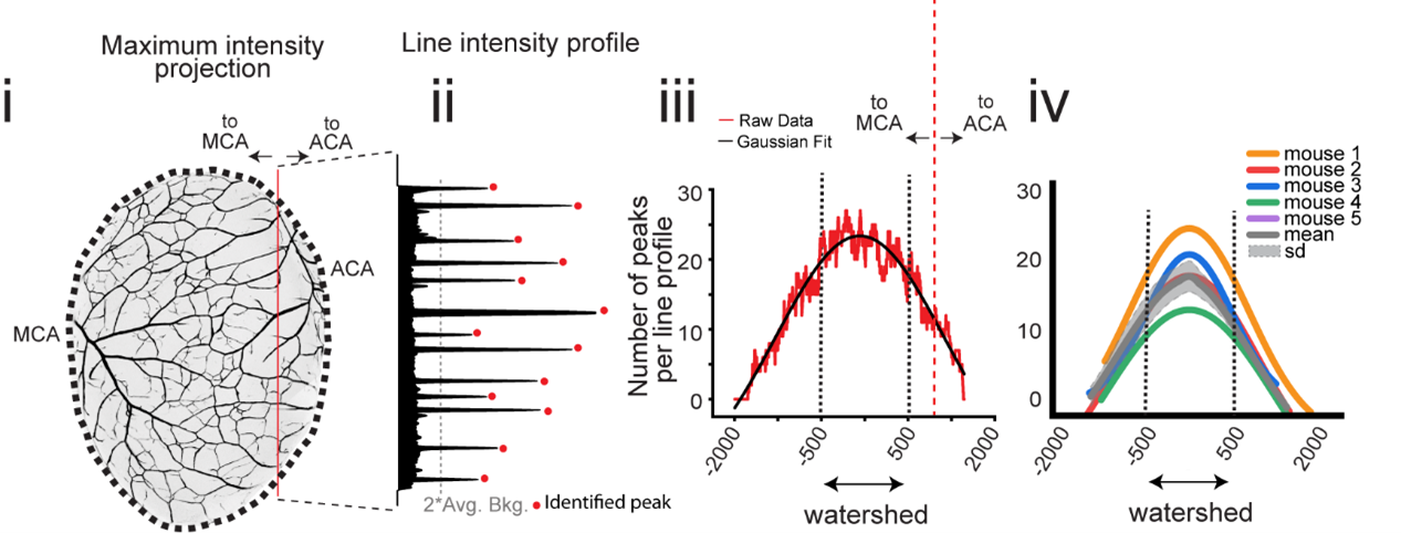
None of our (older) tactics allowed us to reliably visualize the collaterals. With time, we realized that we will need to develop new tools to address the questions we were asking. During the late-gestational period, when the pial collateral arteries develop (embryonic day (e) 15.5 to 18.5), the cranium and the skin (with its very own intricate vascular network) grow concurrently. This transformation posed a great obstacle for us to accurately visualize the pial blood vessels across different stages without the interference from surrounding tissue. It took us a year and a half to optimize a method for immunostaining and whole brain imaging of vasculature, with cellular resolution. After many trials and errors, we had a protocol which allowed us to confidently identify pial collaterals in all stages of developing mouse brain. Alongside, we also developed a quantitative method (Figure 5) to distinguish pial arteries from pial collaterals. Eventually, we were able to leverage this quantitative approach to delineate the precise timeline for development of pial arteries vis-à-vis collaterals in the mouse brain. With this, our curiosity deepened. We asked what was the cellular lineage of the pial collaterals. To address these questions, we utilized several transgenic mouse lines which enabled us for genetic fate mapping of the endothelial cells that form collateral arteries. As anticipated, we found a major cellular contribution (77%) from the preexisting artery cells with a smaller but notable contribution (31%) from the capillary cells, resulting in a mixed lineage composition.
Figure 5: A quantitative approach of analyzing complex brain arterial network. (i, ii) Intensity line profile were generated across each hemisphere. (iii) peaks per line profile were plotted along the width of a hemisphere and (iv) fitted with a Gaussian function. The fitted curve shows arterial coverage and the area under the curve within ±500 micron denotes the extent of collateral network. (Figure adapted from Kumar et al. 2024 with modification).
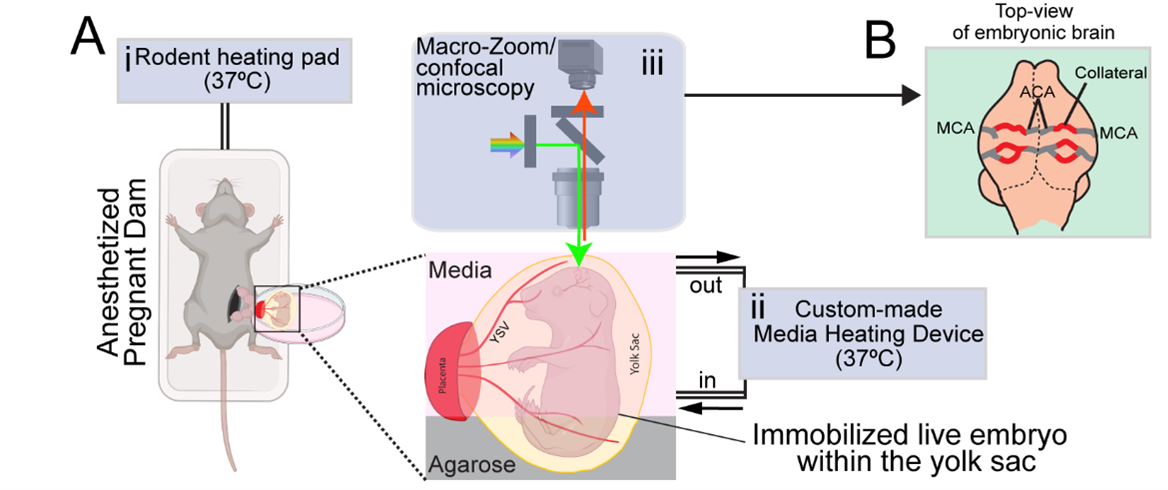
We next asked how pial collaterals form. This led us to embark on a new phase of investigation using in vivo experimentation and whole organ imaging to capture snapshot of the developmental process. Here we encountered one of the most formidable challenges. From immunostaining fixed samples, we realized that pial collaterals develop rapidly during embryogenesis. After a series of unsuccessful attempts to capture the cellular process in action, we refined our work-plan, and pushed the limits. We decided to build a system which allowed us to image brains of live embryos (Kawasoe et al., 2020; Yuryev et al., 2016). From immunostaining whole brains, we already knew that pial collaterals form in a very short time-range of 1-2 days. Hence, we hoped that keeping the embryo alive outside the mother’s womb for 3-4 hours (at the relevant gestational period, i.e., e16-e16.5) and making it accessible to microscopy, would help us capture the cellular dynamics involved in pial collateral artery development. We followed a surgical procedure where the developing embryos were taken out from the anesthetized pregnant dam. The embryos were still connected via umbilical cord and kept within the intact yolk sac. The whole set-up was kept adequately hydrated and temperature was stringently maintained to ensure the survival of the embryos. This set up was coupled with a (stereo and confocal) microscope to capture the vascular dynamics at cellular resolution (Figure 6). By live imaging embryos, we bypassed all biases and concerns, be it technical or biological. And the results were truly surprising and rewarding. The process we captured was distinct from what we observe in the heart. We captured pre-existing artery tips walking on defined microvascular structures (Kumar et al., 2024). It was exciting to observe the collaboration of artery and capillary endothelial cells.
Figure 6: Schematic diagram of the intravital time lapse imaging set up to capture collateral artery development during embryogenesis. (Figure adapted from Kumar et al. 2024)
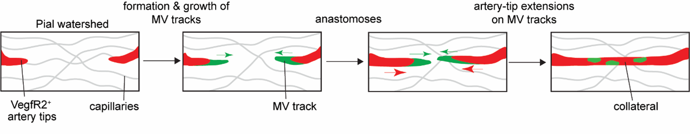
We also systematically tested the role of CXCL12 and VEGF pathways in pial collateral development and remodeling. We chose these molecules as they are already known to perform critical functions during coronary collateral development, post-MI. Remarkably, in contrast to the heart, CXCL12 was dispensable for the development of pial collaterals during embryogenesis. While VEGF pathway was critical, it performed a very different function─helping in artery tip extension. We also performed longitudinal imaging of adult pial layer, and assessed the effects of hypoxia on individual artery cells which make up pial collaterals. Together, using a combination of mouse genetics and intravital/longitudinal imaging we showed organ-specific mechanisms drive collateral development in the brain and heart (compare Figure 3 with Figure 7).
Figure 7: Schematic diagram of the process of artery tip extension involved in the formation of pial collateral artery in mouse brain. VegfR2+ artery tips (in red) grows along microvascular (MV) track (in green) to form pial collaterals. (Figure adapted from Kumar et al. 2024, with modification)
This study was a result of combined output from a post-doctoral work, a part of doctoral research work and three master’s theses. It, indeed, was a team-driven pursuit and a notable example of curiosity-driven science. Many of the authors have moved on to their next stage of careers, continuing to explore multiple aspects of neuroscience and vascular biology. We anticipate amazing outcome from their works in near future.
References:
Arolkar, G., Kumar, S. K., Wang, H., Gonzalez, K. M., Kumar, S., Bishnoi, B., Rios Coronado, P. E., Woo, Y. J., Red-Horse, K. and Das, S. (2023). Dedifferentiation and Proliferation of Artery Endothelial Cells Drive Coronary Collateral Development in Mice. Arterioscler Thromb Vasc Biol 43, 1455–1477.
Augustin, H. G. and Koh, G. Y. (2017). Organotypic vasculature: From descriptive heterogeneity to functional pathophysiology. Science (1979) 357,.
Bam, K., Olaiya, M. T., Cadilhac, D. A., Donnan, G. A., Murphy, L. and Kilkenny, M. F. (2022). Enhancing primary stroke prevention: a combination approach. Lancet Public Health 7, e721–e724.
Das, S., Goldstone, A. B., Wang, H., Farry, J., D’Amato, G., Paulsen, M. J., Eskandari, A., Hironaka, C. E., Phansalkar, R., Sharma, B., et al. (2019). A Unique Collateral Artery Development Program Promotes Neonatal Heart Regeneration. Cell 176, 1128-1142.e18.
Grossman, A. W. and Broderick, J. P. (2013). Advances and challenges in treatment and prevention of ischemic stroke. Ann Neurol 74, 363–372.
Kawasoe, R., Shinoda, T., Hattori, Y., Nakagawa, M., Pham, T. Q., Tanaka, Y., Sagou, K., Saito, K., Katsuki, S., Kotani, T., et al. (2020). Two-photon microscopic observation of cell-production dynamics in the developing mammalian neocortex in utero. Dev Growth Differ 62, 118–128.
Kumar, S., Ghosh, S., Shanavas, N., Sivaramakrishnan, V., Dwari, M. and Das, S. (2024). Development of pial collaterals by extension of pre-existing artery tips. Cell Rep 43, 114771.
McDonald, A. I., Shirali, A. S., Aragón, R., Ma, F., Hernandez, G., Vaughn, D. A., Mack, J. J., Lim, T. Y., Sunshine, H., Zhao, P., et al. (2018). Endothelial Regeneration of Large Vessels Is a Biphasic Process Driven by Local Cells with Distinct Proliferative Capacities. Cell Stem Cell 23, 210-225.e6.
Trimm, E. and Red-Horse, K. (2022). Vascular endothelial cell development and diversity. Nature Reviews Cardiology 2022 1–14.
Yuryev, M., Pellegrino, C., Jokinen, V., Andriichuk, L., Khirug, S., Khiroug, L. and Riverat, C. (2016). In vivo calcium imaging of evoked calcium waves in the embryonic cortex. Front Cell Neurosci 9, 173603.
In this SciArt profile, we find out more about B. Duygu Özpolat, an assistant professor whose research focuses on germ cell regeneration in segmented worms. Duygu enjoys creating pottery pieces of worms, bugs, and other “creepy” animals to show people the beauty and wonders of these organisms.
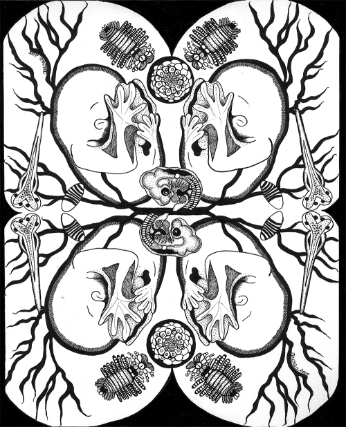
This drawing combines a few embryos and larvae from research organisms used in Developmental Biology! The blood vessels in the background are inspired by chicken embryos.
Can you tell us about your background and what you work on now?
I am an assistant professor of biology and a visual artist currently living in Saint Louis, Missouri. I earned my Ph.D. in Cell and Molecular Biology from Tulane University in New Orleans, and my B.Sc. in Biology from Middle East Technical University in Ankara, Türkiye. I primarily study stem cells and regeneration, with a specific focus on germ cell regeneration, using segmented worms as a model. Here is a great article about my lab’s work.
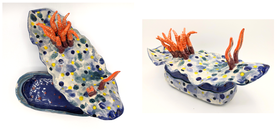
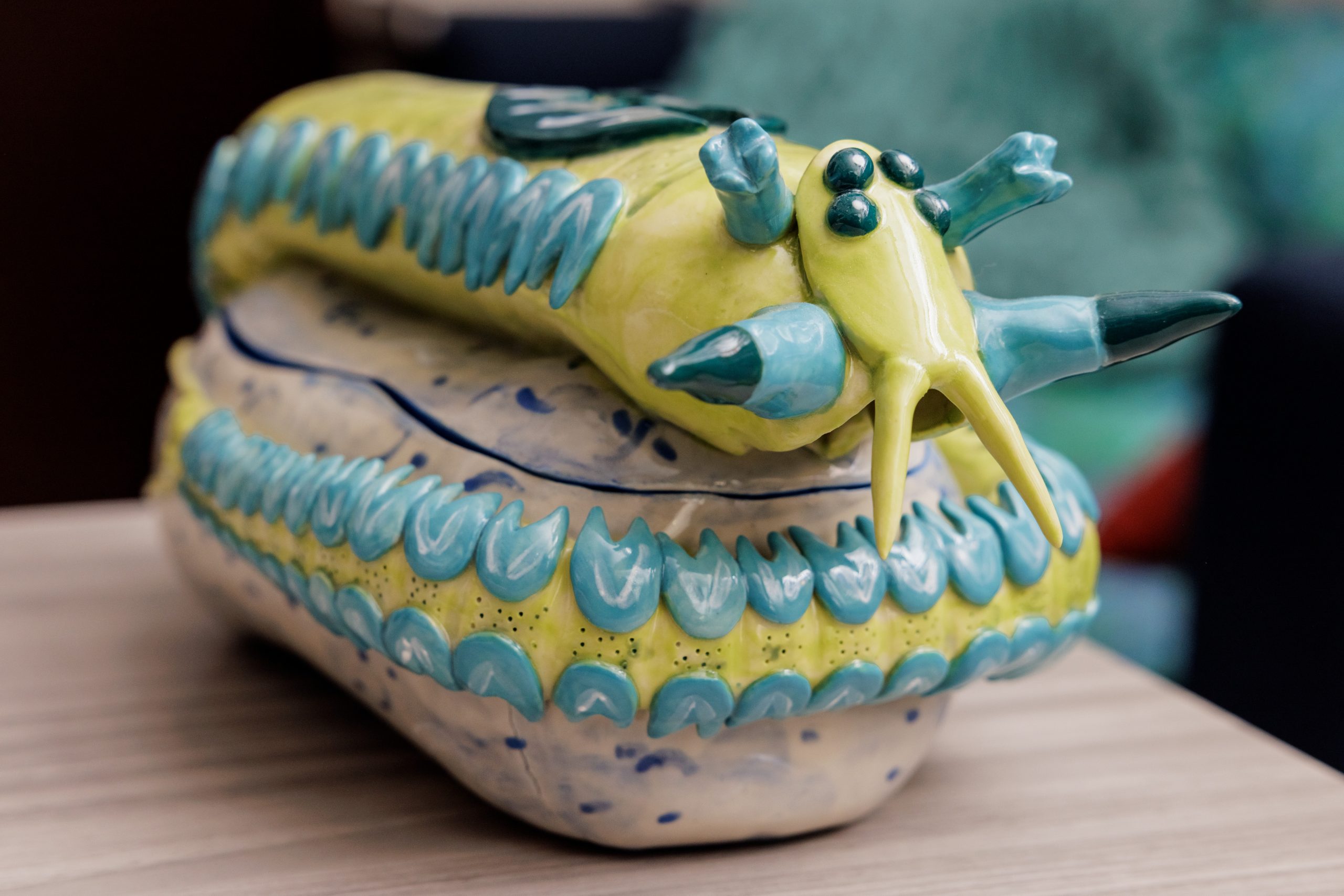
A seaslug (nudibranch) dish. Inspired by the species Hypselodoris confetti.
Were you always going to be a scientist?
Kind of! I grew up with a mother who was an arts teacher. So, art was in my life from early ages. But then when I started primary school and started getting exposure to science topics, and simple home experiments, similar to making a battery from potatoes or baking powder and vinegar volcano experiments, I fell in love with science. My mom loves telling the story that one day while in primary school, I asked her whether I could become a tailor and a scientist when I grow up, to which she replied, “Of course honey!”. I think of this as one of the defining moments in my life because I was so lucky to have a mother who believed I could be whatever I wanted to be. I got no discouraging, gender-stereotyped messages from her.
A membracid insect. Membracids are amazing insects that come in crazy shapes and I love recreating these fun insects using ceramics.
And what about art – have you always enjoyed it?
There are many layers to this for me: Art, just like science, is the exploration of the unknown, which is one of the main reasons I am also drawn to art. But unlike science, art for me has less anxiety and planning around it. I like mindlessly losing myself in a piece of artwork, and not worrying about what people will think about it, whether anyone will like it. I enjoy the process of creating, the tactile aspect of it, and how making art keeps me present in the moment.
What or who are your most important artistic influences?
Like any biologist, I have been absolutely charmed by the German zoologist Ernst Haeckel’s illustrations (but have been disappointed to find out about his racist ideology). When I visited Harvard Museum of Natural History and saw Leopold and Rudolf Blaschka’s glass flowers I was blown away! Some contemporary Scientist-Artists who inspire me are Steph Nowotarski and Bob Goldstein. Bob does screen printing, him and I collaborated on a poster project for a seminar series, which was great fun. It is encouraging for me to see full-time scientists like Steph and Bob make such great art inspired by science.
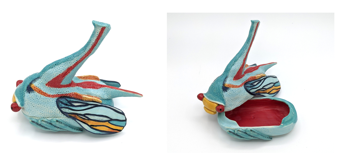
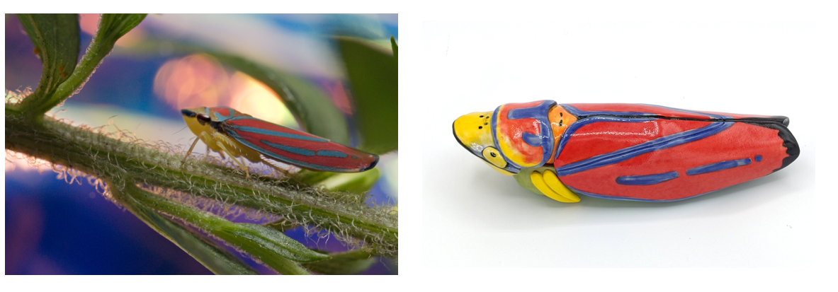
Meanwhile, my work could not exist without people who go out and document these amazing creatures. My husband Ryan Null is the person who taught me so much about insects and other arthropods. It is amazing to have somebody so knowledgeable around you. He is able to find all kinds of insects just in our backyard and photograph them, and sometimes I help him with his photography. We took the candy-striped leafhopper photo together while living on Cape Cod and later I made the ceramics piece inspired by it.
I also follow quite a few macro and wildlife photographers’ works such as Markus Kam and Alexander Semenov (who is also a scientist).
Finally, I am a huge fan of contemporary art and related museums. When I lived in Paris, I visited Palais de Tokyo often and was introduced to Marguerite Humeau’s work there for the first time. The exhibition at the time included her work called FOXP2, which is a gene associated with language ability in vertebrates. Another contemporary art museum experience that I cannot forget is Mark Dion’s “Misadventures of a 21st-Century Naturalist” exhibition at ICA in Boston, which left me absolutely speechless. I would love to be able to make artwork at the scale Mark Dion works one day.
Since moving to Saint Louis, I have been mostly making my pottery in my basement, and I take the pieces to be fired in a kiln at a pottery shop. I would like to go back to taking classes at a pottery studio at some point because I like the communal aspects of studios, and having made a commitment to taking a class makes it harder to procrastinate on my arts projects.
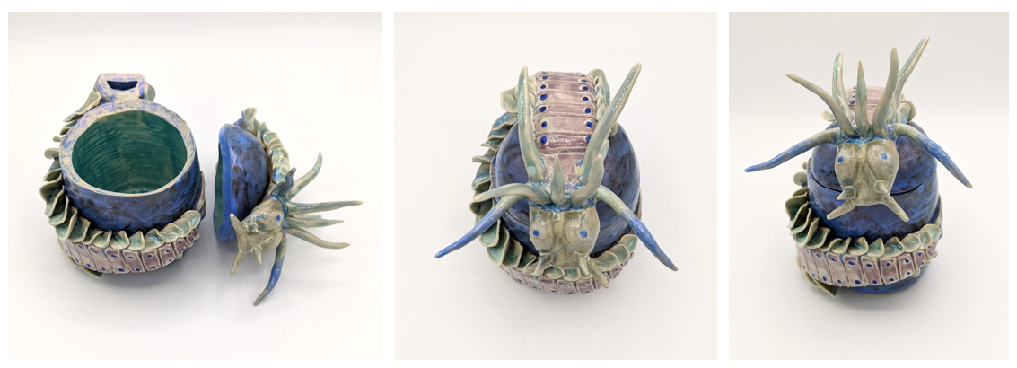
A nereidid polychaete worm with some artistic license. Ceramic jar at Duygu’s office (it usually has candies inside). Photo credit: Whitney Curtis. (video: https://www.instagram.com/p/Cb5SaX6lKhY/)
Does your science influence your art at all, or are they separate worlds?
Yes, my science influences my art very much. I am on a mission to get worms, bugs, and other “creepy” animals more appreciated! It is very important to me to invite people to leave their preconceived notions about these animals aside and open their minds to the beauty and wonders of them. We see only a handful of animals (usually vertebrates) celebrated in popular culture when there are literally millions of amazing species out there. I have given SciArt talks at conferences, and would like to use my pieces for science outreach projects in the future.
The photo of the actual animal is by my husband Ryan Null. The second photo is the ceramics piece I made inspired by this colorful bug.
What are you thinking of working on next?
I am very excited about building my own plaster molds to use in slip casting. This is a ceramics technique where clay in liquid form is used to cast a piece using a mold. I trained with Mary Rhein (an amazing local Saint Louis area artist). The process of making molds is very technical, you definitely need a “protocol” for this practice which makes me feel like I am at the lab bench. I am currently working on making molds for some of the insect figures such as the membracids and candy-striped leafhopper I made in the past, so I can focus on painting and decorating pieces.
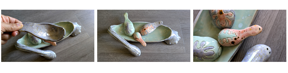
I also love my most recent collaboration with my husband Ryan Null, who is also a scientist. Ryan loves leeches (so do I!). Leeches have been an amazing developmental biology model, and we finally got some from David Weisblat’s lab. Ryan came up with the design of a leech ice cream dish, with small baby leeches as the ice cream spoons. I want to make a few more variations of this piece.
This is a collaboration with my husband Ryan Null, who adores leeches. He designed this plate with the spoons (the baby leeches), and I built it from clay. We picked the glazes together. This is Helobdella austinensis. They are hermaphrodites, and when they have babies, the babies live attached to the parent’s belly by their suckers. Whenever the parent goes feeding, the babies can reach for the food and feed as well. This species doesn’t suck blood. It eats other animals like insect larvae etc.
Find out more about Duygu:
www.ozpolatlab.org – mostly science and my lab’s news, academic self-help resources, and some science-art
In 2022, Biology Letters launched the Early Career Researcher Competition to highlight the best research papers published in the journal by early career researchers (ECRs). We are delighted to announce that the 2025 competition is now open until Wednesday 30 April. The overall winner will receive £1000 and two runners-up will receive £500 each (or currency equivalent). We hope the prizes are particularly helpful for funding new research and/or attending conferences. Please see our terms and conditions before entering or contact the editorial office with your questions.
You don’t have to be an ECR to submit to the journal! Take a look at our Information for Authors page for more information on what we publish and how you can get involved.
Back in the summer of 2011, I was just finishing up work for the day. Sitting in my office at EMBO in Heidelberg, Germany, I happened to open up the Node for a quick scroll before heading home. And there I spotted a post advertising the vacant position of Executive Editor at Development. I wasn’t looking for a new job – I had been at The EMBO Journal for less than three years and I was very happy there, but this was too good an opportunity to miss. I’d published my first ever paper in Development, I knew and loved the journal and I missed thinking about developmental biology – we didn’t get many submissions from the field at EMBOJ. Plus my sister had just had a baby and there was an attraction to moving back to the UK and being closer to my family. The application deadline was just a few days away, but I scrabbled to brush up my CV, write a decent cover letter and get them sent off. The rest, as they say, is history, and I’ve now been fortunate enough to hold the position of Executive Editor at Development for over 13 years.
I can honestly say that it’s been the best job in the world – I get to work with a fantastic in-house team of dedicated editors, administrators, production staff and more, and with a wonderful group of Academic Editors who somehow manage to juggle their editorial work alongside their full-time academic responsibilities. I’ve worked a lot on (hopefully!) improving some of our policies, processes and workflows at the journal, and I’ve been able to kick-start new initiatives from the journal, like our series of human development meetings and the Pathway to Independence programme. I’ve developed a wide network of contacts and friends in the developmental biology community, and I’ve loved hearing about all the fantastic science going on in the field.
Soon, however, it will be time for me to move on. Fortunately, I’m not going far – I’ll be taking up a new role at The Company of Biologists that has arisen due to some internal structural reorganisation, where I’ll be working strategically across all our journals. I’m delighted to be able to further my career in publishing while staying with an organisation in whose aims and ethos I deeply believe, though I’m gutted to be moving away from the developmental biology community.
All this means that my job is up for grabs. So, on the off-chance that someone like the me-of-13-years-ago happens to be reading this post, we’re looking for an experienced professional editor with a love of developmental biology to take on this role. Full details can be found in the job advert, and please feel free to reach out if you have any questions about this position.
While I’m going to find it hard to ‘let go’ of the journal after so many years, I’m excited to see what new ideas my replacement will bring to Development so that the journal continues to flourish going forwards.
The Latin American Society for Developmental Biology(LASDB) calls for nominations for these two prizes for outstanding researchers in different stages of their careers.
LASDB New Investigator Award 2025. The LASDB New Investigator Award honors early-career independent scientists conducting exceptional research in Developmental Biology in Latin America. Information on Eligibility and Nomination can be found at: https://lasdb-development.org/pages/young_investigator_award.html Deadline EXTENDED: March 9, 2025
LASDB Prize 2025 The LASDB Prize recognizes an outstanding scientist who has made a major contribution to Developmental Biology. Information on Eligibility and Nomination requirements can be found at: https://lasdb-development.org/pages/prize.html Deadline EXTENDED: March 9, 2025
These award ceremony will take place at the LASDB-ISDB-SDB joint meeting in Puerto Rico in June 2025 , where both winners will be invited to give a plenary lecture.
As part of our ‘first issues’ series on the Node, Development’s in-house team are researching the authors of articles published in the first issues of Journal of Embryology and Experimental Morphology (JEEM) and its reincarnation, Development.
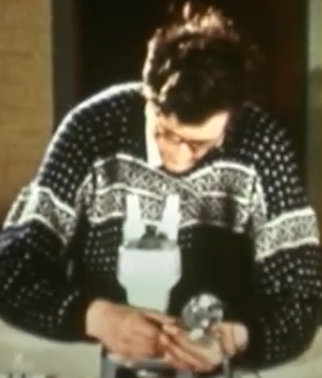
In this final post, we meet Tom R. Elsdale, a former embryologist at the Medical Research Council Human Genetics Unit in Edinburgh, UK, (Fig. 1) and an author in the first issue of Development, with an interest in understanding one of life’s big philosophical questions: time.
Born in 1929 in Somerset, UK, Tom Elsdale was raised by opera singer Vivian (née Miller) Elsdale and World War I marksman Henry Elsdale. Unusually, his dad also had a second home and family, fathering many children – one of whom accidentally blinded Elsdale in one eye by shooting him with an arrow (Bard, 2014). Despite this unorthodox upbringing, Elsdale would go on to be a remarkable developmental biologist and have fantastic taste in jumpers (Fig. 1).
Fig. 1. Tom Elsdale performing a nuclear transfer experiment in Xenopus. Still from a video posted by Denis Duboule (https://youtu.be/HhGvk5Aqh5k).
Following his undergraduate degree at the University of Reading, UK, Elsdale undertook his PhD studies with the renowned Conrad Waddington (Bard, 2014), a founding Editorial Board member of JEEM and likely one of its early supporters who pitched the journal to The Company of Biologists (Eve, 2025). Waddington published in the second-ever issue of the journal (Waddington and Carter, 1953) and several times thereafter, although Waddington and Elsdale never did publish together.
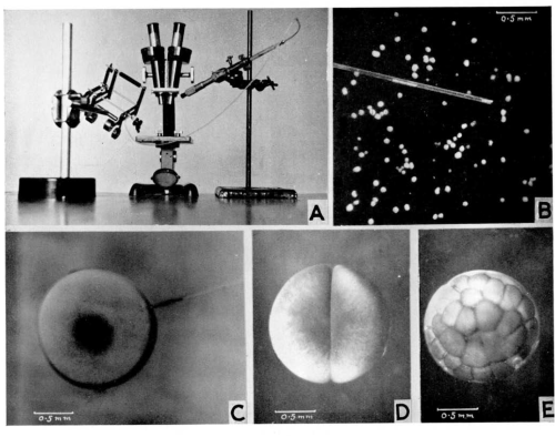
As a postdoc at the University of Oxford, UK, with Russian-born biologist Michail “Michael” Fischberg, Elsdale taught John Gurdon how to perform nuclear transfers in Xenopus laevis, following in the footsteps of Audrey Muggleton-Harris‘s work on amoebae. They adapted their approach from Briggs and King (1952), which involved taking nuclei from embryonic cells and transferring them into enucleated eggs, successfully cloning animals that survived into adulthood (Gurdon et al., 1958). Their technique was later published in JEEM (Elsdale et al. 1960), and you can watch Elsdale performing it as part of the Node’s ‘show and tell’ series (see Fig. 1, 2). Gurdon would use this approach to publish an article in 1962, also in JEEM, where he demonstrated that nuclei from differentiated cells (epithelia) could give rise to new embryos, successfully cloning an adult frog (Gurdon, 1962). It was for this work that Gurdon was awarded the 2012 Nobel Prize for Physiology or Medicine (Gurdon, 2013a,b), together with Shinya Yamanaka and his discovery of reprogramming cells to produce induced pluripotent stem cells (Takahashi and Yamanaka, 2006). Before being awarded the Nobel Prize, Gurdon also served as the Chair of the Board of Directors for The Company of Biologists from 2001 to 2011, demonstrating another indirect link Elsdale had with the Company (Bray et al., 2025).
Fig. 2. Figure from Elsdale et al. (1960) showing the apparatus for nuclear transplantation (A), performing the experiment (B, C) and the resulting cloned embryos (D, E).
Following his work on cloning amphibians, Elsdale started his own lab in the seemingly close-knit community at the Institute of Animal Genetics (IAG) in Edinburgh, UK, where he was colleagues with Ruth Clayton, and links him to The Company of Biologists yet again via the IAG’s founder, Francis Crew (Hankins and Rutledge, 2023; Knight, 2023). Elsdale became interested in optimising in vitro biology, demonstrating that frog embryonic cells could be cultured as aggregates. These studies revealed insights about gastrulation, germ layer formation and differentiation (Jones and Elsdale, 1963). One might also argue that these were prototype ‘xenobots’ – as published 60 years later to much media fanfare (Kriegman et al., 2020). Like the Wolffs, Elsdale was also a pioneer for culture conditions, using collagen rather than glass or plastic to aid fibroblast cell maintenance well before the days of Matrigel™ (Elsdale and Bard, 1972).
By the point of the latter study, Elsdale had moved from the IAG to the MRC Clinical & Population Cytogenetics Unit in Edinburgh, UK, where he then published a series of papers addressing somitogenesis. Returning – once again – to frogs, Elsdale and colleagues frequently used heat shock, which was known to cause developmental phenotypes in rats (Škreb and Frank, 1963), as a method of understanding more about developmental timing and robustness in morphogenesis (Elsdale et al., 1976; Pearson and Elsdale, 1979a,b; Cook and Elsdale, 1980; Elsdale and Davidson, 1983). Elsdale’s first paper on somitogenesis followed the landmark ‘clock and wavefront’ model of segmentation by Cooke and Zeeman, published the previous year, which became textbook in the field (Cooke and Zeeman, 1976). Today, although the model has not been immune from scrutiny (e.g. Murray et al., 2011; Klepstad and Marcon, 2024), it has formed the basis for many significant studies revealing the molecular mechanisms behind somitogenesis in vivo and, more recently, in vitro systems that undergo segmentation-like processes (reviewed in Miao and Pourquié, 2024). Collectively, these studies on the segmentation clock are a wonderful illustration of one form of timekeeping in development.
Before retiring in Spain, Elsdale published his final paper in the first issue of Development, ‘Timekeeping by frog embryos, in normal development and after heat shock’, in which Elsdale and Duncan Davidson explore how a population of embryos within a species develop at the same rate (Elsdale and Davidson, 1987). If you’ve ever seen a clutch of frog eggs developing, such as the Xenopus embryos undergoing cleavage in the video below (Fig. 3), you’ll appreciate that the synchrony between the individuals is extraordinary.
Fig. 3. Video of the synchronous cleavage of Xenopus laevis. From Xenbase.
In their paper, Elsdale and Davidson collected Rana temporaria spawn from a local pond and incubated the embryos at 15°C. By recording the development of three spawns from different locations, they report relatively little variation in developmental rate. Next, they use Elsdale’s favourite manipulation (heat shock by limited exposure to 37°C) to perturb the embryos, being careful to ensure that the extent of the heat shock does not induce developmental phenotypes that occlude accurate embryo staging through counting somites. They reveal that heat shock delays embryonic development, with longer exposures to high temperatures increasing the delay, roughly equivalent to one hour of delayed growth per minute of heat shock. Interestingly, though, the embryos go through a ‘rest and recover’ process: embryos stop developing during the heat shock during a ‘rest phase’ and then continue development at the same rate as controls. In other words, the delay in development caused by heat shock is not due to an overall slowing of the pace of embryogenesis, nor do embryos speed up following heat shock to recover lost time. Elsdale and Davidson speculate that some exceptionally stable ‘relaxation oscillator’ could account for this ‘rest phase’, resulting from the degradation of a factor during heat shock that must be replenished before development can continue; they propose that DNA might be the factor involved. Finally, they pursue an earlier observation that short exposure to heat shock-inducing temperatures protects against subsequent shocks (Pearson and Elsdale, 1979a). They show that embryos exposed to two shocks of 8 minutes each, given two days apart, have less developmental delay than embryos exposed to a single 16-minute shock. In the former condition, embryos have a shorter rest phase.
I think this is a really interesting paper that raises many questions relevant to modern research but, with only six citations, it seems to have largely gone unnoticed. Understanding how embryos ensure a steady developmental pace, perceive and interpret time for the synchronous development of organs and tissues, and modify regulatory mechanisms for heterochronic development in or between species is pertinent today. The field of developmental time has grown in recent years with regular meetings on the topic and is a flourishing area of research, both conceptually and experimentally (e.g. Garcia-Ojalvo and Bulut-Karslioglu, 2023; Ebisuya et al., 2024). The heat shock pathway Elsdale used to induce developmental phenotypes was later coopted for inducible transgenic expression systems in several models, including Xenopus (Wheeler et al., 2000), and temperature-sensitive manipulations remain important tools for studying development that continue to be developed (e.g. Benman et al., 2025). Similarly, the relationship between temperature and development is as relevant now as 40 years ago, particularly in the context of climate change (e.g. Moriyama et al., 2023; Kale et al., 2023, preprint). I think it’s safe to say that, in many ways, Tom Elsdale was ahead of his time.
And thus, this post concludes our ‘first issues’ series in honour of The Company of Biologists’ 100-year anniversary. We hope you’ve enjoyed another look at JEEM/Development’s archives, and maybe we’ve inspired you to take a look yourselves. If you find a hidden treasure, why not tell the community about it here on the Node?
Benman, W., Huang, Z., Iyengar, P., Wilde, D., Mumford, T. R. and Bugaj, L. J. (2025). A temperature-inducible protein module for control of mammalian cell fate. Nat Methods. https://doi.org/10.1038/s41592-024-02572-4
Bray, S. J., Royle, S. J., Shiels, H. A. and St Johnston, D. (2025). The Company of Biologists: celebrating 100 years. Development; 152 (1): dev204567. doi: https://doi.org/10.1242/dev.204567
Briggs, R. and King T. J. (1952). Transplantation of living nuclei from blastula cells into enucleated frogs’ eggs * Proc Natl. Acad Sci USA; 38 (5) 455-463, https://doi.org/10.1073/pnas.38.5.455
Ebisuya, M., Rayon, T., Diaz-Cuadros, M., Chalut, K. J., Wu, G., Dodd, A. N., Torres-Padilla, M. E., Levine, M. and Gladyshev, V. N. (2024). Understanding how cells and organisms keep time during development. Dev Cell; 59(13):1623-1627. doi: 10.1016/j.devcel.2024.05.029
Elsdale, T. R., Gurdon, J. B. and Fischberg, M. (1960). A Description of the Technique for Nuclear Transplantation in Xenopus laevis. Development; 8 (4): 437–444. doi: https://doi.org/10.1242/dev.8.4.437
Elsdale, T. and Davidson, D. (1987). Timekeeping by frog embryos, in normal development and after heat shock. Development; 99 (1): 41–49. doi: https://doi.org/10.1242/dev.99.1.41
Garcia-Ojalvo, J. and Bulut-Karslioglu, A. (2023). On time: developmental timing within and across species. Development; 150 (14): dev201045. doi: https://doi.org/10.1242/dev.201045
Gurdon, J., Elsdale, T. and Fischberg, M. (1958). Sexually Mature Individuals of Xenopus laevis from the Transplantation of Single Somatic Nuclei. Nature; 182, 64–65. https://doi.org/10.1038/182064a0
Gurdon, J. B. (1962). The Developmental Capacity of Nuclei taken from Intestinal Epithelium Cells of Feeding Tadpoles. Development; 10 (4): 622–640. doi: https://doi.org/10.1242/dev.10.4.622
Gurdon, J. B. (2013b). The egg and the nucleus: a battle for supremacy. Development; 140 (12): 2449–2456. doi: https://doi.org/10.1242/dev.097170
Hankins, L. E. and Rutledge, C. E. (2023). Class of 1923: looking back at the authors of JEB’s first issue. J Exp Biol; 226 (1): jeb245424. doi: https://doi.org/10.1242/jeb.245424
Jones, K. W. and Elsdale, T. R. (1963). The Culture of Small Aggregates of Amphibian Embryonic Cells in vitro. Development; 11 (1): 135–154. doi: https://doi.org/10.1242/dev.11.1.135
Kale, G., Agarwal, P., Diaz-Larrosa, J. J. and Lemke, S. (2023). Elevated temperature fatally disrupts nuclear divisions in the early Drosophila embryo. bioRxiv; 2023.09.17.558127; doi: https://doi.org/10.1101/2023.09.17.558127
Klepstad, J. and Marcon, L. (2024). The Clock and Wavefront Self-Organizing model recreates the dynamics of mouse somitogenesis in vivo and in vitro. Development; 151 (10): dev202606. doi: https://doi.org/10.1242/dev.202606
Knight, K. (2023). Journey through the history of Journal of Experimental Biology: a timeline. J Exp Biol; 226 (22): jeb246868. doi: https://doi.org/10.1242/jeb.246868
Miao, Y. and Pourquié, O. (2024). Cellular and molecular control of vertebrate somitogenesis. Nat Rev Mol Cell Biol; 25, 517–533. https://doi.org/10.1038/s41580-024-00709-z
Moriyama, D. F., Makri, D., Maalouf, M.-N., Adamova, P., de Moraes, G. F. A., Pinheiro, M. de O., Bernardineli, D. L., Massaia, I. F. D. S., Maalouf, W. E. and Lo Turco, E. G. (2022). The effects of temperature variation treatments on embryonic development: a mouse study. Scientific Reports; 12(1), 2489. https://doi.org/10.1038/s41598-022-06158-y
Murray, P. J., Maini, P. K. and Baker, R. E. (2011). The clock and wavefront model revisited. J Theor Biol; 283(1):227-38. doi: https://doi.org/10.1016/j.jtbi.2011.05.004. Epub 2011 May 27. PMID: 21635902
Škreb, N. and Frank Z. (1963). Developmental Abnormalities in the Rat Induced by Heat Shock. Development; 11 (2): 445–457. doi: https://doi.org/10.1242/dev.11.2.445
Takahashi, K. and Yamanaka, S. (2006). Induction of pluripotent stem cells from mouse embryonic and adult fibroblast cultures by defined factors. Cell; 126(4):663-76. doi: https://doi.org/10.1016/j.cell.2006.07.024
Continuing the ‘First issues’ series, in this post we’ll find out more about Audrey Muggleton-Harris, who published in Development’s first issue in 1987.
Audrey Muggleton-Harris was born in London, England, in 1932. Throughout her prolific career spanning over 40 years, she moved across the Atlantic several times while continuing her research into in vitro systems, early human embryonic development and improvements in assisted reproductive technologies.
While the reasons behind Muggleton-Harris’ multiple relocations are not clear (but probably involved a combination of professional opportunities and personal motivations), moving abroad is still very common, and at times challenging, for academic researchers in the present day. For Muggleton-Harris, the UK and the USA no doubt played a vital role in shaping her professional and personal lives.
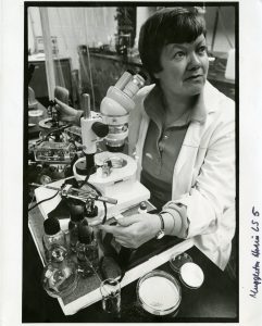
Photo of Audrey Muggleton-Harris while she was working at Worcester Polytechnic Institute in the 1970s (From WPI Historical Images, made available under the CC BY-NC 4.0 license.)
Early experiments on nuclear transfer
Muggleton-Harris was originally trained as a teacher, but after reading about the work of cell biologist James Danielli, she joined his team as a research assistant at King’s College London in 1952 [1]. James Danielli was a Director of The Company of Biologists, and helped find a publisher for the previous incarnation of Development – Journal of Embryology and Experimental Morphology (JEEM).
Together, Muggleton-Harris and Danielli performed some of the earliest cloning experiments, first in amoebae [2] and later in frog embryos [3]. They transferred nuclei between cells to test the ability of the transferred nucleus to direct development in a different cytoplasmic environment. These early experiments paved the way for future cloning techniques.
In the early 1960s, Danielli was invited to lead the Center for Theoretical Biology at the State University of New York. Most of Danielli’s group at King’s College London, including Muggleton-Harris, crossed the Atlantic to join him. This move foreshadowed the multiple relocations between the USA and the UK that Muggleton-Harris would make over the next few decades.
In the late 1960s, Muggleton-Harris was a Research Associate at the Institute for Cancer, and then became the Research Director of the Cell Biology program at the Wills Eye Hospital. Muggleton-Harris and her family went back ‘home’ to the UK in 1972 and she worked in Cambridge University. But after just 2 years, they returned to the USA, where Muggleton-Harris was a Research Fellow and Professor of Biology and Biotechnology at Worcester Polytechnic Institute from 1976 to 1983. From 1983 till her retirement in 1997, she would remain in the UK, working at the MRC Experimental Embryology and Teratology Unit in Surrey [1].
Developing in vitro systems to understand development and disease
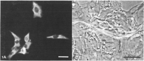
In her paper in Development issue 1, Muggleton-Harris and co-author N. Higbee found that fragments of the lens capsule, a thin membrane around the eye, could promote mouse lens epithelial cells to synthesize the collagen, glycoproteins and proteoglycan components required for further lens cell differentiation in vitro. This paper was part of a body of work by Muggleton-Harris on culturing mice lens epithelial cells to understand normal lens development and congenital cataracts. Since this Development issue 1 paper, Muggleton-Harris published four more papers in Development during her time working at the MRC in the UK.
Left: A clone of cultured mouse lens epithelial (MLE) cells; Right: A monolayer of MLE cells grown from cloned cells. Figures from Muggleton-Harris & Higbee, 1987 [4]
Preimplantation development and genetic testing
Her work in lens development was in fact only one part of Muggleton-Harris’ significant contribution to in vitro systems and early development. Almost a decade prior to the Development issue 1 paper, during her two-year stint in Cambridge Univeristy, she published a paper in JEEM in 1976 on ‘surface alloantigens on preimplantation mouse embryos’, investigating immune recognition and compatibility during early development, which is important in understanding maternal-fetal interactions.
In 1982, Muggleton-Harris et al. reported the first successful mammalian transfer of cytoplasm in mice, demonstrating that an unidentified cytoplasmic factor(s) can prevent embryo arrest at the two-cell stage in vitro [5]. In 1988, together with Marilyn Monk and colleagues, Muggleton-Harris developed the trophectoderm biopsy technique in mice [6]. This involved extracting cells from the trophectoderm layer of a blastocyst-stage embryo for preimplantation genetic diagnosis, while minimising damage to the embryo. A few years later, Muggleton-Harris and Findlay tested this method in ‘spare’ human preimplantation embryos in culture [7].
These are just a few examples of Muggleton-Harris’ effort into developing and refining methods for genetic analysis of biopsied cells from preimplantation embryos. Her work has contributed significantly to improving outcomes in assisted reproductive techniques and enhancing the accuracy and safety of preimplantation genetic testing.
An active member of scientific and local communities
Apart from her research, Muggleton-Harris was also a valued member of the scientific community, serving as a committee member of the British Society for Developmental Biology (BSDB) and organising the 1992 BSDB Autumn meeting on the developmental basis of inherited diseases [8].
After many prolific years at the MRC in the UK, Muggleton-Harris retired in 1997 and moved across the Atlantic one last time to Cape Cod, Massachusetts. During her retirement years, according to her obituary [1], she “volunteered as a docent at the Cape Cod Museum of Art… and will be remembered by visitors to the museum as the very proper Englishwoman who led museum tours and the annual historic house and garden bus tours” – a testament to how she left a mark on both sides of the pond, scientifically and socially.
[2] Muggleton A, Danielli JF. Inheritance of the “life-spanning” phenomenon in Amoeba proteus. Exp Cell Res. 1968;49(1):116-120. doi:10.1016/0014-4827(68)90524-7
[3] Muggleton-Harris AL. Cellular changes occurring with age in the lens cells of the frog (Rana pipiens) in reference to the developmental capacity of the transplanted nuclei. Exp Gerontol. 1970;5(3):227-232. doi:10.1016/0531-5565(70)90042-2
[4] A. L. Muggleton-Harris, N. Higbee; Factors modulating mouse lens epithelial cell morphology with differentiation and development of a lentoid structure in vitro. Development 1 January 1987; 99 (1): 25–32. doi: https://doi.org/10.1242/dev.99.1.25
[5] Muggleton-Harris A, Whittingham DG, Wilson L. Cytoplasmic control of preimplantation development in vitro in the mouse. Nature. 1982;299(5882):460-462. doi:10.1038/299460a0
[6] Monk M, Muggleton-Harris AL, Rawlings E, Whittingham DG. Pre-implantation diagnosis of HPRT-deficient male and carrier female mouse embryos by trophectoderm biopsy. Hum Reprod. 1988;3(3):377-381. doi:10.1093/oxfordjournals.humrep.a136711
[7] Muggleton-Harris AL, Findlay I. In-vitro studies on ‘spare’ human preimplantation embryos in culture. Hum Reprod. 1991;6(1):85-92. doi:10.1093/oxfordjournals.humrep.a137264
As part of our ‘first issues’ series to mark The Company of Biologists’ 100th anniversary, Development’s in-house team are researching the authors of articles published in the first issues of the Journal of Embryology and Experimental Morphology (JEEM) and its reincarnation, Development. In this post, we meet Ruth Clayton, a biologist who worked at the University of Edinburgh and who published an article in the first issue of JEEM.
Ruth Clayton was born in London in 1925 [1], the same year that our publisher, The Company of Biologists, was founded. Since Clayton would have turned 100 this year, it feels appropriate to honour her as part of the Company’s own 100th birthday celebrations. Clayton studied Zoology at Oxford before moving to the Institute of Animal Genetics (IAG) at the University of Edinburgh. Interestingly, this links Clayton to The Company of Biologists, since the IAG was initially established as the Department of Research in Animal Breeding by Francis Crew, who happens to have been the Managing Editor of our sister journal, Journal of Experimental Biology (JEB). I found out a bit about Crew and the institute a couple of years ago when researching the authors from JEB’s first issue [2]. The journal, which was founded in 1923 as the British Journal of Experimental Biology, quickly ran into financial difficulties, and George Parker Bidder III founded The Company of Biologists to safeguard its future [3].
By the time Clayton joined the IAG, it had come under the leadership of C. H. Waddington. Waddington is of course best known for his iconic epigenetic landscape [4], but he was also a member of JEEM’s first Editorial Board [5]. Clayton helped produce a gift for Waddington’s 50th birthday: a commemorative photo album [6]. This is worth a mention, mostly because it features candid shots of other IAG researchers [7] including Charlotte Auerbach and Tom Elsdale (who we’ll hear about on Friday, in the final post of this series), but also because the album documents the institute’s “Drosophila ballet”, which looks just as bizarre as it sounds [8]. The photo album was presented to Waddington at his birthday party, which seems to have been a similarly eccentric event, apparently featuring a pinball machine modelled on Waddington’s landscape [9]. Sadly, I could not find photographic evidence of the original machine, but during my search I did stumble across a paper that redraws Waddington’s epigenetic landscape as a pinball machine [10; the idea seems to be that the machine’s flippers can promote dedifferentiation]. So, maybe the party organisers were onto something.
Clayton’s article in JEEM’s first issue focuses on antigen specificity in the alpine newt (Triturus alpestris) embryo [11]. She begins her paper with a statement that still rings true for developmental biologists over 70 years later: “The mechanism of differentiation presents one of the central problems of embryology.” Clayton’s study aimed to address one aspect of this problem by investigating how antigens in the early embryo differ from those in the adult, to give hints as to what changes during the process of differentiation. To do this, she dissected different sections of newt embryos (e.g. ectoderm, mesoderm and tailbud) at different stages (e.g. blastula, gastrula) and homogenised them for injection into rabbits, to produce antisera.
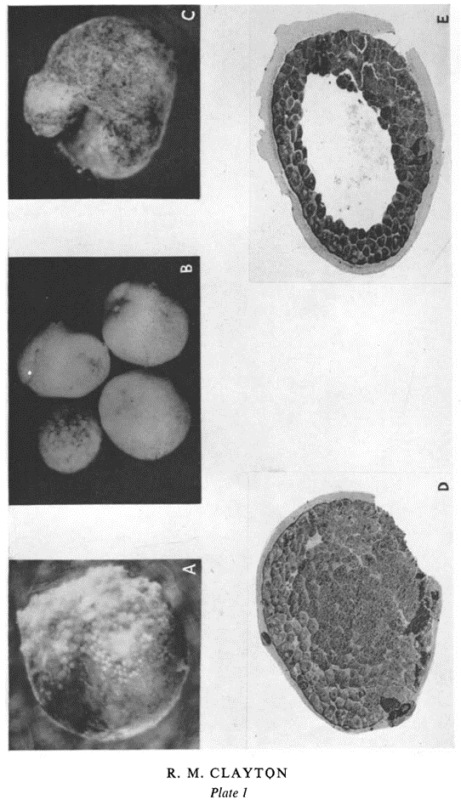
Fig 1: Plate from Ruth Clayton’s paper in the first issue of JEEM [11] showing examples of embryos placed in different combinations of antiserum.
The use of antisera in biology goes back a remarkably long way; at the turn of the 20th century, Emil von Behring pioneered ‘serum therapy’, injecting mammals including guinea pigs with diphtheria and tetanus toxins and ultimately using the resulting antiserum to treat patients [12, 13]. Nowadays, developmental biologists might be most familiar with using antibodies for Western blots, or for immunostaining. In her paper, Clayton mixed the antisera and antigens produced from different embryonic structures and recorded whether they cross-reacted. She went on to test the effects of placing embryos (Fig. 1) and explants in sera, antisera or absorbed antisera (i.e. the antigen and antisera mixes from earlier in the paper). The results are all a bit of a puzzle to tease out, because the experiments are done in multiple combinations, but they allow Clayton to deduce the existence of common antigens (which are present throughout development) and antigen fractions that arise at specific timepoints. For example, she notes that blastulae die or exhibit perturbed development when placed in “anti-gastrula serum absorbed with blastula” and suggests that this “is due to antibodies left in the medium after removal of antibodies to blastula antigens, i.e. that at gastrulation a new fraction appears”.
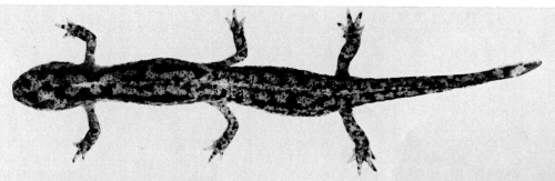
Clayton explains that she chose to work with the alpine newt for a couple of reasons: she needed “an embryo whose dissection might prove relatively easy and large numbers of which were available”. Today, other salamander species, such as the axolotl, are widely used to study regeneration. By contrast, the alpine newt doesn’t seem to have endured in developmental biology research. Indeed, a very quick search of Development’s archives suggests that it’s been nearly 50 years since we last published a research paper about alpine newts (to be fair, it’s a memorable article, featuring a chimeric newt with six legs (Fig. 2, 14). However, there is evidence that JEEM used to feature a pleasing range of newt species, including the California newt [15], the Iberian ribbed newt [16] and the Japanese fire-bellied newt [17]. Clayton’s rationale is a reminder of the importance of selecting the most appropriate organism to address a particular research question, so maybe the alpine newt will make a comeback when the circumstances are right. We highlighted the use of unconventional model organisms in our 2024 Special Issue [18].
Fig. 2: A six-legged alpine newt featured in a later JEEM paper [14]
Clayton published in JEEM a total of four times. She continued to make use of antisera in her investigations, but her subsequent work focused on cell differentiation in the context of the developing chick eye. This included research into the transdifferentiation of neural retina cells into pigment and lens cells [19]. Today, transdifferentiation is an active area of research that may hold promise for stem cell-based therapies. Clayton remained at the IAG, where Waddington later won funding to establish an Epigenetics Research Group. However, according to an obituary written by Alan Robertson, much of the project’s resources were used for molecular biology research that had no clear implications for development [9]. Indeed, Robertson mentions Clayton as “a notable exception” whose work on avian lens development “fitted into Waddington’s original concept”. Later in her career, Clayton’s expertise in lens development saw her lead an interdisciplinary collaboration to investigate risk factors for cataract formation [1]. She retired in 1993 and was appointed Reader Emeritus. Clayton’s obituary was published in the University of Edinburgh’s eBulletin in February 2003. It states that “though never formally a Director of Studies, she was often the member of staff to whom students turned for guidance”, suggesting that she was a dedicated mentor as well as a great scientist.
[2] Hankins, L. E. and Rutledge, C. E. (2023) Class of 1923: looking back at the authors of JEB’s first issue. J Exp Biol; 226 (1): jeb245424. doi: https://doi.org/10.1242/jeb.245424
[3] Knight, K. (2023) Journey through the history of Journal of Experimental Biology: a timeline. J Exp Biol; 226 (22): jeb246868. doi: https://doi.org/10.1242/jeb.246868
[9] Robertson, A. (1977) Conrad Hal Waddington, 8 November 1905-26 September 1975. Biogr. Mems Fell. R. Soc; 23, 575–622. doi: https://doi.org/10.1098/rsbm.1977.0022
[10] Sareen, D. and Svendsen, C.N. (2010) Stem cell biologists sure play a mean pinball. Nature biotechnology; 28(4), 333-335. doi: https://doi.org/10.1038/nbt0410-333
[11] Clayton, R. M. (1953) Distribution of Antigens in the Developing Newt Embryo. Journal of Embryology and Experimental Morphology; 1 (1): 25–42. doi: https://doi.org/10.1242/dev.1.1.25
[12] Behring, E. V. (1890) Ueber das zustandekommen der diphtherie-immunität und der tetanus-immunität bei thieren. Dt. Med. Wochenschr; 49, 1113–1114.
[13] Behring, E. V. (1913) Ueber ein neues Diphtherieschutzmittel. Dt. Med. Wochenschr; 39: 873-876.
[14] Houillon, C. (1977) Tractus uro-génital des chimères chez l’amphibien modèle Triturus alpestris Laur. Journal of Embryology and Experimental Morphology; 42 (1): 15–28. doi: https://doi.org/10.1242/dev.42.1.15
[15] Tucker, R. P. and Erickson, C. A. (1986) The control of pigment cell pattern formation in the California newt, Taricha torosa. Journal of Embryology and Experimental Morphology; 97 (1): 141–168. doi: https://doi.org/10.1242/dev.97.1.141
[16] Deparis, P. and Jaylet, A. (1984) The role of endoderm in blood cell ontogeny in the newt Pleurodeles waltl. Journal of Embryology and Experimental Morphology; 81 (1): 37–47. doi: https://doi.org/10.1242/dev.81.1.37
[17] Matsuda, M. (1980) Cell surface properties of amphibian embryonic cells. Journal of Embryology and Experimental Morphology; 60 (1): 163–171. doi: https://doi.org/10.1242/dev.60.1.163
[18] Extavour, C., Dolan, L., Sears, K. E. (2024) Promoting developmental diversity in a changing world. Development; 151 (20): dev204442. doi: https://doi.org/10.1242/dev.204442
[19] De Pomerai, D. I. and Clayton, R. M. (1978) Influence of embryonic stage on the transdifferentiation of chick neural retina cells in culture. Journal of Embryology and Experimental Morphology; 47 (1): 179–193. doi: https://doi.org/10.1242/dev.47.1.179
 (No Ratings Yet)
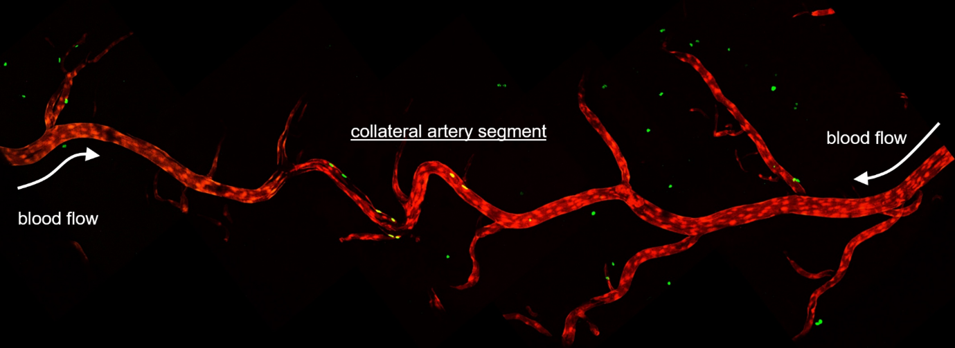
 (2 votes)