A postdoctoral position is available in the laboratory of Philippe Soriano to study FGF and PDGF regulated craniofacial morphogenesis in the mouse embryo. Possible projects focus on intracellular signaling pathways using receptor point mutant mice and proteomics; the impact of ERK1/2 and PI3K signaling dynamics on morphogenetic movements; or differential signaling mechanisms underlying ligand specific responses. A strong background in cell signaling or developmental biology is desired. Expertise in molecular/cell/systems biology, imaging technology, or mouse genetics are valuable but not required.
For more information, please visit our laboratory website at labs.icahn.mssm.edu/sorianolab. We benefit from a highly interactive scientific environment in the Department of Cell, Developmental, and Regenerative Biology at Mt. Sinai (www.mssm.edu/drb), and an outstanding developmental biology community within the city. In addition, New York City offers unequaled cultural and recreational opportunities.
Creative, interactive but independent, and highly motivated applicants are in particular invited to apply. Please send a letter of application including a brief description of previous research experience and interests, a CV, and the name and contact information of three references to Philippe Soriano at philippe.soriano@mssm.edu
The vertebrate lymphatic vascular network provides crucial circulatory and immune functions but its developmental origin has been a contentious issue, in particular the question of whether lymphatic endothelial cells have an exclusively venous origin. A new paper in Development addresses this issue in the dermis of the mouse embryo. To find out more about the story behind the work, we caught up with first author Cathy Pichol-Thievend and last authors Natasha Harvey (Associate Professor at the Centre for Cancer Biology, SA Pathology and University of South Australia) and Mathias Francois (Associate Professor at the Institute for Molecular Bioscience, The University of Queensland).
Mat and Natasha, can you give us your scientific biographies and the questions your labs are trying to answer?
MFIn 2004, I was awarded a PhD in molecular genetics from the University PARIS VI (Pierre et Marie Curie). During this period I worked on nuclear receptors PPARs and their role in cartilage inflammatory response (INSERM UMR-S747- Paris V). At the end of 2004, I moved to Australia to gain postdoctoral experience with Professor P. Koopman, an expert in the field of sex determination and SOX transcription factors (2005-2011, The Institute for Molecular Bioscience, The University of Queensland). During that time the key discovery from my work identified the transcription factor SOX18 as a molecular switch that induces the lymphatic differentiation program by turning on Prox1 expression in pre-existing veins.
In 2012, I set up my own independent research group at the IMB. Since then we have designed a novel molecular strategy to pharmacologically manipulate developmental transcription factor activity. We successfully identified and characterised small compounds that interfere with SOX18 activity. This gives us a unique handle to complement classic genetic methodologies with pharmacological approaches to study vascular development. The research interest of my group revolves around on how endothelial cell fate is dynamically regulated at a transcriptional level. To decipher how transcription factors orchestrate lymphangiogenesis and subsequent assembly of vessel networks we rely on the utilisation of mouse and zebrafish model systems.
NH After a PhD with Sharad Kumar focussed on defining the molecular mechanisms underlying caspase-2 activation during apoptosis, I wanted to investigate the role of caspase activation during embryonic development. To this end, I cloned a novel Drosophila caspase, Damm, and in collaboration with Helena Richardson’s group, investigated the role of Damm during Drosophila development. This work ignited my interest in developmental biology and stimulated me to embark on postdoctoral training with Guillermo Oliver at St Jude Children’s Research Hospital. When I joined Guillermo’s lab, the team had just published a landmark paper in Cell demonstrating that the homeobox transcription factor Prox1 is crucial for development of the lymphatic vasculature during embryogenesis in the mouse. The scene was set to investigate how Prox1 controls embryonic lymphangiogenesis and that was the focus of my project. My postdoc in Guillermo’s lab was transformative, it was my introduction to both mouse embryogenesis and transcription factors and I was hooked! I remain completely fascinated by developmental biology!
After four years in Guillermo’s lab I returned to Australia to the Centre for Cancer Biology in Adelaide, to establish my independent laboratory focussed on understanding how the lymphatic vascular network is built during development. A really rewarding aspect of our recent work has been our synergy with Hamish Scott’s genetics team at the Centre for Cancer Biology. Together with Hamish’s group, we work to dissect the genetic and developmental mechanisms underlying human lymphatic vascular diseases including primary lymphoedema and non-immune foetal hydrops. It’s an exciting time to be a developmental biologist!
My research program aims to understand how the lymphatic vasculature is constructed during development and how defects in this process result in human disease. We are particularly interested in defining the mechanisms by which endothelial cell identity is transcriptionally programmed and in understanding how lymphatic vessel valves are built.
I understand this is not your first collaboration with Natasha?
MF Tash and I have been long standing collaborators since 2005; at this time we were developing our own independent research programs focussed on embryonic lymphangiogenesis. Given our shared interest in the transcriptional control of lymphatic endothelial cell identity, it made immediate sense to join forces in order to develop this exciting new area of research. We have continued to collaborate since then; we regularly discuss ideas, have published many papers together and often apply together for grant funding to develop our synergistic approach. For this particular paper, our common interest in understanding how the lymphatic vessel network is assembled during development led to our teams making independent but complementary observations while studying the process of lymphangiogenesis in embryonic mouse skin. We therefore decided to join forces and combine research tools to push the investigation further. Our initial observations and discussions regarding clusters of lymphatic endothelial cells in embryonic skin date back to 2008!
And Cathy, how did you come to be involved with this project?
CP-T When I joined the Francois lab, Mat and I discussed several projects to work on. One of them was about the cellular origin of the dermal lymphatics. Mat and Natasha previously observed the presence of isolated lymphatic endothelial clusters in the skin that were not connected to the sprouting lymphatic vessel emerging from deeper tissues suggesting a potential distinct source of lymphatic progenitors. I was really excited about this observation and the biological question behind it so I embraced the challenge of investigating the cellular origin of dermal LECs.
Why has the origin of the lymphatic vasculature been so hard to pin down?
MF & NH Despite the advances that have been made to date, tools and technology have been limiting factors in completely defining the embryonic origins of lymphatic vessels. A major limitation in studying embryonic mouse development is the lack of live imaging capability, so even though we have a good understanding of morphogenetic events as observed in fixed tissues, we have not yet had the capacity to image lymphangiogenesis in real time in the developing embryo. We’re sure to be amazed once the technology is available to do this!
Whole-mount immunolabelled skin at 14.5dpc, from Fig. 1 in the paper
Can you give us the key results of the paper in a paragraph?
CP-T, MF& NH Our manuscript identifies a novel source of lymphatic endothelial progenitor cells that is employed during construction of the lymphatic vasculature in embryonic mouse skin. While it is well established that the majority of lymphatic endothelial cells originate from progenitor cells located in the cardinal and intersomitic veins, we identify here the dermal blood capillary plexus as a new source of progenitor cells. These progenitors are first observed as isolated Prox1-positive cells within the capillary bed in the midline region of dorsal embryonic skin around embryonic day 13 of development. These cells bud off from the capillary bed and proliferate to generate clusters of lymphatic endothelial cells (LECs) which sprout to meet up with LECs sprouting either from other clusters or the venous derived lymphatic plexus. Like the exit of LEC progenitors from veins, the exit of progenitors from the capillary bed is dependent on Ccbe1 and regulated by VEGF-C. Our data suggest that progenitor cell sources including the capillaries might contribute to the development of new lymphatic vessels both in other tissues and in pathological settings.
Tie2-Cre:tdTomato skin at 14.5dpc, from Fig. 2 in the paper
What might be upstream of Prox1 expression in the cells that become LEC progenitors, and why do only certain cells in the capillary plexus adopt the LEC fate?
MH & NF This is really fascinating question and one we don’t yet have an answer to. Endothelial cells within the capillary plexus appear relatively plastic and display markers of both arterial and venous identity. It is therefore likely that many of these cells have the potential to become LEC, however only a discrete population is programmed at particular times and locations in the skin. Whether this is due to spatially restricted cues that turn on Prox1, or to other cellular events remains to be established. We think that the emergence of LEC clusters might act as anchor points throughout the skin to guide the actively sprouting lymphatic plexus towards the midline. During this stage of development the embryo is growing rapidly, so it seems logical that a dual mechanism of vascular growth would allow the plexus to be built and connected more rapidly than one relying on sprouting growth alone.
What might be the functional relevance of building the lymphatic system in this way?
CP-T, MF& NH It is well established that the signalling pathways important for embryonic lymphangiogenesis are reactivated and face dysregulation in pathological states. This is the case during tumour induced lymphangiogenesis for instance, where the production of VEGF-C and VEGF-D by tumour cells promotes the growth of new lymphatic vessels within the tumour microenvironment and facilitates tumour metastasis. Our finding that the capillary plexus harbours lymphatic endothelial progenitor cells during embryogenesis raises the possibility that this, or a similar progenitor source might be called into play during pathological lymphangiogenesis, in contrast to new lymphatic vessels sprouting entirely from pre-existing lymphatics as current dogma suggests.
Prox1-CreERT2:tdTomato embryonic skin at 13.5dpc, from Fig. 4 in the paper
When doing the research, did you have any particular result or eureka moment that has stuck with you?
CP-T Probably one of my best moments while I was working on this project was when I studied the Ccbe1 mutant mouse embryos. We had this hypothesis that if the capillary plexus in the skin is a source of lymphatic endothelial cells, then LEC progenitors should be unable to leave the capillary plexus in signalling mutants that interfere with LEC mobility. We were able to capture images of some lymphatic progenitors “stuck” inside the capillary plexus, and this was a key advance to support our hypothesis. I remember screening a skin sample from a CCBE1 mutant embryo under the confocal microscope and then seeing it. This observation got me really excited!
And what about the flipside: any moments of frustration or despair?
CP-T What was really challenging about this project was the fact that we were dealing with a rare event so it was hard to study it. As every scientist I guess, I had many moments of frustration and despair, but that make the “eureka moments” event more pleasant!
LEC progenitors in the blood vascular capiliary plexus, from Fig. 5 in the paper
What next for you Cathy – I hear you’ve moved to a new lab?
CP-T Yes, I have moved back to France where I am working now at the Curie Institute. I will still work on the vascular system but related to brain tumour biology.
Where will this work take the Harvey and Francois labs?
MF At present, the lab is focussing on new genes that we have identified as key regulators of lymphangiogenesis. We will use the identification and characterisation of LEC clusters as a readout to characterise subtle phenotypic responses.
NH We would love to build our imaging capacity! I’m fascinated at the prospect of watching lymphatic vessels grow in real time.
Finally, let’s move outside the lab – what do you like to do in your spare time?
MF At the moment I am doing under-water rugby, lots of fun.
NH Weekends involve time at the beach or the park with my kids, together with enjoying the fantastic food and wine that Adelaide has to offer. My early morning boxing class is as good for my brain and my energy levels as it is for my physical health!
CP-T Back in my home country just recently, I am enjoying spending time with my family and old friends.
A postdoctoral position is available in the group of Taija Mäkinen at the Department of Immunology, Genetics and Pathology (IGP) at Uppsala University in Sweden.
IGP provides a well-developed infrastructure for advanced molecular sciences. Several prominent vascular biology groups have recently been recruited to the department, creating an inspiring frontline environment for vascular research.
Research in the lab focuses on discovering fundamental mechanisms controlling vascular morphogenesis and functions. The selected candidates will focus on studying organ-specific mechanisms of lymphatic vessel formation by utilising and developing advanced mouse genetic tools and cell and molecular biology techniques (e.g. single cell RNA sequencing, flow cytometry, confocal, light-sheet and super-resolution microscopy). Available projects include: 1) identification and functional characterisation of vascular-bed specific genes, and 2) functional characterisation of tissue-specific lymphatic endothelial progenitor cells.
Qualifications needed:
We are looking for a highly motivated individual with a PhD in a relevant area and a proven track record of successful scientific work. Strong background in molecular/cell biology, mouse genetics and/or imaging is required.
Please send you CV together with the names of three references and a short description of yourself and the motivation to join the group to: taija.makinen[at]igp.uu.se
In this post we report the backstories behind our recently published paper. It was an enjoyable research adventure driven by discussions, readings, exciting experiments and unexpected discoveries. As a result, we described a novel molecular mechanism underpinning stem cell and progenitor maintenance during development. Here is the sequence of the main events that inspired us in making this story.
The foundations; it’s good to have a side project
The project has started from the following basic developmental biology question: How are the Drosophila flight muscle progenitors (MPs) kept undifferentiated throughout much of the animal development? To solve this question, we initially embarked on a genome wide approach to define their unique characters. But after many rounds of dissections and ChIPs, the ChIP-seq results that came back (over the Christmas break!) they were basically meaningless and we were lucky that we had a side project in parallel with one of our favourite genes. Our enthusiasm for the zinc finger homeodomain transcription factor Zfh1/ZEB came from its identification in a previous genome wide screen for direct Notch targets in different Drosophila cell lines, including Drosophila DmD8 cells (MP-like cells) (Krejcí et al., 2009). Also, it was well established that Zfh1, as well as its mammalian homologues ZEB1/2, had roles in maintaining stem cells. Together these elements prompted us to investigate the role of zfh1 in MPs maintenance and we ended up showing that indeed Notch pathway activates directly zfh1 expression in vivo in the MPs and this cascade is required to keep them undifferentiated. This regulatory mechanism set the basic foundations for our subsequent work.
Figure 1. Zfh1 expression is directly activated by Notch to maintain muscle progenitors and prevents premature differentiation.
How the microRNA miR-8 entered the scene
Having described the Notch/Zfh1 regulation, we realised that one important question was how the progenitors cells get rid of Zfh1 in order to differentiate. This led us to a paper from Maria Dominguez lab, which linked zfh1 to the micro RNA miR-8 in the intestinal stem cells (Antonello ZA et al., 2015). Indeed we discovered from there that miR-8/zfh1 regulation orchestrates many developmental process in either Drosophila or vertebrates so it seemed natural to check whether it plays any role in the muscle differentiation program … as turns out to be the case. We found that miR-8 down regulated zfh1 in the MPs to facilitate the transition into functional flight muscles. Thus, we added miR-8 to the Notch/Zfh1 regulation and started to fill the gaps, strengthen the data and think about how to complete the story.
Figure2. The micro RNA miR-8/miR-200 antagonizes zfh1 to promote the muscle differentiation.
Satellite cells do exist in Drosophila ?
So far the focus was on the muscle progenitors prior to the muscle differentiation. But might some of these cells persist in the adult? At that time, I had no idea of how to do antibody stainings on adult muscles. Thankfully, Hannah Green and Juanjo Perez-Moreno kindly agreed to show me how to dissect and stain adults and pupal muscles. Excitingly there were Zfh1-positive cells associated with the adult muscles. But this did not immediately mean they were persistent muscle precurors…. the lab (aka my PI!) was not convinced! They pointed out that blood cells were also likely to be cruising around and that the zfh1 enhancer I used overlapped with a powerful blood cell enhancer identified by Alex Stark group (Zabidi M A et al., 2015). So next, I was tasked with double staining with blood cell markers to rule out this possibility. After a few trials I could finally show that indeed there was a population of cells expressing Zfh1 that were distinct from the plasmatocyte blood cells. So it seemed I was right! There is a persistent muscle progenitor cell population in adult flies similar to the satellite cells in mammals.
Very excitingly, another group had reached the same conclusions. In early 2016, we came across an intriguing pre-print paper from VijayRaghavan’s Lab describing muscle Satellite Cells (SCs) in adult flies (Now published as Chaturvedi D et al., 2017). In this manuscript the authors described and characterised a SC population, resident beneath the basal lamina of the adult muscles, and had found that they are lineal descendant of the larval MPs. So we carried out some similar lineage analysis to confirm that indeed the Zfh1-expressing cells had the same properties as their satellite cells. Furthermore, we could show, using one specific zfh1 enhancer to drive expression of a pro-apoptotic gene, that their elimination affected muscle homoestasis. Good news never comes alone: while we were writing our paper, an updated version of the VijayRaghavan Lab SC pre-print was released, they included new data with Zfh1 stainings in the SCs. It was reassuring to see that our data converged together!
Figure 3. From Figure 2 of the paper. Zfh1 expression (Green) indicates the existence of satellite cells (SCs; arrows) associated with the muscle fibres (Phalloidin (Cyan)) and expressing low level of the myogenic transcription factor Mef2 (Red).
Short vs Long zfh1 isoforms: Why does length matter?
The discovery of these Zfh1-SCs in the adult flies placed us in an unprecedented position to address the exiting question of how such SCs escape the differentiation program during muscle development. Indeed, It is quite intriguing that this small subset of MPs behaves differently from the rest of the main pool of myoblasts that normally initiate the muscle differentiation program. In a discussion after my lab meeting presentation we ended up looking at the different zfh1 transcripts on Flybase (The online Drosophila database) and realised that there is a second zfh1 isoform, which is characterised by a shorter 3’UTR. Importantly, and unlike zfh1-long isoforms, zfh1-short isoform lacks the miR-8 seed site so it could be invisible to miR-8 detection. We thought this could be an exciting mechanism to protect the SCs and so we set out to test the hypothesis that a switch in zfh1 isoforms could be the trick to prevent SCs from differentiation. We quickly designed RNA-probes and checked the expression profile (by smFISH) of these isoforms during development. The results were striking and consistent with the initial hypothesis, since we found that zfh1-short is accumulated to high level specifically in the SCs. A series of experiments followed up and the manuscript started to have a pretty nice shape especially after the first round of elife reviewing process where the reviewers gave us many valuable comments and suggestions.
Figure 4. From figure 7 of the paper. Schematic representation of zfh1 isoforms. zfh1-short (zfh1-RA) is initiated from a different transcription start site and has shorter 3’UTR that lacks the target site for miR-8 (Green; Antonello et al., 2015) present in zfh1-long isoforms (zfh1-RB,zfh1-RE); the position of the miR-8 seed sites in zfh1-long 3’ UTR are depicted. Non-coding exons and coding exons are depicted by grey and black boxes respectively, red lines indicate the probes used for FISH experiments.
Conclusion
Our voyage ended in a very different place than we started it! Key to its success was challenging ourselves to go beyond the simple results and seizing the ideas from the literature. Bringing these together meant that we could delve into the mechanisms, but only with the additional inspiration from the data in Flybase did we make the final leap, which shows how important these resources are! In the end, we discovered a mechanism that explains how stem cells can escape the differentiation program and survive until the adult stage where they contribute to tissue homeostasis. At the root of it is a switch in RNA isoforms to produce RNAs that are insensitive to miRs. We believe that this is a powerful mechanism that may be of widespread significance.
Intrahepatic bile ducts (IHBDs) are epithelial tubular structures that transport bile from the liver to the intestine, but the molecules and mechanisms controlling IHBD morphogenesis have remained largely unclear. A a recent paper in Development reports an investigation into IHBD development and the role the tumour suppressor and cytoskeletal regulator Merlin plays in the process. We caught up first authors Samira Benhamouche-Trouillet and Evan O’Loughlin, and their supervisorAndrea McClatchey, Professor of Pathology at Harvard Medical School and PI at the Massachusetts General Hospital Cancer Centre, to find out more.
Evan, Andrea and Samira
Andrea, can you give us your scientific biography and the questions your lab is trying to answer?
AMcC I have always been drawn to scientific discovery and decided early in my training that to apply that to discovering causes and cures for human disease was the most fulfilling thing I could imagine. I completed my PhD training in human genetics in the laboratory of Dr. James Gusella at Mass General Hospital (MGH)/Harvard Medical School (HMS) when DNA sequencing was done by hand and sequencing the human genome was just beginning. During that time, I realized that I was particularly lured by spatial patterns in science – for example, in DNA and protein sequences, the layout of genes on chromosomes or the three-dimensional topography of folded proteins. When I finished, I took on the challenge of cancer research in the new laboratory of Dr. Tyler Jacks at MIT. I decided to focus on the neurofibromatosis type 2 (NF2) tumor suppressor, merlin, that had just been identified by Dr. Gusella’s laboratory. I was fascinated by the fact that merlin was unique among known tumor suppressors as a member of a family of membrane:cytoskeleton linking proteins (the ERMs; ezrin, radixin and moesin). I imagined how these proteins might organize topographical patterns on the surface of cells and tissues and how that could be important for preventing inappropriate cell behavior. In studies that began then and carried forward to my own laboratory at MGH/HMS, we have come to appreciate the important functions that merlin and the ERMs have in organizing the interface between the plasma membrane and underlying cortical cytoskeleton during tissue morphogenesis and tumorigenesis. My lab is trying to understand the molecular and cellular mechanisms by which merlin/ERMs organize this critical cellular compartment in normal tissues and how it is defective in tumors, with the ultimate goal of harnessing that information to develop new therapies for NF2-mutant and other cancers. This work by Samira and Evan is an important example of that strategy. We believe this work sets the stage for understanding how aspects of biliary morphogenesis, and NF2-deficiency specifically, contribute to biliary tumorigenesis, and for developing ways to interfere with those processes therapeutically.
Samira and Evan: how did you come to work in Andrea’s lab and be involved with this project?
SB-T I met Andi, in 2006, after a seminar she gave at the Curie Institute in Paris about the role of ezrin in intestinal development and morphogenesis and its implications in diseases. I was very intrigued by her first slide mentioning the role of Nf2/merlin in liver cancers – both hepato- and cholangio-cellular carcinomas. Andi’s work was extremely elegant and I understood quickly that I wanted to work with her in understanding the role of Nf2/merlin and the membrane-cytoskeletal interface in controlling cell polarity and cell fate/plasticity. When I joined the lab in 2008, I had the chance to learn from Marcello Curto, a postdoctoral fellow in the lab who had developed the mouse model of Nf2 loss in the liver (Alb-cre;Nf2lox/lox). We handled together the phenotypic characterization of the model and showed that Nf2 loss leads to neoductulogenesis in adult mice, and eventually liver cancer (Benhamouche, Curto, et al 2010). I further wanted to understand the origin of the defects in Nf2 null livers and took advantage of recent data from Dr. Lemaigre’s group showing the role of Sox9+ ductal plate cells as the cell of origin for adult liver progenitor cells, biliary cells and periportal hepatocytes (Antoniou, et al 2009, Carpentier et al, 2011). This led us to study how merlin controls bile duct development.
EO’L I joined the McClatchey lab because I have a longstanding interest in understanding development alongside the perspective of cell biology. How cells undergo dramatic, choreographed rearrangements with respect to each other to shape complex tissues like a branched network of tubes is a fascinating question for me. I think it’s an issue that requires us to both examine the behavior of individual cells (For example, how are they oriented? What molecules are required for cells to appropriately “sense” their neighbors?), as well as the organization of cell collectives over time. When I arrived in Andi’s lab, Samira and a former postdoctoral fellow, Marcello Curto, had done beautiful work demonstrating that NF2 is required to restrict cell fate in the liver, and that its loss can lead to liver cancer. More recently, Samira, wondering if this cell fate change was a developmental phenomenon, investigated the origins of this phenotype, describing how biliary morphogenesis goes awry in the Nf2-mutant embryonic liver. When I joined the lab after Samira left, I was immediately drawn to this project mainly because liver development just seemed so remarkable to me. How do cells go from existing as an undifferentiated “mass” of hepatoblasts to building tubes within a tissue, that have to be the correct proportions and align properly with adjacent tubes?
Biliary cells during liver development in wild type and mutant conditions, from Fig. 1 in the paper
What was known about biliary tube morphogenesis before your work?
SB-T Not much was really known regarding morphogenesis. Implications of signalling pathways such as Notch and Wnt/beta-catenin had been reported but lack of good tracers made it difficult to image the in vivo process of bile duct morphogenesis. The use of lineage reporter mice, improvement of microscopy techniques and the discovery of Sox9 cells as the earliest event of bile duct development with the formation of asymmetric primitive ductular structure (Antoniou et al, 2009, Zong et al, 2009), brought up new insights for me to ask whether merlin was involved in limiting the distribution of ERM apical complexes and Sox9+ ductal plate cell fate.
Can you give us the key results of the paper in a paragraph?
AMcC We wanted to understand how Nf2 mutation causes massive biliary neoductulogenesis and tumorigenesis in the mouse. In tracing the origin of the phenotype back to the developing liver, we realized that the defect begins at a remarkable but poorly understood stage of biliary morphogenesis, and if we were to understand how Nf2-deficiency caused it we would need to understand how the earliest stages of normal biliary morphogenesis proceeds. Samira and Evan discovered that the formation of a ‘tree’ of biliary tubes within a bed of hepatoblasts proceeds via a self-organizing process that is initiated by a single cell that adopts a biliary fate, becomes polarized and creates a tiny apical microlumen at a boundary with an adjacent undifferentiated hepatoblast. Adjacent cells are then recruited to the developing biliary tube via a process involving cell-cell communication, lumen expansion and apical constriction. Importantly the formation of the biliary tree is entirely inductive and occurs without proliferation. Samira and Evan’s studies suggest that the recruitment of cells into the developing tubes ceases for mechanical reasons – when apical constriction limits lumen expansion. In the absence of merlin this recruitment continues unabated, yielding an abundance of disorganized biliary structures without proliferation.
Stages of de novo biliary morphogenesis, from Fig. 3 in the paper
It’s quite remarkable that a complex morphogenetic can be initiated by a single cell. Do you have any idea how this one cell convinces its neighbors to join in?
EO’L Yes, we were really struck by the observation that we consistently observed single biliary cells with luminal “puncta” on their lateral boundary, and this appears to be the “starting point” for generating whole tubes. I have some theories and we would definitely like to investigate these hypotheses further in future studies. I think it’s important to consider that deposition of luminal material on the boundary of a cell means that cell’s neighbor now also has an apical domain. And, after this event, the neighbor seems to also turn on Sox9, a biliary marker. So, we believe that this initial polarization cue may be instructive to the fate of the neighboring cell. We don’t yet know what that signal is, although it has been shown in other model systems like the zebrafish lateral line primordium that the acquisition of luminal architecture can serve to spatially restrict growth factor signaling, which can then feedback and promote cell fate (Durdu et al, 2014). Especially given that our lab has shown that a key function of NF2 is to restrict the membrane distribution of EGFR, we speculate that having spatially-restricted RTK signaling within this nascent lumen may promote the biliary differentiation of cells that contact the lumen. Thus, as the lumen itself grows, more and more biliary cells are incorporated.
Ultrastructural analysis of de novo lumen formation, from Fig. 4 in the paper
What do you think the most important implications of your work are for liver disease and regeneration?
EO’L I think that a lot of disease processes probably involve the inappropriate activation of developmental programs in mature tissues. In particular, many liver diseases such as cholangiocarcinoma feature “neoductulogenesis”, the rampant formation of new ducts. I think that a mechanistic understanding of this phenomenon will require both deciphering the specific signals that trigger duct formation and the factors that serve to limit the expansion of these tubes during development. I think our work provides novel insight into how biliary tubes “get started” and how a tumor suppressor such as NF2 (which has recently been found to be mutated in a subset of human biliary cancers, by the way) can halt the growth of these ductular lesions.
SB-T This work highlights the importance of coordinating lumen formation and cell fate for liver homeostasis. I hope that this work will open new insights to approach liver cell plasticity in liver diseases but also how to control cell behavior for regenerative medicine.
Altered lumenal expansion in the Nf2 mutant liver, from Fig. 5 in the paper.
When doing the research, did you have any particular result or eureka moment that has stuck with you?
SB-T I was really excited when I observed for the first time that defects in the Nf2-deficient livers were proliferation independent and associated with excessive lumen expansions. This suggested that the latter could be responsible for the over-recruitment and over-conversion of Sox9 positive cells at sites of de novo tubulogenesis.
EO’L I think a big one for me was the observation of single polarized cells, which we touched on above. A second was when I started measuring the sizes of the apical and basal surfaces in the bile ducts of livers lacking NF2. I knew the lumens were much too large, but I wasn’t expecting so many cells to have such a distorted apical:basal ratio – often the apical surface is several-fold larger than the basal side! It’s as if, in the absence of NF2, the apical surface just “springs open”!
And what about the flipside: any moments of frustration or despair?
SB-T Research is a journey with ups and downs but I was always confident with the results. Despite some frustration concerning the lack of in vitro tools to test our model and bring further mechanistic explanations, we are showing for the first time how bile duct tubulogenesis proceeds and how this impacts our understanding of liver cell differentiation.
EO’L I think I initially didn’t appreciate the full complexity of liver development and so Andi and I spent a lot of time going back and forth about how to make sure we were capturing everything that goes on. It’s especially hard when you’re looking at fixed tissues, which can only be a “snapshot” in time. For example, it’s evident that not all portal veins are created equal: larger ones, closer to the “base” of the tree, are sites of biliary differentiation well before veins on the periphery. Also, not everything happens synchronously – so you can have a portal vein, for example, with early, middle, and late stages of biliary differentiation happening simultaneously. Finally, it’s important to recognize that everything we describe happens in three-dimensions. Thanks to our wonderful collaborator Bill Polacheck in Chris Chen’s lab, we tried to provide some insight into how the initial shapes of the lumens differ in 3D in the mutant, but how these lumens expand along the z-axis later in development, and anastomose, are outstanding questions.
What next for you two after this paper?
EO’L I am currently interested in the three-dimensional culture of liver cells, and in using these methods to answer some of the more molecular questions that the current study left unanswered – such as whether there are specific signaling pathways, downstream of NF2, that promote biliary expansion. I have found ways, for example, to coax primary hepatoblasts into making tubes in vitro. I believe 3D culture can complement both traditional cell culture, which has the disadvantage of being a highly artificial environment for cells, and in vivo work, which can be expensive and time-consuming.
SB-T After this tremendous training in Andi’s lab, I came back to France to develop my own group. We have uncovered the physiological role of an AAA+ ATPase protein called Reptin in liver metabolism and homeostasis (Javary et al, 2017). Now I am currently developing a project to understand cell heterogeneity in the liver and how this can affect liver regeneration.
And where will this work take the McClatchey lab?
AMcC We are already focused on follow-up studies in several areas. First, with the help of our bioengineering colleagues, we are working to develop an in vitro model that ultimately recapitulates the self-organizing process of biliary tube formation from within a mass of hepatoblasts. This will allow us to dissect the molecular mechanisms by which this process normally occurs as well as those that interfere with it. This has important implications for tissue engineering, liver regeneration and biliary tumorigenesis. Second, we are working to understand these cellular and molecular mechanisms may be aberrantly re-enacted to drive or contribute to tumorigenesis. For example, how do the neoductular patterns that are seen in human biliary tumors arise? Can an aberrant biliary cell recruit normal neighbors into a developing tumor, creating an important source of heterogeneity? How does aberrant proliferation ultimately arise? How do known mutational drivers of biliary tumorigenesis affect this self-organizing process? Can we interfere with specific aspects of this process as a therapeutic strategy? Finally, how can this information inform our understanding of other types of Nf2-mutant tumors?
Finally, let’s move outside the lab – what do you like to do in your spare time?
SB-T Outside the lab, I love spending time with my family. We travel and visit places all around; I enjoy hiking and reading thriller and history books.
EO’L I grew up in the beautiful mountains of Boulder, Colorado, so getting outdoors and into nature is sort of my lifeblood. It’s definitely a bit harder in Boston, but I try to find time to go to nearby parks and nature reservations for a walk. I especially enjoy birdwatching. I actually think watching birds is a bit like developmental biology, making careful observations and looking for patterns, and it also satisfies my curiosity about the world around me.
AMcC Believe it or not I love to curl up and read a cool science paper with a little bit of hard-to-come-by spare time, and to let my mind be free to consider scientific connections, with our own work or maybe with something else I read. Outside of science I am a big music buff (particularly blues, jazz and rock) and a big sports fan, and I have one or the other on the radio a lot of the time. I also grew up hiking and backpacking in the mountains (of New England), and like nothing better than to be on a nice long hike, maybe with my son and my dog Fenway, particularly in the Maine wilderness where my family has a log cabin.
We are currently seeking applications for the role of Reviews Editor for the journal Development, our flagship journal for the developmental biology and stem cell community. This is a permanent, full-time position.
Joining an experienced and successful team, this is an exciting opportunity to make a significant contribution to one of the major journals in the field of developmental biology. Development publishes outstanding primary research articles, reviews and other front section content across the breadth of the developmental biology and stem cell fields.
Applicants should hold a PhD in developmental or stem cell biology. Post-doctoral and/or previous editorial experience is desirable, although we will provide full training on the role in-house. The successful candidate will have a broad interest in science, the scientific community and publishing. Excellent interpersonal and literary skills, enthusiasm and commitment are also essential requirements for the position.
Core responsibilities include:
Commissioning, handling peer review and developmental editing of material for the front section of the journal
Representing the journal at international conferences and within the wider scientific community
Writing press releases, article highlights and material for Development’s community website ‘the Node’
Creative involvement in the journal’s development
Additional responsibilities may be available for the right candidate. The Reviews Editor will work alongside an experienced in-house team, including the Executive Editor and one other Reviews Editor, as well as with our international team of academic editors. This position provides an excellent opportunity to gain experience on a highly successful life-science journal, and offers an attractive salary and benefits. The position will be based in The Company of Biologists’ attractive modern office on the outskirts of Cambridge, UK.
The Company of Biologists (biologists.com) exists to support biologists and inspire advances in biology. At the heart of what we do are our five specialist journals – Development, Journal of Cell Science, Journal of Experimental Biology, Disease Models & Mechanisms and Biology Open – two of them fully open access. All are edited by expert researchers in the field, and all articles are subjected to rigorous peer review. We take great pride in the experience of our editorial team and the quality of the work we publish. We believe that the profits from publishing the hard work of biologists should support scientific discovery and help develop future scientists. Our grants help support societies, meetings and individuals. Our workshops and meetings give the opportunity to network and collaborate.
Applicants should send a CV to recruitment@biologists.com, along with a covering letter that summarises their relevant experience, why they are enthusiastic about this opportunity, and their current salary level. Please direct any informal enquiries to Development’s Executive Editor, Katherine Brown: katherine.brown@biologists.com
Applications will be considered on an ongoing basis and should be sent as soon as possible. Application deadline: June 24th.
This year is the 70th anniversary of the BSDB – an obvious occasion to look back at the society’s history. As the BSDB’s communications officer I felt a need to become proactive and started, a good three years ago, to investigate the society’s past – only to find a rather blank sheet, with the laudable exception of a published article by Jonathan Slack (Slack, 2000). In his article, Jonathan provides an overview of the early decades of our society: starting as “(London) Embryologists’ Club” in 1948 (notebook #2, p.2f.; Fig.1), renamed in 1964 into “Society for Developmental Biology” (SDB; notebook #1, p.28f.) and, eventually, into “British Society for Developmental Biology (BSDB)” in 1969 (to avoid confusion with the American partner society SDB). Apart from Jonathan’s article, there was no organised information about BSDB chairs, let alone officers or committee members of the last 70 years, nor about its conferences or potential educational or political activities. I therefore started a BSDB history project trying a number of strategies to unearth some of its past, which eventually led to the launch of the BSDB Archive.
Fig. 1 Extracts from page 2 and 3 of the digitised notebook #2 documenting the birth hour of “The Embryologists’ Club” on February 19th 1948, with E.A. Fraser as chairman, Alan Fisk as secretary, Margaret Tribe as treasurer, and two further committee members.
A story of discovery
My first attempt to dig into the BSDB’s history was to contact former publications/communications officers of the last two to three decades. This yielded PDF files of newsletters covering the period since 2000. However, little newsletter information appeared to have been held for the period before that. Also former BSDB meetings officers could not help with sufficient relevant documents to reconstruct the conference history.
I also enquired at history archives. For example, the Wellcome Library holds conference documents of the years 1973, 1975, 1976, 1983, 1986-88. But these could only be viewed on-site and, if digitised, could not be made publicly available – hence another dead end.
Furthermore, I followed up on a hint that some old conferences were published as issue supplements in JEEM (precursor of the journal Development), but lengthy searches through Development’s online archive of the 70s and 80s revealed only one such issue (BSDB, 1984) – and this exact issue was also the only one we later found as a hard copy (1984-2). However, I learned that in the 80s and 90s it was a requirement for all invited speakers to provide a paper for publication in a JEEM or Development Supplement, something which all speakers seemed to have willingly agreed to. As Phil Ingham commented to me: “When you look at the calibre of speakers that provided these papers, you get a sense of the very high esteem in which the BSDB meetings were held – basically, all the top developmental biologists in the world wanted to be invited to our meetings (I am sure this is still true)“. Some examples of these special issues are linked out from our meeting documents 1987-3, 1988-1+2 and 1989-1+2.
Finally, I searched for former BSDB members and contacted them one by one. But I was usually informed that potentially helpful documents vanished when offices were cleared out upon retirement – a sign that it might in fact be too late for the BSDB history project.
Thomas Stoneman and a glance into the lower drawer of the archive.
Although none of the attempts yielded significant outcome reaching back into the last millennium, new hope arose when contacting Robert Kelsh (Secretary 2003-08) who pointed out that the same BSDB archive used by Jonathan Slack for his history article should still be with Michael Taylor (Secretary 2008-13) in Cardiff. A few days later, the archive was opened and it was agreed with the BSDB committee that Mike’s student, Thomas Stoneman, would be paid to sort through it and provide an overview. His findings revealed that we had struck gold! There were newsletters from 1979-2002 (#1 to #23-2), an almost complete list of meeting programs (and partly even abstracts) dating back to 1964, and hand-written or printed minutes of committee meetings starting with the foundation meeting in 1948, apart from membership indices of many different years, plenty of correspondence, meeting planning documents, financial statements and contemporary information about other societies, in particular ISDB and EDBO (Fig.1; for more details see the archive list and the archive-curiosities document).
Fig.2 Logo design for the digital BSDB archive (Box 1).
Following our initial euphoria, in came the sobering thoughts of what to do with these materials – and proper archiving and digitisation was (and remains) the obvious ultimate goal. Upon consultation with Carsten Timmermann, science historian here at Manchester, I contacted libraries and history archives, but none of these attempts provided a satisfactory way forward. Pragmatic solutions had to be found, and it was eventually decided that Thomas would continue his work for a while and start digitising key documents including the notebooks of the (London) Embryologists’ Club, all newsletters, and part of the conference documents. I organised the files as they became available, uploaded them as “BSDB Archive” (BSDB comms account) on the free and indexed online repository figshare.com, and designed a specific logo for future branding (Fig.2). To close remaining gaps, Mike Taylor kindly hosted me for a day in June 2018 and, together with Thomas, we searched through the archive using rapid photo-documentation to digitise further interesting finds. Through all these efforts, an important fraction of our documents has now been made publicly available (Box 1).
Clearly, I am not a historian, but even a lay person can sense the value of the BSDB archive. Here, I share some of my own thoughts and observations that arose when archiving the materials and browsing through them. And I will also explain some of my ideas of how to make practical use of the documents.
The BSDB newsletters
Newsletters are an essential pillar of scientific societies or communities (Kelty, 2012). They reveal a lot about a society’s nature and areas of engagement, and this perspective is now provided for the BSDB reaching back four decades. As shown in Box 1, BSDB newsletter #1 was published in 1979, and we hold an almost complete list of issues since then, with only 3 issues missing from this entire period! Initially, the newsletters were numbered individually up to issue #45 in 2002. From then on, the numbering occurred as single volume per year with summer and autumn editions sub-numbered as 1 and 2; consequently issue #46 was replaced by #23-2, as if newsletters had been numbered by year from start, thus making it possible to calculate the publication year and issue of previous newsletters in retrospect (A. Furley, pers. comm.; footnote in Newsletter #23-2, 2000, p.1). Since 2013, during my time as communications officer, publication has been reduced to only one newsletter per year. This latter change reflects the fact that information is now made available in more timely fashion on the BSDB website and on The Node, so that newsletters have changed into legacy items rather than carriers of urgent news and information (see editorial of Newsletter #37/38, 2016/17).
Another obvious trend is the dramatic improvement of editing and printing technology (from type writer to computer, from black-and-white print to colour; Fig. 3), as well as the advent of the internet which becomes obvious during the first two years of the millennium. The first BSDB website address (http://www.ana.ed.ac.uk/BSDB) is mentioned in issue #44 (2000, p.5), which then changed into today’s bsdb.org in issue #23-2 (2002, p.2), followed by several issues alerting people to the fact that the new website exists. To my surprise, I could not find any dedicated article introducing the BSDB website to the community. Shortly after, a website co-ordinator was introduced on the committee (see Appendix), first represented by Kate Storey (2003-4) followed by Andrew Jarman (2004-5), who then became publication secretary and website co-ordinator rolled into one (2005-10); this combined task was thereafter renamed into “communications officer”. After the turn of the millennium, web links were increasingly used in many contexts, paralleled by the disappearance of paper versions of important forms (e.g. to register for conferences, apply for travel grants or submit abstracts – which had often been grouped together in the so called “centre section”). Before the advent of the internet, it made a lot of sense to provide these forms in newsletters because it helped to reduce the burden of postage; as Phil Ingham explained to me of his time as publication officer (1991-95): he needed to get ~700 newsletters printed, stuffed into envelopes and sent out one-by-one.
Fig. 3 Two examples of newsletter cover images
When browsing through the newsletters, there are obvious phases where the emphasis lies on different forms of contents, likely due to the personal preferences of officers and chairs in charge. For example, there are periods where meeting reports are a regular feature, whereas they are completely absent at other times. Constant elements that feature in almost all newsletters throughout four decades include: (1) meeting announcements (see below for more detail); (2) lists of officers and committee members of the time (see Appendix); (3) obituaries and book reviews – although they gradually vanished during the last years likely due to the advent of The Node as a “modern newsletter” for the wider community of cell and developmental biologists covering much of the more general news (Vicente et al., 2017; see editorial of Newsletter #37/38, 2016/17); (4) reports (or at least mentions) of the winners of society awards, i.e. the Waddington medal since 1998, Beddington medal since 2004, Cheryll Tickle medal and Dennis Summerbell Lecture award since 2016.
To start capitalising on newsletters as a unique source of information, I have linked BSDB medal winners listed on our website to the newsletters which contain the respective reports/mentions of awardees. I feel that this does not only provide evidence for the otherwise anecdotal lists, but it also enriches them with contemporary views about the awardees’ achievements. Furthermore, I extracted a list of officers on the committee (see Appendix), as well as meeting information (see below for more detail); the latter includes existing meeting reports which are now part of the documents listed under “Meeting programs” (Box 1) and provide insightful contemporary views about those events. Obituaries are further valuable documents of the time which should be made accessible; for example I inserted Ed Lewis’ obituary (Newsletter#25/2, 2004, p.8) in a link collection of Drosophila research history articles, or Rosa Beddington’s obituary (Newsletter #43, 2001, p.13) on the BSDB’s Beddington medal page.
But there are many examples of interesting further discoveries that I came across. First of all, the newsletter does not restrict to BSDB-specific information, but it provides a wider insight by reporting community-relevant news of the time, such as new editors of subject-related journals, prizes in the field, conferences of other societies, etc. (i.e. the kind of information that is well covered these days by The Node; Vicente et al., 2017). Some contents touch on topics of political or societal relevance still debated these days. For example, Newletter #23-2 (2002, p.6) mentioned a plenary lecture at the BSCB/BSDB Spring meeting in Warwick in 2003 entitled “Government support for world class science” given by Lord Sainsbury of Turnville (then Government Minister for Science and Innovation); Kate Storey reported on a meeting entitled ‘“Women on Top”, Reflections on Women in Science’ (Newletter #23-2, 2002, p.10); or some pieces reflected on the newly emerging PLoS journals and the wider question of open access (Newsletters #24-1, 2003, p.3 and #27-2, 2006, p.8).
Also relevant in this context are contributions about science communication and advocacy. For example, the article entitled ‘The headless tadpole affair” (Newsletter #36, 1997, p.1) described a misleading press release about the rather forward-looking topic of mammalian organ cultures, and touches on the dangers of interacting with the media (“never get entangled with the media without some training“). Newsletter #41 (2000, p.3) contains a report about the UK Life Sciences Committee (UKLSC), a joint forum for UK bioscience societies aiming to speak “with one voice to the media, the government and the research councils, for the things we need and want”. Paul Martin acted as the BSDB liaison followed by Guy Tear. One of its legacies was the speakers database (www.biology4all.com) which still continues to lead a cryptic life. More importantly, the UKLSC merged into the Bioscience Federation in 2002 (Newsletter #23-2. 2002, p.3f.), which then combined with the Institute of Biology into the (Royal) Society of Biology in 2009 – and the BSDB became a member society in 2016 (Newsletter #36-2, 2015, p.23). In spite of all these efforts to promote biology in society, the above mentioned report in Newsletter #41 about the UKLSC reads rather sobering from today’s perspective: “There is an ongoing programme to aid quality science education in schools and the UKLSC sponsors school level videos on topics like genetic engineering so that in 10 years time we’ll all have PhD students who know significantly more than we do before they even start their bench work!” That the topic of education was taken with great enthusiasm at the time, is also reflected in the creation of education officer posts on the BSDB committee which were held by David Wilkinson (2002-2006) and Corinne Houart (2003-2007) – but these posts were not continued thereafter.
Another highlight of science communication is Ann Lackie’s article “Taking the anoraks out of fiction” (Newsletter #26-1, 2005, p.5), about the need to establish dialogue and collaboration between fiction writers and scientists, later formalised in the SciTalk initiative and website which is still live. Also the various experiments with sci-art fall into this category. Newsletter #42 (2000) shows a glass window termed “Window on Life” with Developmental Biology motifs. It was a collaboration between the MRC Centre for Developmental Neurobiology at Guy’s, Jim Cohen (Kings’ and St. Thomas’ Hospital) and the glass artist Carole Nunes. The window was installed in the north wing link corridor at St Thomas’ (any news of its present existence?). Other examples are the inclusions of sci-art exhibitions at the York Spring Meeting in 2002 (Newsletter #45, 2002, p.2 & back cover; announced in the previous newsletter) and at the Autumn Meeting 2016 in Edinburgh, where chimaera- and embryo-inspired artwork was on display (Newsletter #37/38, 2016/17, p.14). The sci-art exhibition in York prompted Phil Ingham to comment on science in society: “If the public are prepared to put their hands in their pockets to support the arts or sport, why then should they not be persuaded to support science in a similar way?” (Newsletter #23-2, 2002, p.10). Unfortunately, this topic remains as important today as it was then, and this is clearly demonstrated by the current advocacy campaign which was initiated by the BSDB together with the Company of Biologists (Maartens et al., 2018; Prokop, 2018; several articles in Newsletter #37/38, 2016/17).
An internal communications issue observed over the decades is the attempt to integrate graduate students into society life (Fig.4). For example, a graduate student meeting at the Edinburgh conference in 1981 is mentioned in Newsletter #5 (1981, p.7), and there was an announcement of a PhD student conference in Autumn 1999 – although I could not spot any traces thereafter confirming that this meeting had taken place or reflecting on its success. Furthermore, there were attempts to animate PhD students to write for the newsletter (Newsletter #23-2, 2002, p.5), which then happened for a short while starting in 2003 (Newsletter #24-1, 2003, p.6). This was further promoted also by an announcement to pay students a £50 reward for writing meeting reports (Newsletter #23-2, 2002, p.10; Fig.4). An important step was taken with the inclusion of student representatives on the committee starting in 1987, joined by a postdoc representative in 2015, which eventually led to the introduction of a separate webpage for young members (Newsletter #37/38, 2016/17, p.35). The importance of uncovering this particular part of the society’s history became clear to me during an engaging conversation I had with our newly elected postgraduate representative, Jessica Forsyth (2018-21); learning the mere fact that she has become part of a thirty year history sparked enormous enthusiasm and the immediate idea to get in contact with former graduate representatives.
Fig. 4 Attempts to animate students to actively contribute to BSDB newsletters and activities.
A further interesting read are the articles announcing and explaining the introduction of new society awards and medals, such as the Waddington medal (Newsletter #36, 1997, p.9; Fig.5 – to recognise contributions both to the field and to the Developmental Biology community in the UK), the Beddington medal to honour Rosa Beddington who had tragically died of cancer aged 45 in May 2001 (Newletter #23-2, 2002, p.2 – to recognise the achievements of PhD students), the Gurdon Summer Studentships (Newsletter #35, 2014, p.10 – to allow undergraduates to work in a lab over summer), the Cheryll Tickle medal (Newsletter #36, 2015, p.14 – to recognise achievements of female researches at their mid-stage career), and the Dennis Summerbell Lecture award (Newsletter #37/38, 2016/17, p.47 – to honour good work by postdoctoral rsearchers).
Fig. 5 First presentation of the Waddington medal design on the cover page of Newsletter #36 from 1997.
New to me was the origin of the BSDB logo, announced as a competition in 2001 (Newsletter #43, 2001, p.7). Out of 18 submissions, the design by Jeff Christiansen was the clear winner (Newsletter #44, 2001, p.25; Fig.6; logos). As Christiane Ruhrberg’s explained at her Cheryll Tickle medal lecture at the 2018 Spring meeting, she was the first person ever to have seen the logo, since Jeff stayed at her house when he designed it. – and it is little anecdotes like this that bring history to life.
Fig. 6 Jeff Christiansen’s designs that won the BSDB logo competition (Newsletter #44, 2001, p.25)
Each time I look at an issue, I stumble across other interesting features, and there will be many other topics and events worth reporting, such as the gradual development of scientific questions, themes and methodologies, the BSDB’s financial history, the development of its close and unique relationship with the Company of Biology, or little anecdotes such as the food poisoning at the York Spring Meeting in 2001 (Newsletter #45, 2002, p.2) which are also part of our society’s history. This said, the newsletters are now publicly available, and new discoveries can be made by anyone taking an interest!
The BSDB Meeting programs
The current archive contains digitised meeting programs covering most of the meetings, from the inaugural conference of the newly formed SDB in 1964 (meeting #1) through to the present. Where no meeting schedules were encountered (although they may still be hidden somewhere in the materials!), I could retrieve a lot of detailed information from the newsletters covering the period after 1996 (although relevant conference information became less detailed in newsletters after 2002, likely due to the availability of programs online that are now lost). For conferences since 2007, I could often retrieve further information from organisers who still kept relevant files – and it might be a worthwhile effort to write to former conference organisers more systematically to unearth the occasional stock of documents collecting dust on shelves or in filing cabinets.
Fig. 7 Newsletter covers from 1993, 1995 and 1996 showing illustrations for meetings of the time which might reflect the actual meeting posters.
Meetings often start featuring in newsletters about 2-3 years leading up to the conference, and interesting planning developments can be observed during this period. For example, initial announcements of the Spring Meeting 2000 were entitled “Cell death and proliferation” which then refined into “Pattern formation and control of cell number”; or the Spring Meeting of 2002 started off as “Evolution & development” which then turned into “Evolution of developmental mechanisms”. Reconstructing the meeting history from newsletters also led to the addition of insightful meeting reports to the “Meeting programs” section of the archive, and also a number of poster designs resurfaced in this way (Fig.7).
Fig. 8 Phil Ingham and Jonathan Cooke, organisers of the 1988 BSDB Spring Symposium, pose together at the 70th anniversary Spring meeting in 2018.
In general, the recovered documents will be an exciting resource for those who attended or organised those meetings, and part of that excitement came across at the 70th anniversary Spring meeting in 2018 (Fig. 8). But the conference documents also provide an important insight into the scientific topics that dominated the field at any given time. They offer an opportunity for young developmental biologists to understand the historical roots of their specific sub-fields – and perhaps the surprising revelation that many questions they address today were already asked long before they got into science. The documents also make transparent which other societies the BSDB collaborated with over the years.
Fig. 9 Nick Hopwood showing an image of the early committee meeting notes during his talk about the history of Developmental Biology at the BSDB Spring meeting 2018
The early meeting notes
The hand-written meeting notes are the archive documents which are least accessible to a lay person, but might likely be the most interesting ones for historians (Fig.9). The two note books of the “The (London) Embryologists’ Club” digitised so far, cover meeting minutes of the time from its foundation in 1948 to its transition into the SDB in 1964 (Box 1). Book #1 covers mainly committee meetings, and book #2 predominantly scientific symposia and their associated general society meetings. The precise dates for the various minutes tend to be provided at the start of each entry, and meetings are usually signed off with date on the next meeting, providing a complementary means to deduce or confirm dates. Using these materials, Jonathan Slack extracted a brief overview of this period (Slack, 2000), but more work will be required. For example, they will enable us to partly reconstruct the early conference history before 1964 (i.e. the period not covered by current meeting documents; Box 1; archive list), and I already extracted a list of early presidents/chairs, officers (Appendix). Another interesting addition is the little rule book of the newly founded SDB (SDB-1964), to which we added some documents illustrating the transition process and the thoughts leading up to it.
Conclusions, your contributions, final home for the archive
Here I have explained the story behind the digital BSDB Archive and provided my personal view of its contents, relevance and potential applications. The BSDB will likely not go further with the digitisation. But I hope that the “open source” nature of the BSDB Archive will attract wider interest and inspire others to join in and help develop its full potential – be it biologists browsing around, or (hobby) historians making systematic scientific use of it (Fig.10). Importantly, also the hard copies of our archive have now found a home: they will form part of the Historical collections of the John Innes Centre (collections.jic.ac.uk) where they will be kept together with the archive that covers 100 years of history of the Genetics Society, thus allowing comparative studies across two societies.
I hope that the BSDB archive will be of value to our community and those studying the history of science. Carsten Timmermann wrote from his perspective as science historian: “Your archive is a little treasure trove and will enable us to understand the history of Developmental Biology in this country much better. I wish other societies would follow your example. If we had a whole set of similar archives at our disposal, this would help us to study the way the life sciences overall have developed, comparing and contrasting sub-disciplines and understanding trends. For example, one could look at conference programmes in different fields within the life sciences and study how molecular methods have transformed biology.”
Fig. 10 Tweet by Cheryl Lancaster historian from Durham working on the history of stem cell science
I would like to finish this blog post by asking your help: if you read or study documents of the archive and gain any new insights, recall anecdotes, have additional background knowledge that complements available information, or realise that you hold any additional documents that might help to fill remaining gaps, please be so kind to contact comms@bsdb.org and let us know – as a contribution to the wider understanding of the history of our society and field in general.
Andreas Prokop
————————————————————-
References
British Society of Developmental Biology (BSDB) (1984). European Developmental Biology Congress (abstracts). J Embryol Exp Morphol Suppl. 1, 1-271 – [LINK]
Kelty, C. M. (2012). This is not an article: Model organism newsletters and the question of `open science’. BioSocieties 7, 140-68 – [LINK]
Maartens, A., Prokop, A., Brown, K., Pourquié, O. (2018). Advocating developmental biology. Development 145 — [LINK]
Prokop, A. (2018). What is Developmental Biology – and why is it important? Open Access Govern 17, 121-123 – [LINK]
Slack, J. M. (2000). A short history of the British Society for Developmental Biology. Int J Dev Biol 44, 79-83 — [LINK]
Vicente, C., Maartens, A., Brown, K. (2017). The Node and beyond – using social media in cell and developmental biology. Sem Cell Dev Biol — [LINK]
————————————————————-
Appendix:
BSDB chairs & officers since 1948
This list was extracted from the digitised newsletters, note books and committee/general meeting minutes (Box 1; early officers). The precise years of transition between consecutive officers were not always easy to establish and might need further refinement.
Fig.11 Description of the roles of BSDB officers (Newsletter #1, 1979, p.2)
Chairs and presidents
Ottoline Leyser (chair, 2014-2019)
Elizabeth Robertson (chair, 2009-2014)
Matthew Freeman (chair, 2004-2009)
Phil Ingham (chair, 1999-2004)
Jim Smith (chair, 1994-1999)
Michael Akam (chair, 1989-1994)
Martin Johnson (chair, 1984-1989)
Chris Graham (chair, 1979-1984) – president: David R. Newth
Anne McLaren (1975-1979)
Michael Abercrombie (1970-75 – thereafter Honorary President)
David R. Newth (chair 1963-69)
William J. Hamilton (chair 1951-63)
John Dixon Boyd (chair, 1950)
E.A. Fraser (chair; 1948-50)
James P. Hill (first president; 1948-54)
Secretaries:
Megan Davey (2018-23)
Kim Dale (2013-18)
Mike Taylor (2008-13)
Robert Kelsh (2003-08)
Ivor Mason (1998-2003)
Jonathan Slack (1993-98)
Peter Thorogood (1988-93)
Chris Ford (1984-88)
Michael Balls (ca. 1978-1983; last 4 years Secretary/Treasurer)
Postdoctoral position in the research group of Dr. Nikolay Ninov at the Center for Regenerative Therapies Dresden and the Paul Langerhans Institute Dresden (PLID) (of Helmholtz Zentrum München and the German Center for Diabetes Research (DZD e.V.).
Our goal is to understand beta-cell regeneration and function in vivo in order to develop innovative cures for diabetes. We use the zebrafish as a model organism. The zebrafish is ideal to observe the behavior of beta-cells in their endogenous environment using live imaging. To do so, we have developed new tools to visualize beta-cell function and proliferation while performing genetic and lineage-tracing analysis (see Janjuha et al., 2018; Singh et al., 2018, Alfar et al., 2017; Spanjaard et al. 2018).
Currently, we are focusing on the following projects:
At the last AGM, held at the 2018 Spring Meeting in Warwick, five new BSDB committee members were elected to take term in autumn. They will replace the five leaving members: our Graduate Representative Alexandra Ashcroft, Postdoc Representative Michelle Ware, Secretary Kim Dale, Meetings Officer Josh Brickman, and Communications Officer Andreas Prokop (see a complete list of current committee members here). Please, read below about the new committee members, their careers, research interests and plans for their time on the committee.
Jessica Forsyth – the new Graduate Representative
I’m extremely happy to be acting as the new Graduate Representative for BSDB, following Alexandra Ashcroft who has worked to represent graduate students at meetings and enhance the student experience. I hope to further this work, and make sure the BSDB meetings continue to meet the needs of students at various stages within their academic careers (see Newsletter #37/38, 2016/17, p.30ff.).
As a Physics with Medical Physics graduate, I’m relatively new to the field of Developmental Biology. I made this switch when I applied for the Quantitative and Biophysical Biology programme at The University of Manchester. Now in my first year of my PhD, I’m completing two rotation projects within the department. In my first rotation project, I worked on the pre-implantation mouse embryo with Berenika Plusa, and started to develop a mathematical tool to match single cells across imaging modalities, together with Simon Cotter from the Mathematics department. Now I am currently working with Martin Baron, and attempting to develop a mathematical model which encompasses the role of Notch in Drosophila wing vein formation, and to inform this model with live imaging studies.
Changing fields for my PhD seemed daunting when applying, but having been a part of two labs, I realise that there is a huge role for Mathematics and Physics to play in Developmental Biology. This was confirmed in my recent attendance to the BSDB Spring Meeting, where numerous talks described their collaborations with more theoretical labs. I hope to encourage the attendance of more theoretically based labs to BSDB meetings to encourage collaborations across disciplines.
If you have any questions or suggestions please feel free to contact me by email. I look forward to hearing from you and meeting you at the next BSDB meetings.
Charlotte Sophie Louise Bailey – the Postdoc Representative
Having completed my PhD in the field of vertebrate somitogenesis in the lab of Kim Dale at the University of Dundee, I am now a Marie Curie postdoctoral fellow in the lab of Elke Ober at the Novo Nordisk Center for Stem Cell Biology (DanStem) in Copenhagen. I am interested in determining the cell behavioural dynamics underpinning liver regeneration in zebrafish.
I am honoured to have been elected to serve on the BSDB committee as postdoc representative. In this role, I aim to draw on my experiences in event management and public outreach to build on the fantastic work of my predecessor Michelle Ware to support postdoctoral scientists within the BSDB community (see the PhD/postdoc website and Facebook group).
For the budding young developmental biologist, the highlight of the scientific year has to be the BSDB Spring meeting – which I encourage every postdoc to attend! With an unfailingly engaging scientific and societal programme (Newsletter #37/38, 2016/17, p.30ff.), this annual meeting consistently stands apart as the forum to network within the Developmental Biology community and beyond, as well as offering exposure to a broad range of exciting, cutting edge science and ideas. As part of my role as BSDB postdoc representative, I aim to tackle the increasing demand by postdocs for interdisciplinary training and discussion by introducing workshops at the annual Spring Meeting with a focus on introducing and developing cross-disciplinary skill sets and network connections, such as Python/Matlab programming, big data mining and biophysics. These workshops could also be used as a bridge for discussion of career choices both inside and outside of academia and the development of transferable skills.
Undoubtedly, one of the strongest attributes of the BSDB is its great sense of community and inclusion. Following Brexit, sustaining a strong feeling of unity within the scientific community will be more important than ever to preserve the UK’s reputation as a welcoming and international environment for research excellence (see also ‘Chair’s welcome note’ in Newsletter #37/38, 2016/17, p.4f.). In conjunction with the The Company of Biologists, the BSDB offers amazing support to its early-career members both financially through travel grants to attend scientific meetings in the UK and abroad, and personally at the many meetings and workshops organised annually and through multimedia such as ‘the Node’, Facebook and Twitter (Vicente et al., 2017). I implore all postdocs to take advantage of these fantastic opportunities to engage with the BSDB and other subject-specific international societies to help us preserve and nurture our supportive global scientific community.
Take part and develop your potential as a developmental biologist! Become a member of the BSDB to receive all of these great benefits. Don’t forget to follow ‘The Node’ on their website, Twitter or Facebook and check the BSDB website regularly for many interesting posts and discussions.
Got an idea for a great workshop or event? Don’t hold back – get in touch with me by email.
Tanya T. Whitfield
Tanya is Professor of Developmental Biology at the University of Sheffield, where she is a member of the Bateson Centre and Department of Biomedical Science [LINK].
Tanya studied early Xenopus development for her PhD at the University of Cambridge, under the supervision of Chris Wylie. In 1994, she was an EMBO short-term fellow in the lab of Christiane Nüsslein-Volhard in Tübingen, Germany, where she contributed to analysis of mutations affecting ear development isolated in a large-scale zebrafish mutagenesis screen for embryonic phenotypes. She continued to work on these mutants as a postdoc in the lab of Julian Lewis, first at the Imperial Cancer Research Fund Developmental Biology Unit in Oxford, and later in London.
Tanya established her lab in Sheffield in 1997 to continue work on the developing vertebrate inner ear, using the zebrafish as a model system. The ear is a fascinating system for study, due to its complex three-dimensional arrangement of interlinked ducts and chambers, and multitude of different cell types, including neurons, sensory hair cells, supporting and secretory cells. An enduring interest in the lab has been the analysis of signalling events that pattern the anteroposterior axis of the otic placode, precursor of the inner ear. More recently, a major focus has been on the dynamic epithelial rearrangements that generate the three semicircular canal ducts in the ear, and the use of light-sheet microscopy to image these events in real time in the live embryo. Additional recent highlights from the lab include the identification of glycoproteins required for otolith tethering in the ear, and use of the zebrafish as a screening tool for drug discovery.
Tanya is a committed teacher of Developmental Biology, running courses at both undergraduate and postgraduate levels at the University of Sheffield. Her lab also makes regular contributions to outreach events, introducing the public to the beauty and logic of embryonic development.
Shankar Srinivas
Shankar is Professor of Developmental Biology and a Wellcome Senior Investigator in the Department of Physiology Anatomy and Genetics at the University of Oxford [LINK].
He completed his BSc in Nizam College in Hyderabad, India. He then joined the group of Frank Costantini in Columbia University, New York, where he received a PhD for work on the molecular genetics of kidney development. Following this, he moved to the NIMR in Mill Hill, London, where he worked as a HFSPO fellow in the groups of Rosa Beddington and Jim Smith on how the anterior-posterior axis is established. Here, he developed time-lapse microscopy approaches to study early post-implantation mouse embryos, characterising the active migration of cells of the Anterior Visceral Endoderm that is essential for the correct orientation of the anterior posterior axis of the embryo.
In 2004 Shankar started his independent group at the University of Oxford as a Wellcome Trust Career Development Fellow. His group has shown that the coordinated movement of AVE cells requires Planar Cell Polarity signalling and that a stereotypic multicellular-rosette arrangement of cells in the visceral endoderm is essential for normal AVE migration. Currently, the research in Shankar’s group focuses on two main areas. The first is to understand how the coordinated cell movements that shape the mammalian embryo prior to and during gastrulation are controlled. The second, more recent area is to understand how the heart starts to beat. Shankar’s group has shown that, during cardiogenesis, the cellular machinery for calcium oscillation matures before the sarcomeric machinery for contraction. Shankar’s group takes a multidisciplinary and collaborative approach to address these questions, using techniques such as light-sheet and confocal time-lapse imaging, single cell approaches and embryo explant culture.
Shankar is also passionate about science outreach. His group participates regularly in science festivals, for which they have developed 3D printed models of developing embryos and a virtual reality based embryo and microscopy image volume explorer. For more information see Shankar’s public engagement page.
Jens Januschke
Jens is a Sir Henry Dale fellow at the School of Life Sciences at the University of Dundee running his lab in the division of Cell and Developmental Biology [LINK].
He did his undergraduate studies at the University of Cologne and moved for his PhD to the University Paris 7 where he got his degree in Genetics in the lab of Antoine Guichet, working on mRNA localization and microtubule-based transport in Drosophila oocytes trying to understand how the anterior posterior axis is specified in this system.
After his PhD he moved to the Institute for Biomedical Research (IRB) in Barcelona to start working with neural stem cells, called neuroblasts in the developing fly brain in the group of Cayetano González. During this time, he worked on asymmetric centrosome segregation and discovered that mother and daughter centrioles are differently distributed during asymmetric neuroblast division and shed light on the molecular mechanisms controlling this process. This work identified the first daughter centriole specific protein in Drosophila, called Centrobin.
In 2013, Jens started his own group in the cell and developmental biology division of the school of life sciences at the University of Dundee, for which he obtained a Sir Henry Dale Fellowship funded by Wellcome and the Royal Society. Currently, his group focusses on the cell biological mechanisms that control neuroblast asymmetric cell division, which includes studying the establishment of cell polarity, fate determinant localisation and spindle orientation. Jens has been involved in organizing the Scottish Developmental Biology group meeting twice in Dundee and is currently a co-organiser of the UK Workshop on Developmental Cell Biology of Drosophila.
 (No Ratings Yet)
 During that time the key discovery from my work identified the transcription factor SOX18 as a molecular switch that induces the lymphatic differentiation program by turning on Prox1 expression in pre-existing veins.
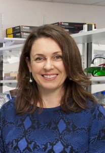
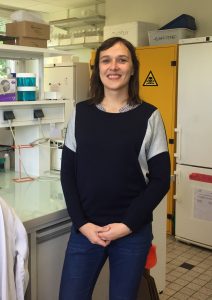
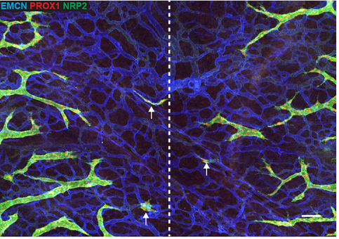
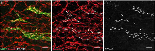
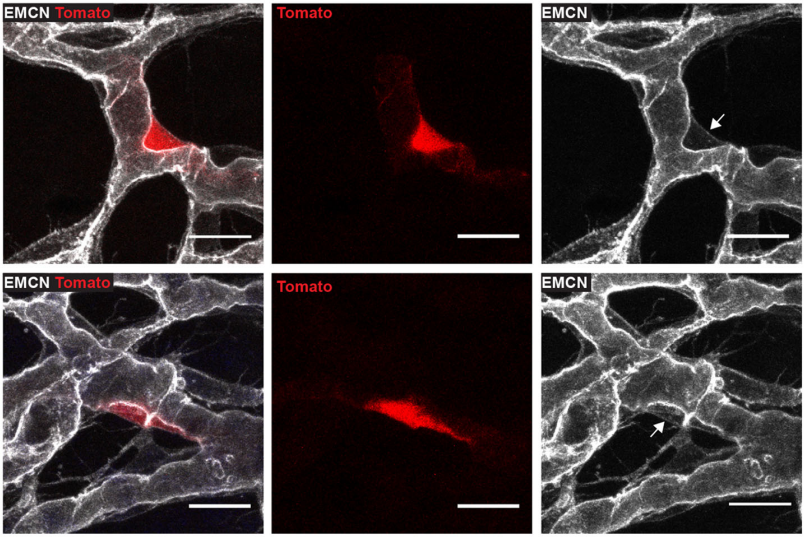
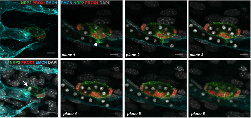
 (2 votes)
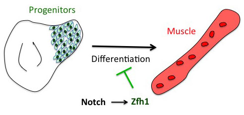
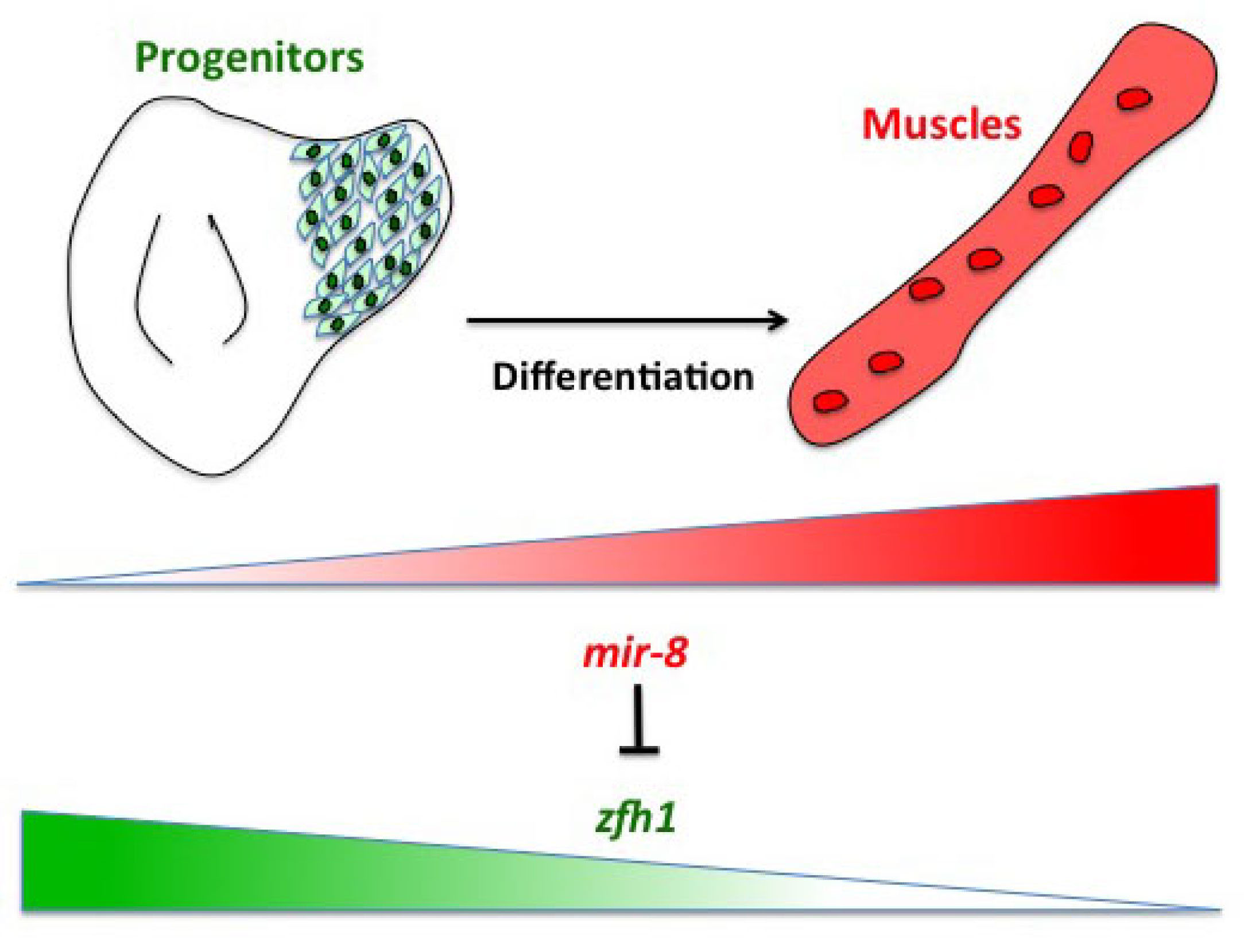
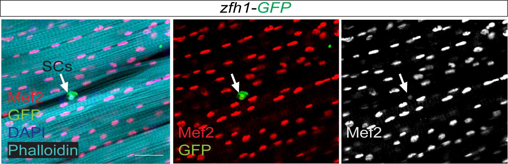
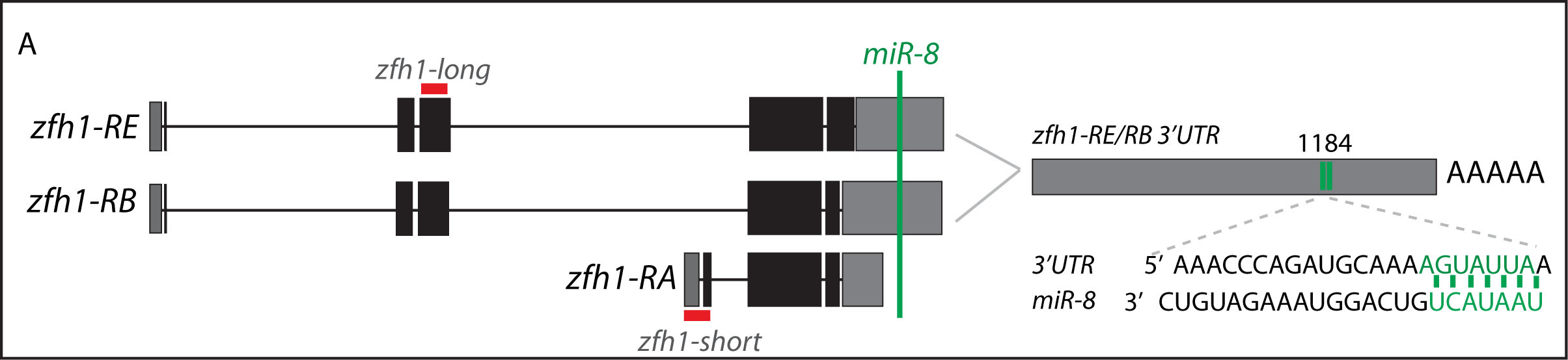
 (4 votes)
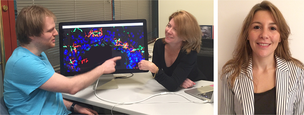

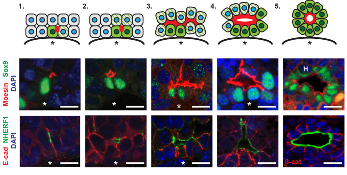
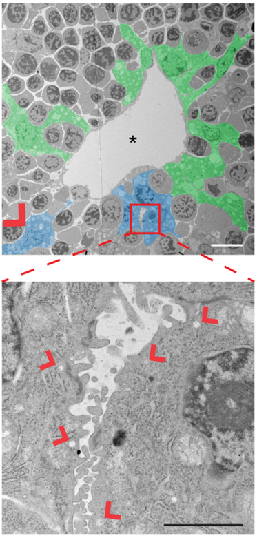
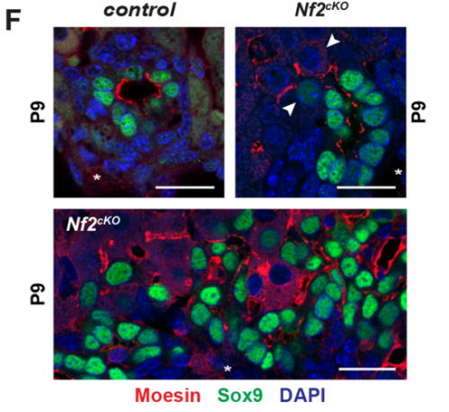

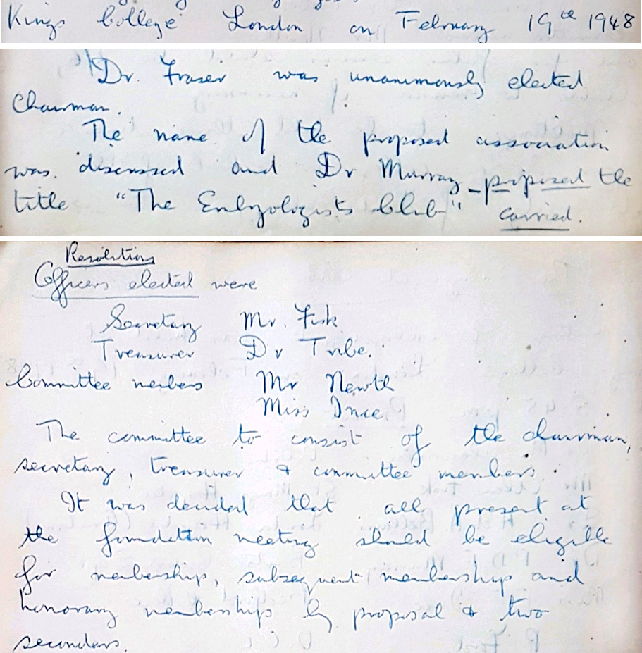
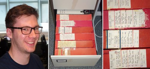

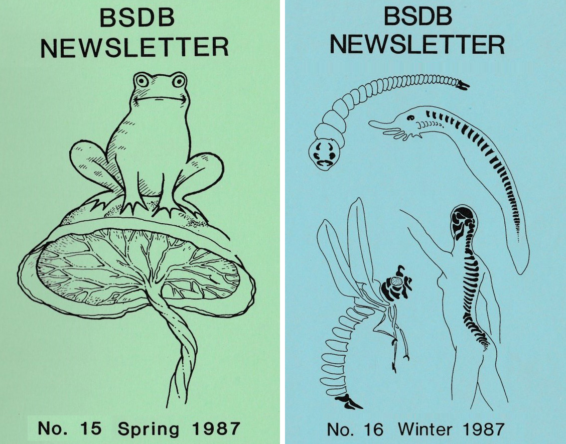
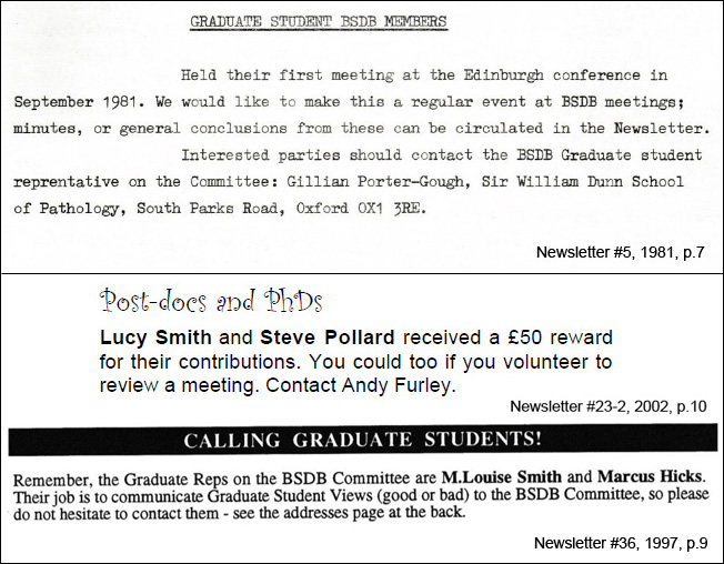
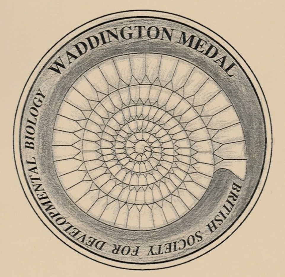

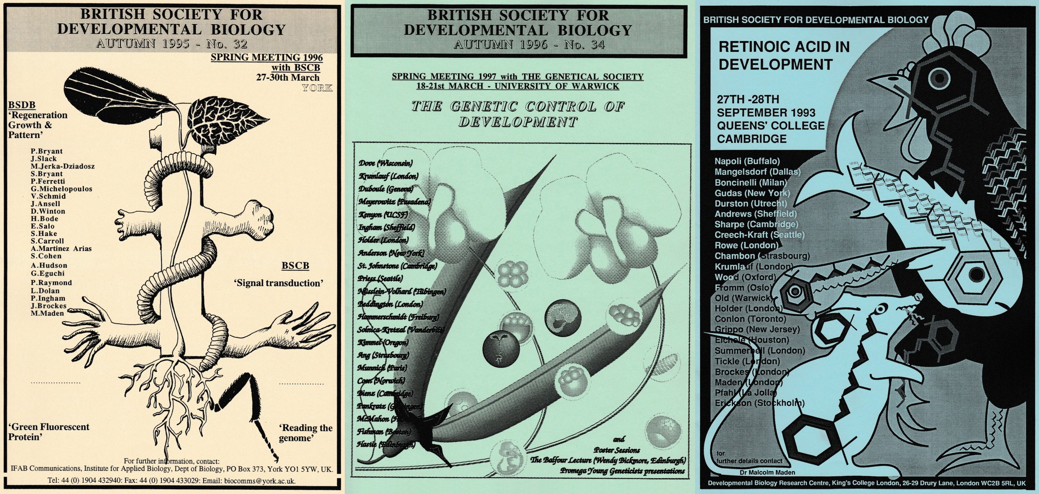
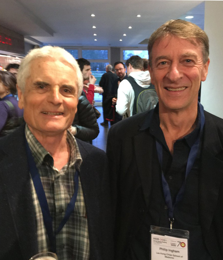
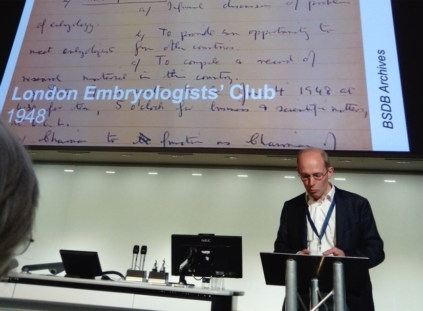
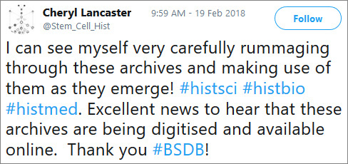
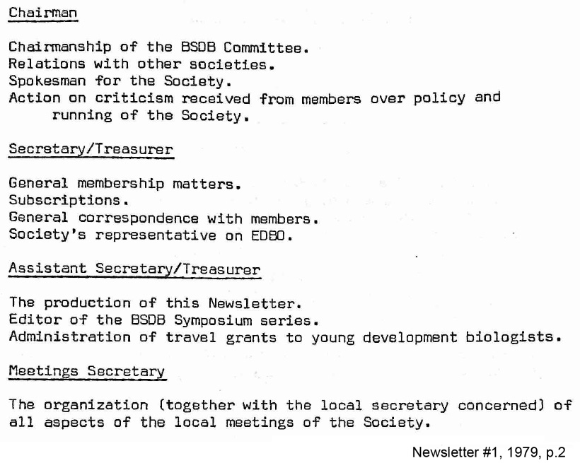
 I’m extremely happy to be acting as the new Graduate Representative for BSDB, following
 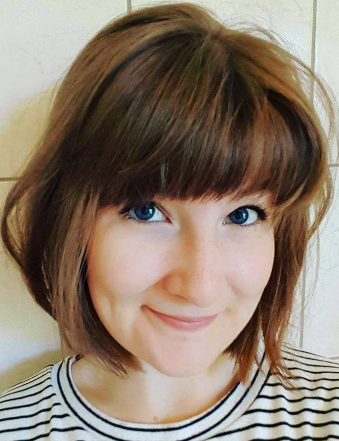 Having completed my PhD in the field of vertebrate somitogenesis in the lab of
 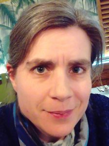 Tanya is Professor of Developmental Biology at the University of Sheffield, where she is a member of the Bateson Centre and Department of Biomedical Science [
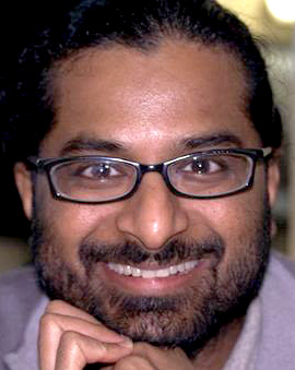 Shankar is Professor of Developmental Biology and a Wellcome Senior Investigator in the Department of Physiology Anatomy and Genetics at the University of Oxford [
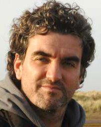 Jens is a Sir Henry Dale fellow at the School of Life Sciences at the University of Dundee running his lab in the division of Cell and Developmental Biology [