The University of Copenhagen, DanStem, seeks to appoint a Tenure-Track Group Leader Position in Pancreatic Cancer Biology to the Novo Nordisk Foundation Center for Stem Cell Biology (DanStem) to commence April 1, 2018 or after agreement. The position is for six years with possible extension depending on the outcome of a peer review.
Background
The Novo Nordisk Foundation Center for Stem Cell Biology (DanStem) is an international research center at the University of Copenhagen. The overall scientific goal is to develop new stem cell-based therapeutic approaches, currently in the area of diabetes and cancer addressing basic questions in stem cell and developmental biology and seeking to identify the factors that govern the development of different cell types in the body or new targets for anti-cancer therapy. Read about DanStem at www.danstem.ku.dk/
Job description
We are interested in recruiting a group leader with important and original contributions and the ability to develop a strong research programme in basic and disease-oriented pancreatic cancer biology. Space will be available at the main center location at the Panum institute (www.danstem.ku.dk) or at BRIC (www.bric.ku.dk). At Panum, there is an outstanding research environment for basic and translational stem cell and developmental biology research, and the main experimental platforms are localized here. At BRIC, there is an outstanding research environment for cancer biology, and two of the research groups at DanStem are located here. Both locations offer up-to-date lab and core facilities for stem cell and cancer research, competitive research support and dynamic and international environments.
The group leader is expected to:
Develop a strong research programme
Attract external research funding
Be interested in collaborating with other DanStem scientists and contribute to common activities at DanStem such as seminars and PhD courses
Be interested in training and supervision of young researchers
To contribute to teaching and educational activities
Terms of employment
Appointment will be as associate professor for six years. Salary and other terms and conditions of appointment are set in accordance with the Agreement between the Ministry of Finance and AC (Danish Confederation of Professional Associations) or other relevant professional organizations. The position is covered by the Job Structure for Academic Staff at Universities 2013.
Applicants recruited from abroad may be eligible for a special researcher taxation scheme.
Further information
For inquiries, please contact the DanStem Director, Professor Henrik Semb, semb@sund.ku.dk For questions regarding BRIC and the cancer research at BRIC, please contact the Director at BRIC, Professor Kristian Helin, kristian.helin@bric.ku.dk.
International applicants may find the university’s International Staff Mobility unit useful: www.ism.ku.dk.
Application
The University of Copenhagen encourages all interested applicants to apply for this position. Please submit the application with the required attachments by clicking on “Apply online” below or via this advertisement found on http://employment.ku.dk/faculty/.
The closing date for applications is February 7, 2018.
The application for the position must be submitted in English and include the following:
Application including reasons for applying for this post
Curriculum vitae
Relevant degree and educational certificates (diplomas)
List of publications
List of 5 publications you specifically wish to be considered in the assessment
Research plan – a concise description of previous research experience and a summary of current and proposed research (max 5 pages)
The online application form requires you to attach a formal teaching portfolio. This is not required for this position. Instead, documentation of your teaching and supervision experience must uploaded
Furthermore, 3 letters of recommendation should be arranged and be sent directly todanstemjob@sund.ku.dk (subject: “GL – name of applicant”) by the recommending person.
Application procedure
After the expiry of the deadline for applications, applicants are selected for assessment on the advice of the Appointments Committee. All applicants are notified whether their application has been passed for assessment by an expert assessment committee. Selected applicants are notified of the composition of the committee and each applicant has the opportunity to comment on the part of the assessment that relates to the applicant him/herself. You can read about the recruitment process at http://employment.ku.dk/faculty/recruitment-process/
Please note that the candidates may be asked to provide additional material. The applicant will be assessed according to the Ministerial Order no. 242 of 13 March 2012 on the Appointment of Academic Staff at Universities.
The Faculty of Health and Medical Sciences comprises app. 7500 students (medical sciences, oral health sciences, pharmaceutical sciences and veterinary medicine and animal science), app. 1500 PhD students and app. 3200 employees. The Faculty creates new knowledge and recognition through its core activities: research, teaching, knowledge sharing and communication. With basic research fields ranging from molecular studies to studies of society, the Faculty contributes to a healthy future through its graduates, research findings and inventions for the benefit of patients and the community.
Neuromesoderm progenitors are a population of stem cells that contribute to the neural tube and somite-forming paraxial mesoderm, and promote axial growth of the vertebrate embryo. In the latest issue of Development, a new paper addresses the transcriptional control of fate determination in this fascinating cell lineage. We caught up with co-first authors Alok Javali and Aritra Misra and their supervisor Ramkumar Sambasivanof the Institute for Stem Cell Biology and Regenerative Medicine in Bengaluru, India, to hear the story behind the work.
Alok, Ramkumar and Aritra
Ramkumar, can you give us your scientific biography and the main questions your lab is trying to answer?
RS It all started with the question how do skeletal muscle stem cells stay mitotically dormant and then wake up to proliferate and differentiate in order to repair damaged muscle. I had worked on this question for my Ph.D. thesis research and identified genes that play a role in return of dormant cells into active mitotic cycling. From addressing the cell biology of muscle stem cells, my interest broadened and I studied skeletal muscle development as well as regeneration in mice as a postdoctoral researcher. Currently, in my laboratory, we study the how the myogenic mesoderm develops and diversifies in vertebrates. Another major interest is on neural crest development, a new addition to my long-standing interest in muscle.
What is the current standing and future prospects for developmental biology research in India?
RS Developmental biologists make a small community in India and there is a need for expansion to derive the benefits of a strong community. However, things appear to move in the right direction. The number of developmental biologists in India has seen a significant spurt in the recent past, which I could gauge from the fantastic biennial meetings of the Indian Society of Developmental Biology. This augurs well.
Alok and Aritra – how did you come to join Ramkumar’s lab?
AJ I joined the graduate program of NCBS and InStem in 2013. Having prior interest in developmental biology, I did my lab rotation in Ramkumar’s lab. Being the first grad student in the lab, I had unique opportunity to be a part of multiple projects, which were still at their inception, ranging from studies on mesoderm patterning to mechanisms of fate choice in neural crest cells. This really excited me to join the lab for pursuing my PhD.
AM Prior to joining NCBS-inStem graduate program, I had trained in evolutionary biology and comparative morphology while studying zoology at bachelors and Masters level. This drove me to pursue PhD in either Evo-Devo or cell communication. Since Ramkumar’s work overlapped with my interest, I chose to join his lab.
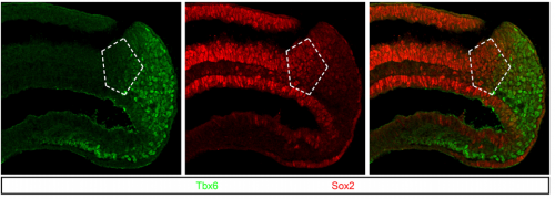
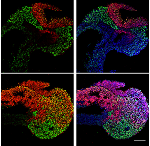
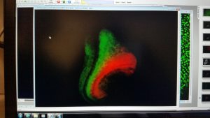
Tail bud NMP co-expression of Tbx6 and Sox2, from Fig 2, Javali, Misra et al. 2017
What’s so intriguing about neuromesodermal progenitors, and what were the questions you were hoping to answer in your study?
RS Neuromesodermal progenitors (NMPs) are key for axial growth in amniotes. I wonder about snakes and whales and the changes in the regulation of these progenitors that enabled such axially extended forms of life. Two different observations by Alok and Aritra took us in the direction of NMP biology and emphasized previous findings that Tbx6 is a key regulator of NMPs. The broad question is Tbx6 function in these progenitors. In this work we laid the foundation to tackle this question by addressing the timing of Tbx6 induction in NMP lineage.
Can you give us key results of the paper in a paragraph?
RS, AJ & AM We show that Tbx6 is expressed in a subset of neuromesoderm progenitors based on spatiotemporal expression pattern in the NMP niche in early mouse embryos. Co-expression of Tbx6 with Sox2, a neurogenic factor in this subset, as well as genetic tracing of Tbx6-expressing cells corroborated our findings of Tbx6 in NMPs. Expression of Tbx6 in NMPs provides a key missing link in favour of the current model in the field, which invokes Tbx6 as mesoderm switch in NMP lineage. In addition, we show that Tbx6 mutant mouse embryos have five developing spinal cords in the tail. This novel aspect of Tbx6 mutant phenotype in the tail further supports the model. Thus, our results strongly support the role of Tbx6 in promoting mesoderm fate choice of NMPs.
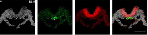
Assaying reporter expression in the caudal lateral epiblast, from Fig. 3, Javali Misra, et al. 2017
Your work supports the notion that Tbx6 as a bistable fate switch – how exactly do you think it plays this role?
AJ & RS Tbx6, being a transcription factor, functions by activating or repressing its transcriptional targets. Published literature already suggests that Tbx6 indirectly supresses the expression of Sox2, a key neurogenic factor, to promote mesodermal fate. An interesting aspect revealed by our study is the existence of a regulatory cell state, wherein Tbx6 and Sox2 are co-expressed. This indicates a biphasic function of Tbx6 in NMP lineage; early role in braking neural fate and subsequent function in mesoderm differentiation. We are currently trying to identify the spectrum of Tbx6 transcriptional targets to elucidate the mechanistic details of its function in NMP fate choice.
When doing the research, did you have any particular result or eureka moment that has stuck with you?
AJ: Looking at five distinct neural tubes arranged beautifully in a transverse section of mutant mouse embryo was special. Not everyday one gets to see such dramatic unreported phenotype. This was not really a Eureka moment as we were not anticipating this phenotype. This observation was accidental. We were trying to get transverse sections of a mutant embryo post in situ hybridization as the staining that we had performed was not very clear. We were not particularly looking into the tail region. Since the embryo is ‘C’ shaped, along with the transverse section of the interlimb region, we got the section of truncated tail of the mutant. And there we saw five distinct neural tubes.
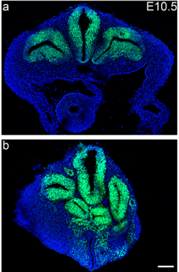
Supernumerary neural tubes in Tbx6 mutants, from Fig. 4, Javali, Misra et al. 2017
AM: Tbx6 is viewed uniquely as paraxial mesoderm factor, but while analysing the confocal images of immunostaining in E8.5 mouse embryo tail regions, I observed specific subset co-expressing Tbx6 and Sox2, a neural marker, within NMP niche. This was indeed a eureka moment for me, because in mouse gastrulation, this expression precedes paraxial mesoderm formation in NMP lineage. This implied that there is more to Tbx6 function than had been appreciated.
And what about the flipside: any moments of frustration or despair?
AM: The most frustrating moment was trying to obtain serial transverse sections of a very specific anatomy of tiny E8.5 mouse embryos for control experiments to demonstrate that our novel Tbx6Cre transgenic line reports for Tbx6 faithfully. I had to ensure that I do not lose any section in the series. This experiment also provided crucial support to our observation of Tbx6 expression in the NMP niche.
AJ The most frustrating time was when I was trying to get non-oblique transverse section of primitive streak and node regions of e8.5 mouse embryos and saggital section of tailbud of e9.5 mouse embryos. The region of interest in both these cases is extremely small. Getting the ideal sections with all the necessary anatomical information to identify the region of interest took multiple attempts.
Sagittal sections of tail buds with Sox2 (r) YFP (g) and Hoescht (b), from Fig. 4, Javali, Misra et al. 2017
What are your career plans following this work?
AM: I want to continue in academia. My interest is to work on the evolution of bilateral symmetry and I want to start by looking into reptiles. But I will also be excited to explore opportunities in facilities such as mouse genome engineering facilities or Jackson Laboratories.
AJ: I am currently working on my PhD project where I am trying to understand the role of micro-RNAs in the development of another interesting cell type called neural crest. As much as I am attracted to academic research, along with it, I would also like to explore a career path in science education, which I believe is a need of the hour in India.
And what next for the Sambasivan lab?
I will address what next for my lab in the context of NMPs. We are curious about the evolutionary origin of NMPs and tracing this in the chordate lineage.
Finally, what do you like to do when you are not in the lab?
AM: I always wanted to be a historian and study the evolution of human civilization. I have not had the opportunity before joining for Ph.D., however, now I use my time out of lab to fulfil this dream. In the past 2 years I have researched on how evolution of Indian civilization can be traced to have more connections to the development of agrarian systems rather than changes in ruling dynasties. Recently I started to work on socio-economic changes in pre-British era driving changes in Indian demography. This began with an amazing book: Rise of Islam and the Bengal Frontiers by Richard Eaton. Currently I have started probing into the concept of Sacred Feminine and evolution of Egyptian society.
AJ: There are few things that I usually do. Swimming or running is my stress buster. I love to play Cricket (famous sport in India), though I don’t get much time for it anymore. I also love to read popular science books
Laboratory: The Arbeitman Laboratory at Florida State University, College of Medicine studies the molecular-genetic basis of complex reproductive behaviors, using Drosophila as a model.
Projects: The laboratory has NIH-funded ongoing projects to understand Drosophila courtship behaviors, using a range of approaches, including neural circuit based studies and cutting-edge, single-cell genomic-scale approaches.
In addition to laboratory funding, Florida State University has initiated several career enhancing programs for postdoctoral scholars, including a Provost Postdoctoral Fellowship and Teaching/Research Postdoctoral Fellowship positions.
We are seeking to recruit a biocurator/ontologist to contribute to two Drosophila databases: Virtual Fly Brain (VFB) and FlyBase. If you are looking for a fulfilling, fly-related career away from the bench, and enjoy the challenge of organizing complex data and presenting it clearly and concisely, then this is the job for you! We will train the new recruit in biocuration and editing of controlled vocabularies (ontologies).
Virtual Fly Brain is a resource that helps biologists make sense of the vast amount of data in this field by enabling scientists to map and visualize neuronal circuits, and find the experimental reagents to target them.
Virtual Fly Brain works closely with FlyBase, the primary community resource for Drosophila genetic, genomic and functional data. The main responsibility of the FlyBase-Cambridge site is to identify relevant data from scientific articles and to record and organize these data in a systematic manner.
The work will include:
Synthesising information from the literature to extend the representation of Drosophila anatomy and phenotypes in VFB and FlyBase, with a particular emphasis on neuro-anatomy and behavioural phenotypes
Curating image, connectomics and phenotype data into VFB and FlyBase
Curation of genotype information from the literature into FlyBase
Contributing to the design and functionality of Virtual Fly Brain and FlyBase
Presenting the work of VFB or FlyBase at conferences and in publications
The end of the year is quickly approaching, and if you are anything like me you are scrambling to try to get as much work done as possible before your holiday break. But while this frequently entails getting papers submitted, committee meetings completed, and experiments wrapped up, I also take the opportunity to reflect on what has happened over the last year, and where I want to be in the upcoming year. This time last year, I was a brand new post-doc. I was just getting settled into my new lab working with Dr. Chenbei Chang, a new city and University, the University of Alabama at Birmingham, and most exciting for me, a new model organism in Xenopus laevis. I was looking forward to new challenges including adding to my science toolkit and interacting with a new community of scientists. I had some general ideas of what I wanted to accomplish in my first year in the lab, but these ideas were still pretty vague as I was just trying to figure out the direction I wanted my research to take.
It was at this time that Chenbei told me about several educational opportunities I might be interested in, including the Cell and Developmental Biology of Xenopus Course run out of the Cold Spring Harbor Laboratory. I have many friends who have taken advantage of these types of specialty courses, from both CSHL and the Marine Biological Laboratory, and heard great things about them. I had always wanted to take part in one of these courses myself, but had never applied due to a myriad of excuses mostly centered around the time it would require out of my normal schedule. But this time, I would not let myself be deterred and was resolved to apply to and attend this course. “What better time to take such a course?” I mused. I am just getting started in the lab so it will be easier to get time away. I will get to learn a bunch of techniques in a short amount of time that will really get me jumpstarted on my own research. And for me, the most intriguing draw to the course was the opportunity to meet all of the amazing instructors that would take part in the course. Who better to learn a technique from than the person for whom the technique is named (Keller sandwich anyone?) I quickly got my application materials submitted and then waited, rather impatiently, for a reply.
Once I heard that I was accepted, I immediately started planning. One part of the current course is to learn how to use CRISPR/Cas9 technology to target your gene of interest in Xenopus embryos. I had just started working on a particular gene in the lab and was excited to try to target it at the course. I submitted the genomic information I had to one of the course TAs and there was a set of custom guide RNAs waiting for me upon my arrival at the course. I also thought about how I might perform experiments at the course relevant to my own research and planned to ship myself some reagents. With great anticipation, I arrived at Cold Spring Harbor on a cold, dreary day in March. Despite the weather, I thought the campus was beautiful and I could just feel the great energy that surrounds this place, where so many great scientific minds have come together.
I won’t bore you with all of the details of my own experience, but suffice it to say that the experience met, and exceeded, all of my expectations. The course directors, Karen Liu and Mustafa Khokha, put together an amazing course. As you can probably guess, my expectation of great experimental data was entirely too high, but what I didn’t bring home in data, I brought in ideas and techniques and enthusiasm. One of the great aspects of a course like this is flexibility. The students attending the course all have different backgrounds and levels of experience with Xenopus. The aim of the course is to provide to the students what they, in particular, feel they need. So while there is a general outline of the techniques that will be taught in the course, you as an individual are free to ignore a technique that you feel comfortable with and instead concentrate your time on the techniques you want to learn more about.
For me, the absolute best part of the course was all of the people involved. Karen and Mustafa bring in a great variety of instructors with all levels of experience. There are classic Xenopus embryologists, such as Ray Keller, who showed me one-on-one how to physically manipulate my embryos. But there are also instructors that use Xenopus in completely different and unexpected ways, such as Peter Nemes who is working to push the limits of in vivo metabolite detection. Another unexpected gift of this course was a new and spontaneous collaboration between myself, another student, and an instructor that spans three different countries and resulted in a manuscript currently in preparation. I made so many great connections with other students, TAs and instructors whom I hope to know as colleagues for many years to come.
And so with these final thoughts I ask you, are you a student, post-doc or faculty looking for a transformative experience that will last for many years to come? Are you currently working with Xenopus as a model system or are you curious about what working with this organism would be like? Are you looking for collaborators and colleagues in this field? This year for Christmas, give yourself the gift of an amazing learning experience and apply for the 2018 Cell and Developmental Biology Course to be held April 4-17 at the Cold Spring Harbor Laboratory. Applications aren’t due until January 31st, 2018 so there is plenty of time to apply! You won’t regret it.
Even the misty rain couldn’t take away from the beauty of the harbor
The stars of this course!
Getting down to business in the lab
Of course there’s some down time to enjoy with new colleagues
One of my fun creations, a GFP to RFP organizer transplant. Mad science at its finest!
The sun came out long enough for a walk on the beach
(1) Associate Professor (or Professor) in Evolution and Development
Department of Zoology, Oxford (in association with Merton College)
Applications are encouraged from candidates with a strong background in evolutionary or comparative developmental biology. https://tinyurl.com/yamkmx3q
Also:
(2) Associate Professor (or Professor) in Comparative Physiology/ Organismal Biology, Department of Zoology, Oxford (in association with Hertford College) Applications are encouraged from candidates with research interests in comparative physiology, neurobiology, biomaterials, biomechanics, bio-inspired technologies or a related field. https://tinyurl.com/y8g9butw
Further particulars, including details of the application procedure and duties, from the above websites. Closing date for applications 19 January 2018.
In the lab we aim to dissect the interplay between metabolism and major signalling pathways, with emphasis on the Notch signalling pathway.
Project: Metabolic stress as regulator of Notch signalling
The Notch pathway has been described in several contexts to regulate glycolytic as well as mitochondrial metabolism (including paper from our lab Slaninova V, Open Biology, 2016). At the same time, recent evidence suggest that Notch pathway activity is sensitive to the cell metabolic parameters, such as the NAD:NADH ratio, amino acid availability or activity of metabolism related signalling pathway such as mTOR (Horvath M, Biochem J, 2016 and our current paper in preparation). We investigate this interplay using Drosophila wing disc, eye disc and immune system as models. The theme of the postdoc/PhD project can be defined according to the postdoc/PhD previous experience and interests, but it could be directed to characterization of mitochondria as regulators of Notch directed proliferation, to identification of binding sites for a Notch targeted transcription factor involved in metabolism or to in vivo analysis of metabolic parameters via genetically encoded metabolic sensors.
Applicant: Applicant should have a degree in molecular or cellular biology. Previous experience with Drosophila, molecular biology techniques and/or confocal microscopy is welcome.
Position: Postdoc offer includes a 2-year contract with the possibility of extension. PhD position is a part of the 4-year PhD program at the Department of molecular biology and genetics, University of South Bohemia. More details of the Departments at http://kmb.prf.jcu.cz
Send your CV including address of two referees to akrejci@prf.jcu.cz
My name is Charlotte Blackburn; I am a Zoology graduate currently studying at the University of Edinburgh for an MSc in Science Communication and Public Engagement … yes, that is something of a mouthful! I recently had the opportunity to participate in the droso4schools science communication project of the Manchester Fly Facility, and would like to explain here how this came about, the experiences I made, and how this aligned with my training in science communication.
I’ve been enthusiastically involved in “scicomm” (as it is known to those in the trade) since my second year of university. I have always found engaging the public with science to be incredibly rewarding, and I chose to apply for the Edinburgh course as I wished to explore this emerging field in more detail. Thus far, I have studied topics such as the relationship between modern science and society, the ins and outs of science-based policy making, and the effects of the media on the public’s perception of science. Interestingly, much of what is discussed concerns getting scientists interested in science engagement, rather than the public, as one might assume. In agreement with this notion, also a recent special issue on science communication in the biomedical sciences emphasises that the low degree of participation of researchers in scicomm is an important issue that needs to be addressed. On the one hand, there might be a lack of understanding of scicomm, but often there simply isn’t a large enough incentive for scientists to get involved, and clearly there is lack of time. Life in academia, with its many different tasks in research, teaching, administration, grant writing, dissemination and industry collaboration, is very busy; it is therefore unsurprising that many seem to view science communication as just another job to throw on their ever-teetering pile.
It is my current ambition to achieve a doctorate, and I would ideally like to be able to combine a career in research with my passion for science communication. However, practical experience is something many graduates struggle to gain once they have left the relative comfort of full-time education. Funded internships and research assistant posts are few and far between, and are not always relevant to one’s interests. So I decided to take a proactive approach and sent a few short emails to neuroscientists at the University of Manchester, enquiring about potential voluntary placements. An unusual request, but one that I am glad I made. Soon after, I received a positive response from Professor Andreas Prokop. Although we could not arrange for a longer-term science project for formal reasons, there was an opportunity to take an active part in the science communication initiatives of the Manchester Fly Facility instead – would I be interested? Of course! I jumped at the chance, and I was offered a one months’ work experience placement, supervised by Andreas and the fly facility manager Sanjai Patel.
The Manchester Fly Facility hosts fourteen research groups, each exploring a different area of biology using, as their research tool and strategy, the model organism Drosophila melanogaster (commonly known as the fruit or vinegar fly). I have to confess, before I began my placement at the Facility, I hadn’t had any significant experience with invertebrate biology, let alone Drosophila – and I was only aware of its use as a model organism in the field of connectomics. Up to that point, I had been much more interested in discussing research involving rodents, or non-human primates. However, by the end of my placement, I could be found happily listing the merits of fruit fly research with anyone who would stand around long enough to listen. What then occurred in this short space of time, to turn a normally vertebrate-oriented biologist into such an enthusiastic cheerleader for an insect? Well, I learned a lot about the power and enormous range of applications of fly research. It turns out that the “simple” fly is actually wonderfully complex – for example, flies can memorise and learn – capabilities I would have thought to be exclusive to higher vertebrates. They can also get drunk, aggressive, and jet lagged … and were the first organisms ever to return safely from a trip to space! And, this year marked the seventh Nobel Prize in Physiology or Medicine awarded for research in fruit flies!
It was a pleasant surprise to find that the researchers at the Manchester Fly Facility are passionate advocators of fruit fly research. As explained in a recent paper, they have developed a comprehensive science communication initiative which not only addresses the public and other non-drosophilist researchers or clinicians, but also recognises the need to communicate with and inspire their own select research community – since the need and importance of science communication and Drosophila advocacy appears not to be recognised by many scientists (as already discussed above).
At the Manchester Fly Facility, however, there is a team of scientists who actively make time. They do so, because they believe firmly and passionately in the importance and benefits of engagement, for both the general public, formal education, and the wider scientific community, and this is clearly spelled out in their “Vision, Mission, Purpose” statement, as well as in a recent PLoS blog.
As part of their work with the public, the Fly Facility also initiated the droso4schools project which aims to introduce Drosophila as a teaching tool in the biology lessons of schools and colleges. Within this project they developed an excellent, regularly reviewed program of biology lessons for schools and colleges, offering a plethora of free, high quality online resources for teachers, as has been explained in a recent publication.
The school biology lessons they generated are the result of long-term collaborative efforts of research staff and placement students together with engaged teachers at partner schools; they link fundamental biology teaching to both past and current fly-based research, and directly address numerous key learning requirements from the national curriculum, often with an end-of-year revision session in mind. There is even a lesson on statistics! Notably, the lesson resources with teacher support materials can be downloaded separately and are being used not only in the UK, but worldwide (see their impact document).
And it doesn’t stop there. The team have also put together a set of complementary resources to be used by other scientists on extra-curricular school visits and science fairs, and they engage in school visits themselves and offer CPD sessions hosted at the university, for teachers interested in using flies as regular hands-on teaching tools in the classroom (see their impressive list of visits and events).
The work Andreas and Sanjai had in mind for me, was lab-based, aiming to develop and document fly experiments for their active outreach programme. When I discovered this at our initial meeting, I became even more enthusiastic – what a great opportunity to combine my interests! My placement allowed me to gain insight into the working behind this initiative which, to my great surprise, is driven in its core by only two people, Sanjai and Andreas.
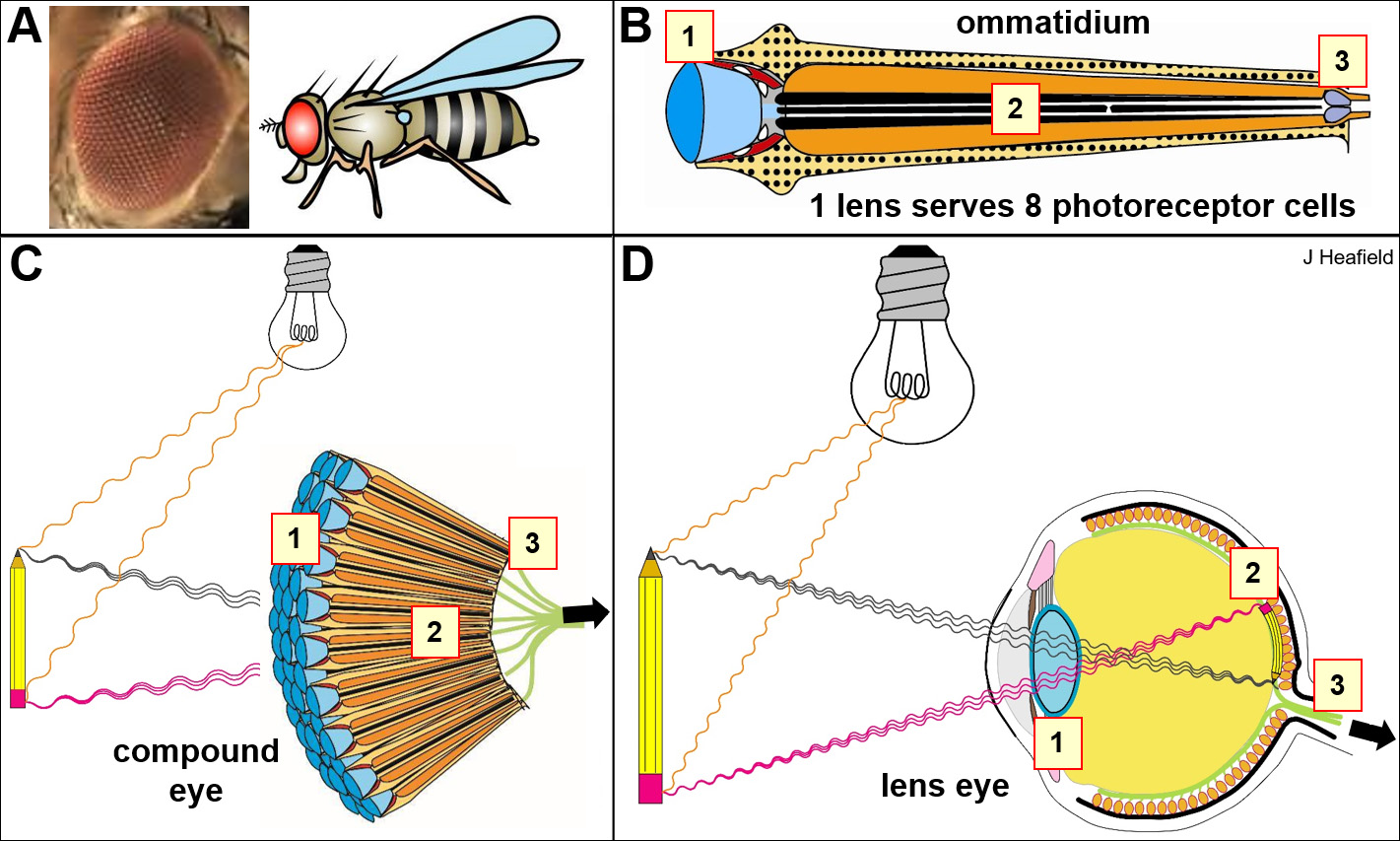
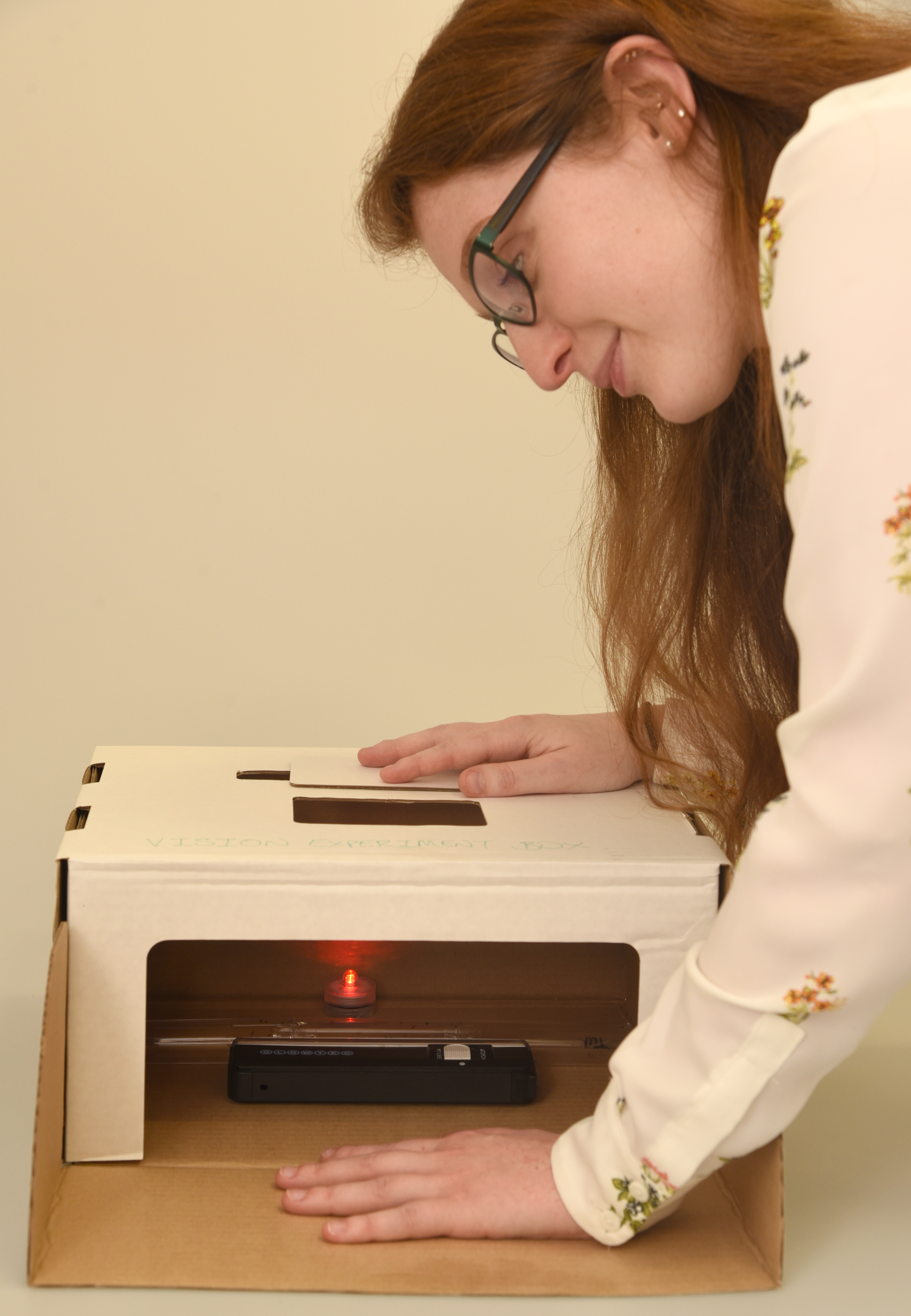
(A) Compound eye of Drosophila. (B) Ommatidium, a single visual unit of the compound eye: 1, lens; 2, photoreceptive cells; 3, exiting nerve leading to the brain. A diagrammatic view of a compound eye (C) compared to a human eye (D); although the eye anatomy is different, the steps in detecting light are the same. For more info, see the vision resource.
My task was to work out and document experiments for an almost completed lesson concerning the science of vision (see the online resource). Drosophila, with their compound eyes, are capable of seeing different wavelengths of light, similar to what we would describe as colour vision in humans. Part of my task was to develop a method of demonstrating this in the form of a hands-on and/or virtual micro experiment for use in classrooms. Initially, I was a little daunted, but I soon settled into the familiar trial-and-error routine of scientific problem-solving. I received training in specialist fly handling techniques, and fantastic support from Sanjai, who was a great help when it came to formulating feasible ideas. It wasn’t too long before I developed an experimental set-up that would allow me to show colour vision and colour-blindness in flies; exploiting the fly’s positive phototaxis behaviour (a natural attraction toward light sources, as is explained on the resource’s web page), I devised an experimental set-up with a series of coloured and UV lights, using materials sourced from my father’s company, which specialises in UV-based hand hygiene training.
After a few sessions of fine-tuning, I began the filming process, aiming to show-case the experiment on YouTube to help teachers set it up, and to complement the web resource. The video footage can also be used in classrooms where teachers do not have the time or capacity to set it up as a hands-on practical. With the fantastic support (and patience) of Nick Ogden and his team at the School of Biology’s Photographics Unit, it took only a few sessions over a number of days to collect some excellent footage of vision-based fly behaviours in action. The art of video editing also took a bit of getting used to at first, but the final cut is something I’m quite proud of.
Apart from having gained an insight into the biology of a truly remarkable organism (which once I would have swatted away without so much as a second thought), this placement has been a fantastic learning experience. I’ve had a chance to synthesise and exercise my theoretical knowledge in practice, and gain hands-on experience in the busy world of academic science communication.
I’ve been able to engage in lively discussions regarding scicomm strategies with my supervisors, and with other researchers at the facility. Yes, scientists are busy, but with a clear, overarching objective, scicomm participation can be woven into even the most hectic of scientific lives. Scicomm strategies don’t just materialise overnight (nor do they need to) – the most successful are formed from a careful step-by-step process of pooling, developing, and evaluating ideas over the long-term.
I have learned some of the ways in which information I’ve generated can be communicated, in order to maximise impact, and capitalise on time invested. For example, the practical experiment I devised could not only be used in school lessons, but also at science clubs, and as a demonstration at science fairs. Furthermore, the video has been posted on YouTube, and can now be shared via other social media platforms. I’ve also been involved in some exciting discussions regarding the potential development of a large-scale engagement initiative, one which could reach thousands of pupils, far more than could ever be reached through scattered school visits. The CusMiBio Project (University of Milan, Italy) has shown how a simple school outreach programme can be cultivated, over time, into a truly remarkable collaborative enterprise. Using this initiative as a blueprint for their own, the Manchester Fly Facility team could help to put Manchester on the map as a national frontrunner in science education outreach. This would not only be a fabulous asset to the university, but also for the schools involved.
The Manchester Fly Facility team are an inspiring example of science professionals who understand that science communication and engagement should be, and indeed is, a mutually beneficial experience. It is often said that enthusiasm can be contagious, and I for one can only hope that this team’s enthusiasm for science engagement continues to spread a buzz throughout the academic community.
Charlotte Blackburn
This blog post was first published on the droso4schools site [LINK]
The Mokalled lab at Washington University School of Medicine is hiring at all levels (http://www.mokalledlab.com/). Our lab uses zebrafish and mouse model systems to study neural regeneration after spinal cord injury or disease. Candidates with enthusiasm for neuroscience, regenerative biology, and zebrafish research are encouraged to forward a cover letter, CV, and list of 3 or more references to mmokalled@wustl.edu.
Here are the highlights from the current issue of Development – the last one of the year! Happy reading…and happy holidays!
Branched actin keeps Nrf2 in check in the skin
The Arp2/3 complex is responsible for the assembly of branched actin filaments. Although its cellular functions are well understood, less is known about the consequence of its disruption in developing animals. To investigate the function of Arp2/3 in the skin, Metello Innocenti and colleagues (p. 4588) have generated mice specifically lacking Arpc4, a core component of the complex, in the epidermis (referred to as Arpc4 eKO). These animals developed progressive psoriasis-like lesions soon after birth, accompanied by characteristic dermal inflammation. Differential gene expression analyses reveal upregulation of inflammatory and dermatological disease-related pathways in the affected Arpc4 eKO tissue. The mutant mice show increased epidermal levels and activity of Nrf2, a master regulator of epidermal homeostasis, the hyperactivation of which was previously found to result in severe keratinocyte abnormalities similar to those observed in the Arpc4 eKO. Furthermore, the authors show that Nrf2 interacts with the actin cytoskeleton, and a functional Arp2/3 complex sequesters Nrf2 to the cytoskeleton, effectively blocking its pro-psoriatic transcription factor activity. Intriguingly, Arpc4 appears to be downregulated in human psoriatic lesions and chemically induced psoriasis-like lesions in mice, thus providing further support to the authors’ model whereby loss of Arp2/3 is involved in the pathogenesis of psoriasis.
Tbx6 seals mesodermal fate in the tail bud
Neuromesoderm progenitors (NMPs) are a distinct stem cell population that express both the mesoderm marker brachyury (T) and the neural marker Sox2, and are required for axial growth in vertebrates. They are capable of binary fate choice, differentiating as either neural or mesodermal progenitors. Tbx6, a downstream target of T, has been proposed to act as a fate switch, stimulating the mesodermal differentiation of NMPs, but the timing of this fate commitment is unclear. On p. 4522, Ramkumar Sambasivan and co-workers report the identification of cells in the mouse primitive streak and later in the tail bud that co-express Tbx6 and Sox2. These Tbx6+/Sox2+ cells represent a novel transient subpopulation of NMPs primed for mesodermal differentiation. Tbx6-null NMPs in mouse embryos are incapable of mesodermal commitment and default to neuronal differentiation, which strikingly results in ectopic formation of neural tubes. The authors show that this phenotype is stronger in more posterior regions, suggesting a differential requirement for Tbx6 in trunk versus tail NMPs. These data confirm the proposed ‘fate switch’ role of Tbx6 in mesodermal commitment of NMPs, and further our understanding of NMP differentiation and their role in body axis elongation.
FGF signalling: making matrix in the lung
Alveologenesis – the repeated division and growth of alveoli to expand the surface area in the lung – is one of the least understood stages of postnatal lung development. Defective alveologenesis results in bronchopulmonary dysplasia, a disorder often observed in premature infants. FGF signalling and the extracellular matrix (ECM) protein elastin are known to be important for alveologenesis, but the underlying mechanisms are poorly understood. On p. 4563, Xin Sun and colleagues dissect the roles of FGFR3 and FGFR4 in alveologenesis. Using both global and conditional Fgfr3;4 double-mutant mouse models, they show that the earliest apparent phenotype – preceding any defects in alveolar organisation – is the improper deposition of elastin fibres. Levels of elastin protein are initially unchanged, but fibre organisation is disrupted, suggesting that this may be the underlying cause of the alveolar simplification observed in the mutants. Furthermore, the phenotype can be partially rescued by depletion of Mfap5, a regulator of elastin deposition that is upregulated in the Fgfr3;4 mutant. Lineage-specific inactivation of Fgfr3;4 demonstrated that normal alveologenesis requires functional Fgfr3 and Fgfr4 in the mesenchyme but not the epithelium, which is consistent with the known role of fibroblasts in ECM organisation. The authors suggest that a defective elastin ECM physically interferes with alveolar septa formation, resulting in simplified alveoli characteristic of bronchopulmonary dysplasia.
Braving the cold with β-Tubulin 97EF
Although mammals have internal mechanisms for regulating body temperature, the vast majority of organisms are ectotherms, meaning that their body temperature is dictated by the external environment. Temperature fluctuations significantly affect cellular homeostasis, but the molecular mechanisms underlying these effects are currently poorly understood. Multiple lines of evidence suggest that microtubules are sensitive to cold, with suboptimal temperatures causing their disassembly. Christian Lehner and colleagues (p. 4573) employed global gene expression analyses of Drosophila S2R+ cells grown over a range of temperatures to identify β-Tubulin 97EF, a previously poorly characterised β-tubulin paralogue, to be among the most temperature-responsive. This upregulation was confirmed in vivo, and exhibited distinct tissue specificity, with expression being most prominent in the gut and the hemocytes. Despite the mild phenotypic consequences of β-Tubulin 97EF inactivation, likely confirming functional redundancy between β-Tubulin paralogues, βTub97EF mutant Drosophila embryos were more sensitive to the cold than their wild-type counterparts. Moreover, although there was no correlation between β-Tubulin 97EF levels and microtubule assembly rates, microtubules containing β-Tubulin 97EF were less prone to destabilisation at lower temperatures. Taken together, these results identify β-Tubulin 97EF as a cold-regulated isoform that promotes microtubule stability, and highlight the importance of mechanisms to allow acclimation to temperature variations.
PLUS:
An interview with Claudio Stern
Claudio Stern is the J. Z. Young Professor of Anatomy at University College London (UCL), UK. His lab studies the processes that regulate patterning and cell diversity in the early embryos of vertebrates, mostly in chick. Claudio, an elected fellow of the Royal Society, the UK Academy of Medical Sciences, and the Latin-American Academy of Medical Sciences, was awarded the 2006 Waddington Medal by the British Society of Developmental Biology, and he also served as President of the International Society for Developmental Biology (ISDB) from 2010-2013. At the 18th Congress of the ISDB (Singapore, June 2017), Claudio was awarded the ISDB’s Ross Harrison Prize, which recognises an individual’s outstanding contributions to developmental biology. We met with Claudio to ask him more about his career, his thoughts on the field, and his advice for early career researchers. Read the Spotlight article on p. 4473.
The TGFβ superfamily in Lisbon: navigating through development and disease
The 10th FASEB meeting ‘The TGFβ Superfamily: Signaling in Development and Disease’ took place in Lisbon, Portugal, in July 2017. Here, Jan Christian andCarl-Henrik Heldin review the findings presented at the meeting, highlighting the important contributions of TGFβ family signaling to normal development, adult homeostasis and disease, and some novel mechanisms by which TGFβ signals are transduced. Read the Meeting Review on p. 4476.
The hallmarks of cell-cell fusion
Cell-cell fusion is essential for fertilization and organ development. Dedicated proteins known as fusogens are responsible for mediating membrane fusion. However, until recently, these proteins either remained unidentified or were poorly understood at the mechanistic level. Here, Javier Hernández andBenjamin Podbilewicz review how fusogens surmount multiple energy barriers to mediate cell-cell fusion. See their Review article on p. 4481
Mechanisms of gene regulation in human embryos and pluripotent stem cells
While the principles that establish and regulate pluripotency have been well defined in the mouse, it has been difficult to extrapolate these insights to the human system due to species-specific differences and the distinct developmental identities of mouse versus human embryonic stem cells. In their Review, Thorold Theunissen andRudolf Jaenisch examine genome-wide approaches to elucidate the regulatory principles of pluripotency in human embryos and stem cells, and highlight where differences exist in the regulation of pluripotency in mice and humans. Read their Review article on p.4496
 (No Ratings Yet)
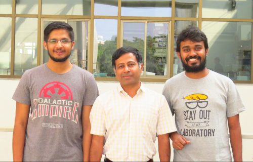
 (17 votes)
 (3 votes)
 The Arp2/3 complex is responsible for the assembly of branched actin filaments. Although its cellular functions are well understood, less is known about the consequence of its disruption in developing animals. To investigate the function of Arp2/3 in the skin, Metello Innocenti and colleagues (p.
  Neuromesoderm progenitors (NMPs) are a distinct stem cell population that express both the mesoderm marker brachyury (T) and the neural marker Sox2, and are required for axial growth in vertebrates. They are capable of binary fate choice, differentiating as either neural or mesodermal progenitors. Tbx6, a downstream target of T, has been proposed to act as a fate switch, stimulating the mesodermal differentiation of NMPs, but the timing of this fate commitment is unclear. On p.
  Alveologenesis – the repeated division and growth of alveoli to expand the surface area in the lung – is one of the least understood stages of postnatal lung development. Defective alveologenesis results in bronchopulmonary dysplasia, a disorder often observed in premature infants. FGF signalling and the extracellular matrix (ECM) protein elastin are known to be important for alveologenesis, but the underlying mechanisms are poorly understood. On p.
  Although mammals have internal mechanisms for regulating body temperature, the vast majority of organisms are ectotherms, meaning that their body temperature is dictated by the external environment. Temperature fluctuations significantly affect cellular homeostasis, but the molecular mechanisms underlying these effects are currently poorly understood. Multiple lines of evidence suggest that microtubules are sensitive to cold, with suboptimal temperatures causing their disassembly. Christian Lehner and colleagues (p.
  Claudio Stern is the J. Z. Young Professor of Anatomy at University College London (UCL), UK. His lab studies the processes that regulate patterning and cell diversity in the early embryos of vertebrates, mostly in chick. Claudio, an elected fellow of the Royal Society, the UK Academy of Medical Sciences, and the Latin-American Academy of Medical Sciences, was awarded the 2006 Waddington Medal by the British Society of Developmental Biology, and he also served as President of the International Society for Developmental Biology (ISDB) from 2010-2013. At the 18th Congress of the ISDB (Singapore, June 2017), Claudio was awarded the ISDB’s Ross Harrison Prize, which recognises an individual’s outstanding contributions to developmental biology. We met with Claudio to ask him more about his career, his thoughts on the field, and his advice for early career researchers. Read the Spotlight article on p.
  The 10th FASEB meeting ‘The TGFβ Superfamily: Signaling in Development and Disease’ took place in Lisbon, Portugal, in July 2017. Here,
  Cell-cell fusion is essential for fertilization and organ development. Dedicated proteins known as fusogens are responsible for mediating membrane fusion. However, until recently, these proteins either remained unidentified or were poorly understood at the mechanistic level. Here,
  While the principles that establish and regulate pluripotency have been well defined in the mouse, it has been difficult to extrapolate these insights to the human system due to species-specific differences and the distinct developmental identities of mouse versus human embryonic stem cells. In their Review,