On Growth and Form at 100: Morphogenesis one century after On Growth and Form
Posted by the Node, on 28 November 2017
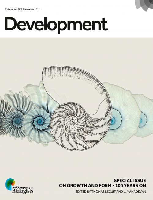
This editorial by Thomas Lecuit and L. Mahadevan originally appeared in Development’s Special Issue: On Growth and Form – 100 Years On
Morphogenesis, the study of how forms arise in biology, has attracted scientists for aeons. A century ago, D’Arcy Wentworth Thompson crystallized this question in his opus On Growth and Formusing a series of biological examples and geometric and physical analogies to ask how biological forms arise during development and across evolution. In light of the advances in molecular and cellular biology since then, a succinct modern view of the question states: how do genes encode geometry?
Understanding this fascinating problem requires insight into how shape emerges when molecular information and physical forces are regulated over many different scales in space and time. To address this requires an appreciation of the enormous ‘morphospace’ of potential shapes and sizes that living forms can take up. In parallel, we need to consider the large diversity in the genetic space of potential regulatory interactions that influence form. While the conceptual framework of developmental patterning explains how cells acquire information and how this defines their behaviours, Thompson’s agenda of describing biological processes in mathematical terms is based on understanding how instabilities and patterns in physical systems might be harnessed by evolution. Consequently, the subjects of morphological (phenotypic) and regulatory (genotypic) diversity that are separated by many orders in length scales, have not been sufficiently coupled intellectually.
100 years after the publication of On Growth and Form, we are in a position to better encapsulate phenotypes and genotypes under a unified conceptual and mechanistic framework
Now, 100 years after the publication of On Growth and Form, we are in a position to better encapsulate phenotypes and genotypes under a unified conceptual and mechanistic framework. This entails a search for a potentially low-dimensional phase space for the description and control of shape over developmental and evolutionary time scales. Any parametrisation of the processes at play must have both physical as well as regulatory bases in terms of biomolecular processes that respond to and control these physical parameters. A fundamental challenge therefore is to connect these different scales while deducing the dimensionality of these ‘morpho-genetic’ spaces underlying the development and evolution of shape.
The past two decades have seen an increasing influx of physicists, mathematicians, engineers and computer scientists into the field of developmental biology, who are all attempting to determine the correspondence between the parameters that describe shape and those that define its generation and transformation. There are three major areas in which they have made contributions. First, they have developed mathematical and algorithmic tools for the quantitative description of shape, i.e. morphometrics, directly inspired by the last chapter in Thompson’s book, titled ‘The Theory of Transformations’. This has led to the modern field of pattern theory and statistical shape analysis. Second, they have contributed theoretical and experimental tools to describe and measure the collective biophysical properties, instabilities and patterns of active living matter. Finally, they have pushed forward the efforts to describe morphogenesis using a limited set of relevant physical or mechanical parameters and relate them to biological regulatory processes, initiating a transition towards a predictive developmental biology.
This Special Issue celebrates this synergy by providing insights into the genetic underpinning of embryo and tissue patterning, the biological basis of cell and tissue dynamics, and a physical framework to capture these processes operating across scales. The issue begins with an interview with Matthew Jarron, curator of the D’Arcy Thompson museum in Dundee (Maartens, 2017). This Spotlight article sets the scene – introducing the reader to Thompson’s life and ideas, as well as his legacy.
The Review and Research papers that follow cover a wide spectrum of topics across developmental biology. Coen and colleagues (Coen et al., 2017) directly address the question laid out at the beginning of this Editorial – how genes regulate geometry – with a particular focus on plant tissues. Continuing the theme of plant morphogenesis, three research papers apply mathematical approaches to phyllotactic patterning (Fal et al., 2017), sepal growth (Tsugawa et al., 2017) and cell packing and topology in the leaf (Carter et al., 2017). Graner and Riveline review chapters VII and VIII of Thompson book, ‘The Forms of Tissues, or Cell-aggregates’, providing an overview of the mathematical and physical principles underlying epithelial cell shape and packing in both historical and modern contexts (Graner and Riveline, 2017).
Also focussing on epithelial tissue, a research paper from Dye and colleagues (Dye et al., 2017) provides a quantitative analysis of growth and patterning in Drosophila wing, which is one of the best-studied epithelial tissues, and Irvine and Shraiman (2017) review our understanding of tissue growth from a mathematical and mechanical perspective, using planar shapes such as wings and leaves as examples.
In their Review, Heer and Martin (2017) discuss how tension and contractility influence morphogenesis. Complementing this, but on a very different scale, Felsenthal and Zelzer (2017) review how the developing musculoskeletal system influences its final form and function. Revisiting the last chapter of Thompson’s book, ‘Theory of Transformations’, Abzhanov (2017) argues that to understand both ontogeny (development) and phylogeny (evolution), an appreciation of the intrinsic ‘laws of growth’ is essential to frame our models of adaptation and speciation across evolutionary time. Complementing this, Sharpe (2017) provides an overview of the kinds of computational tools and problems that Thompson might have used and addressed had he been alive today.
The mechanics of developmental processes involves multiple scales, and a number of papers discuss examples of this: Boselli and colleagues consider the role of fluid flows and shear stress in orienting tissue movements (Boselli et al., 2017), Nelson and colleagues look at the effects of pressure on early branching processes (Nelson et al., 2017), Ruiz-Herrero and colleagues provide a general framework for size control of growing tissue cysts (Ruiz-Herrero et al., 2017), Lefevre and colleagues analyse multi-scale branching in the mammalian kidney (Lefevre et al., 2017), and axis elongation in the avian embryo is the focus of the work of Bénazéraf and colleagues (Bénazéraf et al., 2017).
In their Review, Engler and colleagues (Kumar et al., 2017) provide an overview of the latest advances of our understanding of how such forces can regulate stem cell fate. Several papers directly address the role of mechanics in growth, form and fate, focusing on the role of the extracellular matrix (Chlasta et al., 2017; Vuong-Brender et al., 2017), cytoskeletal dynamics and cell-cell contacts (Sonavane et al., 2017), and the mechanical phenotype of cells during reprogramming and differentiation (Urbanska et al., 2017). In addition to some of those studies already mentioned above, problems ranging from gradient establishment in the Drosophilaembryo (Carrell et al., 2017), eggshell shape in Caenorhabditis elegans (Yamamoto and Kimura, 2017), and patterning of stem cell colonies in culture (Tewary et al., 2017) showcase the variety of physical and mathematical approaches that modern development is beginning to embrace.
Problems yield only when appropriate tools can be developed to solve them
Problems yield only when appropriate tools can be developed to solve them. Thompson’s pioneering vision was exemplified in his statement that ‘[The] problems of form are in the first instance mathematical problems, and [the] problems of growth are essentially physical problems; and the morphologist is, ipso facto, a student of physical science’. Today’s developmental biologists have much more sophisticated tools at their disposal, relative to those a century ago, when we had almost no way to measure gene expression patterns, to image at the subcellular level, to understand physical instabilities and patterns in nonlinear systems, or to use computers to help simulate them. Indeed, looking at the papers in this issue, we can see the influence of quantitative experiments coupled with mathematical modelling and simulations everywhere. For example, the mathematical and computational tools deployed range from topology (Carter et al., 2017; Graner et al., 2017), complex analysis (Irvine et al., 2017), reaction-diffusion theory (Carrell et al., 2017), agent-based models (Yamamoto et al., 2017) and graph theory and lineage analysis (Lefevre et al., 2017) among others.
On Growth and Form raised the question of the origin of biological shape in a physical framework. Since then, advances in our understanding of the biochemical basis of the laws of heredity have provided the modern conceptual understanding for how shapes develop anew at each generation, from a single cell – thus surviving the death of an individual through its offspring. As this Special Issue illustrates, we are now beginning to understand how genes encode geometry. As morphology both enables and constrains function, a natural next question is how biology creates functional (and plastic) shape that begins to link morphology to physiology and behaviour. As you mull this question, we would like to thank all the authors and referees of the articles in this Special Issue for their contributions, and we hope you enjoy reading it!
On Growth and Form at 100 on the Node:
An interview with Matthew Jarron
 (No Ratings Yet)
 (4 votes)