An interview with George Daley
Posted by the Node, on 22 September 2017
This interview by Aidan Maartens appeared in Development, Vol 144 Issue 18
George Daley is Dean of the Faculty of Medicine, Professor of Biological Chemistry and Molecular Pharmacology, and Caroline Shields Walker Professor of Medicine at Harvard Medical School. A former Howard Hughes Medical Institute Investigator and President of the International Society for Stem Cell Research (ISSCR) from 2007-2008, his lab works on the biology and clinical application of stem cells, with a particular focus on hematopoiesis. He was awarded the Public Service Award at the ISSCR 2017 meeting in Boston, where we caught up with him to discuss his move from the lab to the clinic and back again, his quest to derive human hematopoietic stem cells in vitro, and his advocacy for science in uncertain political times.
You’re here to receive the ISSCR Public Service Award, which recognises your role in the formation of the ISSCR guidelines for stem cell research. How did you come to be involved with these guidelines, and what does the award mean to you?
My involvement with the guidelines began around 2005, when we had been living through years of debate about embryonic stem cells (ESCs), and the US National Academy of Sciences (NAS) had established a set of professional guidelines for this work. Although these had some influence internationally, many in the ISSCR leadership felt that there were subtle but meaningful restraints in the US that were unique because of the pressured political environment at the time, and that if the issues were vetted more broadly, global practices might be a bit more liberal. So I appealed to the ISSCR leadership to convene a taskforce that would examine the NAS guidelines and cast them in a broader perspective for international audiences and constituents, and I ended up chairing this ISSCR guidelines taskforce. Ultimately, we endorsed the broad principles of the NAS guidelines but proposed subtle differences as well, for example around payment for egg donation: our language was less restrictive, and this let particular communities evolve their own responses. Subsequently, the ISSCR ethics and policy committee has written a white paper outlining the ethical frameworks on which payments for these kinds of donations can be ethically justified.
Then again, around 2007 it was clear that the issue of premature clinical translation of science and unproven interventions was becoming a real problem. I had been elected ISSCR president, and decided to convene and serve on the clinical translation guidelines taskforce – these deliberations produced a framework for thinking about the legitimate way to translate basic science, and about how to preserve prospects for innovation and flexibility on behalf of practitioners. In the years after the 2006 and 2008 guidelines, the field produced induced pluripotent stem cells (iPSCs) and, by 2014, people started wondering if the existing guidelines were out of date. So I acted as the board liaison to yet another taskforce chaired by Jonathan Kimmelman, which led to the 2016 guidelines. In the end, I’m accepting the Public Service Award on behalf of the many dozens of folks who worked on these guidelines with me for the last 11 years.
In such a fast-moving field, how do the ISSCR guidelines keep pace?
It was specified in the guidelines that they should be re-evaluated periodically – we didn’t specify how frequently, but in response to changing technology. What is challenging about working on those kinds of guidelines is that your goal is to articulate timeless principles – independence, peer review, autonomy of the individual, those kinds of things – so that you don’t necessarily have to tie the guidelines to particular technologies, which we know will evolve. When we wrote the 2006 guidelines, we anticipated various kinds of pluripotent stem cells, without exactly knowing that iPSCs would emerge. In the latest version, we knew CRISPR gene editing was possible, but since we can’t anticipate even more transformative future technologies, we framed a set of principles around the broader concerns of intervening in the germline, whether mitochondrial or nuclear. Like a constitution that is well written, the guidelines can provide principles that can be reinterpreted in different eras but, like the constitution of the US, they are supposed to be subject to amendment.
Let’s go back to the beginning: was there anything that got you into science in the first place?
I always wanted to be a scholar. I was a good student, I liked to read, to think, to solve problems. So I always thought I would end up in academia, but along the way I took a number of crazy turns. When I came to Harvard as an undergraduate, I was as interested in politics as science. I quickly became disillusioned by the anonymity of the large, introductory science courses, so I searched the course catalogue for the smallest class at Harvard and found a freshman seminar on spiders given by a professor and curator of arachnology at the Museum of Comparative Zoology that was limited to two people. I thought that I’d get a lot of attention there, but when I went to interview the professor said he’d already filled the class! It turned out that the two who had already signed up were identical twins! I argued to the professor that he’d never be able to tell them apart, so he should take me as a third; he thought this was quite funny, so let me in. In studying a particular class of animals I was introduced to taxonomy and the underlying principles of evolutionary biology. Meanwhile, I also became very interested in the philosophy of science and evolution, and initially declared my major in philosophy.
I got interested in laboratory science also quite by happenstance. As a freshman on financial aid, I had a work study job washing dishes in one of the dining halls. When I came back for the sophomore year, I figured I would find a more interesting job washing dishes, so this time I began washing glassware in a lab. Very quickly thereafter I ingratiated myself into the lab: once I’d done the beakers and test tubes I would be looking over the shoulders of the scientists to see what they were doing. That was a lot of fun, and I really loved the research angle – you were asking a question that no one else had ever answered. Research wasn’t just being told this is they way it is – you had to discover what the principles were.
How did you first get interested in stem cells?
I had a wonderful early supervisor and mentor, a postdoc named Beth Luna, who told me that if I was really interested in doing graduate work in biology, then I would need more intensive coursework, so I started taking more biology classes, and then ultimately switched my major from philosophy to biology. Then, the summer after my junior year, I decided to spend some time with a clinician just to see what being a doctor was like (initially more to prove to myself I didn’t really want to do it!). I spent time with a neurologist at Massachusetts General and the experience struck me as very compelling – the problems of human biology were deeply entrenched in medicine. This convinced me to apply to the MD PhD programme.
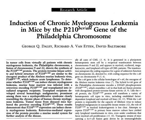
In grad school I ended up working on a problem that had been introduced to me in my hematology class: the molecular basis of chronic myeloid leukaemia (CML). I went to work for David Baltimore, the world authority on ABL, the gene that was disrupted in CML. That was really deep cancer and leukaemia biology, and I ended up making a mouse model of the disease that validated the BCR-ABL fusion gene as the driving molecular lesion in CML and as a target for treatment.
But even then I realised that there were a lot of leukaemias that didn’t have a defined molecular lesion, and that the broad platform for treating leukaemias – if one didn’t have targeted chemotherapy – was bone marrow transplantation. I was heavily influenced by a visiting professor from the UK, John Goldman, who was making his reputation expanding the use of unrelated donors for marrow transplants. We spent a lot of time thinking about hematopoietic stem cells (HSCs), the challenges of finding matched donors, and whether one could either generate a universal ʻoff-the-shelf’ donor cell, or an autologous cell that you could make from some non-leukaemic tissue. I was thinking about stem cell transplants when, once again by happenstance, Martin Evans, who had isolated mouse ESCs, was on sabbatical in the next lab. He introduced me to a paper from Rolf Kemler’s group describing the methodology of in vitrodifferentiation of ESCs to make embryoid bodies – one of the prominent differentiated tissues was blood. So even as a graduate student – and unbeknownst to David – I started culturing ESCs and seeing whether I could coax them to make blood. And I could, and this led to the notion that I could use ESCs as a platform to understand blood development.
But then I had to decide whether I was going stay in science or go back and finish my training in clinical medicine; because I had done work in a human leukaemia I decided to finish medical school and a medical residency and hematology/oncology fellowship. I was asked to spend an extra year as Chief Resident, which delayed my return to the lab but was an amazingly deep and wonderful experience. When I finally came back to the lab, my goal was to see if we could use ESCs as a platform for investigating the mechanisms of blood development. My scientific focus over the last 20-plus years has really been using mouse models in parallel with in vitro models comprising human and mouse ESCs and iPSCs to get at the core pathways driving blood development.
At the end of a recent paper you describe your ultimate goal in this line of research as ‘the derivation of bona fide transgene-free HSCs for applications in research and therapy’. How far are you from achieving that goal?
When I first laid out this goal in my proposal for becoming a Whitehead Fellow back in 1995, my hypothesis was that because pluripotent stem cells give rise to blood, they must transit through an HSC intermediate to get there; the dogma was that all blood cells arose from an HSC. That turned out to be wrong – the HSC isn’t there in the dish, unless you go through a different set of morphogen patterning to make a different type of mesoderm. That’s where developmental biology comes in – to inform directed differentiation in vitro.
The cells that we’ve made recently come from a two-step strategy: morphogen-directed differentiation gets us to what is probably the equivalent of lateral plate mesoderm, from which hemogenic endothelium and HSCs arise; then we direct cell fate specification with transcription factors in the manner of Yamanaka. The cells behave according to principles by which you define HSCs: they self-renew, engage multi-lineage engraftment over a long term (although not as long as a native cell), and they can do this on a single-cell basis. However, functionally we know our iPSC-derived HSCs are way below the standards and performance of freshly isolated human HSCs, and molecularly there are significant differences. I think we can continue to refine our strategy and get ever closer to a fully functional cell; then, the question becomes how native and how normal does it have to be? Does it need to be identical to a native HSC? This sort of goes back to philosophy again – it’s not really science!
Not only does developmental biology inform stem cells, but it also works the other way round
How important has developmental biology been for your research, and how do you see the relationship between developmental and stem cell biology?
I think all of stem cell biology is built upon the foundations of developmental biology. In the absence of the principles by which embryonic polarity is laid down, morphogenesis is induced and gastrulation is regulated, attempts at directed differentiation of pluripotent stem cells in a dish would be pure empiricism, pure tinkering, and not particularly intellectually satisfying. A large amount of directed differentiation is just that anyway, which is part of its well-justified criticism. But what’s so distinctive about stem cell biology is that you are observing every single step of the differentiation pathway – there are very few developmental systems that allow you that degree of direct observation and insight, especially when you get to more complex organisms. We work very closely with Len Zon, in part because the transparent zebrafish embryo allows direct visualisation and experimental manipulation of the earliest stages of commitment to both primitive and definitive hematopoiesis. That’s been enormously powerful, but we’ve had to then make the leap back to mouse and, crucially, to human; many of the principles are conserved, but the detailed mechanisms and specific gene sets might not be.
I’d like to make the point that not only does developmental biology inform stem cells, but it also works the other way round. In our recent work we started with a screen in vitro, taking iPSC-derived progenitors that make primitive forms of hematopoietic cells and asking whether we could identify epigenetic barriers to multipotency. Indeed, we identified a histone-modifying enzyme that broadly inhibits multipotency in embryonic blood precursors. Then, in order to really understand how this negative regulation works, we went back into a developmental system, the mouse. I think the real interest of this work is what it reveals about the regulation of the emergence of hematopoiesis in the embryo.
Back at the start of my lab, I saw an unmet medical need: there were, and still are, lots of patients who could benefit from bone marrow transplants but are never offered the chance, either because they didn’t have a suitable donor or due to the enormous toxicity of the transplant because of immunological dissimilarities. I have for many years thought that we could make a platform for combining gene therapy and cell replacement in a way that could open up a curative marrow transplant approach to dozens of diseases. But I always knew that clinical success would be a very high bar, and so what has really driven me, and the reason I’ve been able to sustain my lab for 20 years on the same model, is that it’s been a fabulous platform for learning about the pathways and mechanisms that contribute to blood formation in the embryo, and blood lineage development.
You were president of the ISSCR a decade ago. What have been the most significant changes in the field in that period, and where do you see the next ten years going?
When I was president, the revolution in reprogramming was just happening. It’s really interesting to see how that evolved: in the later part of the 1990s and early 2000s, many of us were thinking about reprogramming via nuclear transfer as a mechanism for generating customised or individualised patient-derived cells. There were even many who appreciated that genes such as Oct4 and Nanog were almost certainly involved in reprogramming. But Yamanaka’s great insight was that you could identify a manageable set of transcription factors that would change a cell’s fate, contrary to the expectation that reprogramming would be so complex that it could never just involve a small set of factors. There’s been an enormous amount of progress since then, and the number of disease models that have benefitted from reprogramming is staggering.
I think that over the last decade we’ve come to a much deeper understanding of the regulation of the genome, the mechanisms of gene regulation, but also chromatin organisation. Side by side with this, the technological advance that I think is so exciting and likely to dominate the next ten years is organoid biology. It’s just so exciting to see tissues forming because the information is preordained in the cells themselves, but perhaps not surprising given what we know about anatomy and historical experiments on tissue assembly after dissociation in animals such as sponges.
You have been a stem cell researcher and vocal science advocate since the years of George W. Bush. How did stem cell research cope when federal funding for human ESCs was barred?
It was discouraging and profoundly upsetting to be part of a scientific community that was seeing the limitless potential of a new field opening up and then to have it embroiled in a deeply divisive, fundamentally ideological debate around the nature of the human embryo and subject to a form of scientific censorship – the denial of federal funding. Science advocacy was not something I’d planned to get involved in, but the knowledge base that I had and the areas in which I was working made certain individuals in the political sphere seek me out for support and advocacy. I was a junior person at the time and several senior mentors were worried about the effect on my scientific career of being seen as too public; a lot of people told me to keep my head down, focus on the science. It was a struggle and I often wondered about this balance, and did turn down numerous opportunities to speak out more aggressively. But I think it really is a core responsibility of scientists to not simply be withdrawn behind their lab doors but to be willing to speak out on behalf of their science. It’s become obvious once again under the new administration that we have to stand up and justify the value of what we do in the context of lots of other competing priorities.
It really is a core responsibility of scientists … to be willing to speak out on behalf of their science
How do you see the prospects for state-funded research six months into the new administration?
The current President’s suggested budget included a roughly 20% cut in the NIH budget, and cuts for many other areas of discretionary scientific funding. Congress is unlikely to allow these cuts to stand, given that they have a broader base and interest in the importance of scientific research as a foundation not just for the future of human health but as an economic driver, a real creator of jobs, a way of priming the pump of biotechnology and the pharmaceutical industry. So I’m hopeful that the US’s traditional support for scientific research will continue. The other crucial element is that biotechnology is an area of international competitiveness, and the US is arguably the unchallenged leader. But without continued support, that leadership is subject to disintegration, and with the rise of China as a force in science, and the amount of investment they have committed to, international competition can be seen as an existential threat to the US. Trump is nothing if not hugely competitive, and this is an area where we certainly don’t want to lose out.
This year you started as Dean of Harvard Medical School (HMS). What do you hope to achieve during your tenure?
It’s an exhilarating and challenging new role. Before I took it up, I was Principal Investigator of a Howard Hughes-funded lab, a director of a small clinical division of bone marrow transplantation, a teacher of an undergraduate and a medical school course, a consultant to biotechnology and founder of a number of biotech companies. I had a very diverse professional life as it was, but I stayed within a fairly narrow realm of my own expertise. Becoming Dean has forced me to get up to speed with a tremendous array of different areas, both scientific and biomedical, and to some degree political. The Dean of HMS is seen as one of the prominent spokespersons for US medicine broadly defined, so I have to think about things like the economic aspects of healthcare delivery. HMS plays an enormous role internationally in global public health; we’ve got constituencies that are setting the pace in fundamental discovery, we have a community that is organised around translating biology into new medicines and new treatments, and we’re partners with biotech and pharma, which is increasingly concentrated in Boston. Clinical development and delivery of healthcare is an aspect that I now have to think about and influence. It has been exciting, and I hope that I can marshal my deep respect for science and my excitement and enthusiasm for clinical translation, together with a heightened sense of service as a clinician, to help with HMS’s fundamental mission to relieve the considerable suffering that disease inflicts on society.
In 2011 you were awarded the HMS A. Clifford Barger Excellence in Mentoring Award. Is there a ‘Daley’ style of mentorship, and who were your own important mentors in research?
I have to say that was one of my proudest awards, and in fact I was delighted as Dean to preside over this year’s awards just the past week.
I’ve had a phenomenal group of wonderful mentors who have taken an interest in me and helped me to develop throughout my career. I mentioned how important Beth Luna was very early on, and later there were people such as John Goldmann, Sam Lux and David Nathan, physicians who worked with me, and John Potts, who is in his eighties but who still meets and shares wisdom with me.
David Baltimore’s style of mentorship very much influenced the kind of mentor I want to be – the way he organised his lab and ran group meetings, demanding high standards and yet welcoming open candid critical debate about science. He emphasised the importance of the question and not just the technique, which fostered a sense of innovation: in chasing questions you marshalled whatever technology you needed, absorbing established techniques and developing new ones. Like David’s, the best scientific labs have a horizontal structure, so that everyone speaks equally and individually to assert a quest for the truth; that was very influential on me. So I have tried over the years to establish a climate in the lab where people are inspired to ask big questions, are empowered by adequate resources to chase those questions in a relatively unfettered way, and are open to collaboration. I also want to encourage a climate of having fun: we do a lot of social events and sing a lot of karaoke; if I can get up in front of a group, belt out a song and make an ass of myself, I feel it gives others a certain willingness to step forward and to take risks.
Finally: is there anything Development readers would be surprised to find out about you?
Outside of the lab, I’m passionate about cooking and collecting wine: I’m a foodie. It grew directly from travelling for science – going around Europe and being hosted by people who were eager to feed me their local fare and ply me with their local libation, whether wine or beer. My wife and I came together around cooking and we do a lot of that. One has to eat, so it’s a fairly efficient way of exploring new cuisines and experiences while still keeping your time commitments.
 (3 votes)
 (No Ratings Yet)