A postdoctoral position is available in the laboratory of Dr. Martin Basch to study the regenerative potential of the stria vascularis in cases of congenital deafness. Highly motivated, creative and enthusiastic individuals are particularly invited to apply.
Qualifications include a PhD in developmental biology, neuroscience, cell biology or related field. Priority will be given to candidates with experience in cell culture and basic molecular biology. Mouse genetics and/or neonate mouse surgery skills are desired but not required.
Our laboratory is part of the Hearing Research Program, at Case Western Reserve University School of Medicine. Currently the program involves five Research Faculty PIs and Physician Scientists with key areas of research, which include molecular otology, otitis media, congenital/acquired hearing loss, inner ear development, and hair cell biology. The strength of our program is enhanced by an excellent interdisciplinary and collaborative intellectual environment at Case.
Interested candidates should submit their CV and a letter of application (including a brief description of previous research experience and a statement of interests) to: Martin Basch Ph.D. at mlb202@case.edu.
For more information on our laboratory, please visit our website at http://www.baschlab.org
In employment, as in education, Case Western Reserve University is committed to Equal Opportunity and Diversity. Women, veterans, members of underrepresented minority groups, and individuals with disabilities are encouraged to apply.
Case Western Reserve University provides reasonable accommodations to applicants with disabilities. Applicants requiring a reasonable accommodation for any part of the application and hiring process should contact the Office of Inclusion, Diversity and Equal Opportunity at 216-368-8877 to request a reasonable accommodation. Determinations as to granting reasonable accommodations for any applicant will be made on a case-by-case basis
The Beddington Medal is the BSDB’s major commendation to promising young biologists, awarded for the best PhD thesis in Developmental Biology defended in the year previous to the award. Rosa Beddington was one of the greatest talents and inspirational leaders in the field of developmental biology. Rosa made an enormous contribution to the field in general and to the BSDB in particular, so it seemed entirely appropriate that the Society should establish a lasting memorial to her. The design of the medal, mice on a stylised DNA helix, is from artwork by Rosa herself. For further medal and award winners at the 2017 Spring Meeting see here.
The BSDB congratulates the 2017 Beddington Medal winner Erik Clark. Erik did his BA in Biological Sciences at the University of Oxford (1st in his year group) and his MSc in Bioinformatics & Theoretical Systems Biology at Imperial College London where he worked on the project entitled “Evolution of Mutation Rate in Fluctuating Environments”. He then moved on to do his PhD within the BBSRC Genes to Organisms Program supervised by Michael Akam at the Department of Zoology, University of Cambridge, where he worked on his project entitled “The Drosophila Pair-Rule System” and where he continues to work now. Erik won an impressive number of prizes, fellowships and grant awards, including the Gibbs Prize in Animal Biology (Univ. Oxford, 2011), an Isaac Newton Trust Research Grant (2016-17), a Junior Research Fellowship (Trinity College, Cambridge) and he is co-investigator on a BBSRC research grant (2017-20).
His Beddington Medal talk described the outcome of his most successful PhD project. About the background Erik explained: “The Drosophila segmentation cascade is a paradigmatic example of a developmental gene regulatory network, used to gain insight into transcriptional regulation in all animals… Using spatial information from graded domains of “gap” gene expression, seven “pair-rule” genes are expressed in periodic patterns of seven stripes each .. [which] then work in combination to specify precisely-phased 14 stripe patterns of “segment polarity” genes. These output patterns form the template for the segmental organisation of the insect body…[Although] the pair-rule genes have been studied in Drosophila for over 30 years,… there is still no good systems-level understanding of their regulatory interactions“.
In his thesis, Erik used a combination of modelling and experiment to reverse-engineer the structure of the Drosophila pair-rule network and understand how it generates expression dynamics that lead to the patterning of segmental boundaries. He set out to collect a complete time-resolved dataset of relative expression for all pairwise combinations of the 7 pair-rule genes in wild type embryos (see Figure), and a partial dataset for a number of mutant genotypes. Using these data he defined how the regulatory interactions of pair-rule genes change at the mid-cellularisation stage, and identified odd-paired as a temporally regulated factor responsible for these network changes. As an outcome of his work, Erik proposes that that spatial resolution emerges from temporal dynamics, rather than static positional information.
A further part of his thesis proposes that the standard model for how parasegment boundaries are specified, by the interpretation of local gradients of Even-skipped protein, may not be correct. He suggests instead that the shifting of even-skipped stripes across the field of cells in the blastoderm, driven by dynamic gap gene expression, coupled with the temporal control of network interactions, may generate the key offsets in downstream gene expression, and this is an entirely novel idea.
To illustrate Erik’s path to success, Michael Akam writes: “Unlike any other student I have had, Erik spent pretty much the whole of his first year reading. He worked through the entire literature on Drosophila segmentation (spanning 30 years and hundreds of papers), assessing the claims made on the basis of the data presented, and with the advantage of hindsight that the original authors lacked. I suspect he has a more detailed and critical knowledge of this literature than any other researcher.” Michael concludes his support letter with the words: “Erik’s work is strikingly original, and represents a major innovation in thinking about Drosophila segmentation.”
Erik’s publications so far:
Clark, E. (2017) ‘Dynamic patterning by the Drosophila pair-rule network reconciles long-germ and short-germ segmentation’. bioRxiv: 1101/099671
Clark E & Akam M (2016). Odd-paired controls frequency doubling in Drosophila segmentation by altering the pair-rule gene regulatory network. eLife: 7554/eLife.18215
Clark E. & Akam M. Drosophila pair-rule gene double FISH Data (data from Clark & Akam 2016). org:10.5061/dryad.cg35k 2016
BertaVerd, ErikClark, Karl R.Wotton, HildeJanssens, EvaJimenez-Guri, AntonCrombach, JohannesJaeger (2017). A damped oscillator imposes temporal order on posterior gap gene expression in Drosophila. bioRxiv: 10.1101/068072
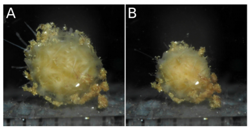
A large-scale study of DMDD data has shown that inactivating the same gene in mouse embryos that are virtually genetically identical can result in a wide range and severity of physical abnormalities. This suggests that the relationship between gene mutation and embryo development is more complex than previously thought.
A comparison of two embryos that are both missing the embryonic lethal gene Coro1c. The embryo on the right has abnormal viscerocranium (facial skeleton) morphology, while the embryo on the left does not.
The study considered 220 mouse embryos, each with one of 42 different genes inactivated. These genes are part of a set known as ‘embryonic lethal’, because they are so crucial to development that an embryo missing any one of them can’t survive to birth. Studying these genes can help us understand how embryos develop, why some miscarry and why some mutations can lead to abnormalities.
Here are the highlights from the current issue of Development:
A new niche for human HSCs
Human haematopoiesis occurs at various anatomical sites throughout development, including the yolk sac, the aorta-gonad-mesonephros region, the liver, the placenta and the bone marrow. Cells marked by high expression of CD34 and low CD45 – suggestive of possible HSCs – have been reported in human fetal membranes; however, their exact niche as well as their functional capacity remain untested. In this issue (p. 1399), Alicia Bárcena and colleagues isolate and interrogate this putative HSC population, and demonstrate for the first time that the human chorion contains transplantable, definitive HSCs. The authors carefully separate the chorion and the amnion, and show via fluorescence-activated cell sorting that only the chorion contains the putative HSCs, and only from 15 weeks of gestation. The cells display markers of HSC and primitive haematopoietic progenitors, such as little CD38 and CD133, low levels of CD117 and CD4, and medium to high levels of HLA-DR, CD31, CD90, CD95, TIE2 and CD71. Cells co-expressing CD34 and CD45 antigens are found either in association with mesenchymal stromal cells or with endothelial cells of chorion vasculature . Using in vivo xenotransplantations, the authors demonstrated that the CD34++ CD45low cells possess multilineage long-term HSC activity specifically between weeks 15 and 24 of gestation. This study reveals novel insight into an unexpected niche for HSCs during human development.
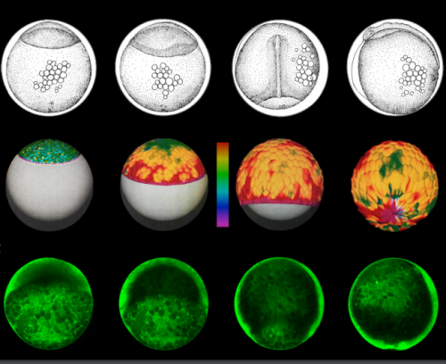
How the stomach gets its curve
Left-right asymmetry is a common feature of many organs, and is crucial for their function. The stomach is one such organ, with marked curvature on the left compared with the right, resulting in a distinctive shape that is highly conserved among vertebrates. Although it is well established that activation of Nodal controls left-right asymmetry of visceral organs, the cell- and tissue-level morphogenetic mechanisms that drive this phenomenon are poorly understood, especially in the stomach. Now, on p. 1477, Nanette Nascone-Yoder and colleagues shed light on the mechanisms that drive left-right asymmetric development of the stomach in both mouse and Xenopus embryos. The authors start with a gross examination of stomach curvature during development and compare their findings with two proposed models: a rotation model and an asymmetric growth model. They find no evidence for the former, and therefore suggest that the stomach acquires its asymmetry by an intrinsic mechanism. In support of this, the authors show that there is an asymmetric thickness of the left and right stomach wall, which depends on intact cilia and Nodal signalling as both Foxj1 mutant mouse embryos and Xenopus embryos treated with a Nodal inhibitor show a loss of this asymmetry. The authors show a role for Pitx2 in this process by overexpressing Pitx2 on the right side or knocking down Pitx2, both of which affect stomach curvature in Xenopus. This study demonstrates that asymmetric morphogenesis of the stomach in frogs and mice is driven by FoxJ1-Nodal-Pitx2-dependent asymmetric remodelling of the gastric epithelium on the left side.
Liverworts breathe easy
The vast majority of land plants regulate gas exchange through their stomata – tiny pores usually found on the underside of leaves. The liverwort plant group Marchantiidae is an exception, as it lacks stomata and instead breathes through air pore complexes. This is an important evolutionary adaptation, and yet the mechanisms that regulate air pore complex development in Marchantiidae remain unknown. In this issue (p. 1472), Victor Jones and Liam Dolan identify the zinc finger protein MpWIP as necessary for the morphogenesis of the air pore complex in the epidermis of Marchantia polymorpha. The gene was first identified through a mutagenesis screen, in which overexpression led to the presence of ectopic rhizoids on the dorsal epidermis. Using a construct containing the MpWIP promoter fused to a reporter gene, the authors show that MpWIP is expressed both ventrally and dorsally and that the dorsal expression pattern is within the developing air pore complex cells. To determine whether MpWIP is required for air pore development, the authors use artificial microRNAs to generate plants with reduced expression of MpWIP, which results in defects in air pore complex morphology. Based on chimeric dominant repressor and activator versions of MpWIP expressed separately in transgenic plants, the authors provide some evidence for the possible role of MpWIP as a transcriptional repressor. This study identifies, for the first time, a gene that regulates the development of the air pore complex, which is an important evolutionary innovation in liverworts as an alternative to stomata.
Specialised fibroblasts maintain the nipple epidermis
The skin is the body’s largest organ, and is the first line of defence against the external environment. The epidermis – the outermost layer of the skin – is highly specialised and often exhibits unique characteristics depending on its anatomical location and the function it serves. It has long been known that this specialisation is dependent on inductive signals that originate from underlying fibroblasts; however, the exact nature of the signals and their role in maintaining the epidermis is only just starting to emerge. In this issue (p. 1498), John Foley and colleagues identify an oestrogen-regulated TGFβ signalling pathway that is crucial for the maintenance of the highly specialised nipple epidermis. Using a series of grafting experiments, the authors show that fibroblasts taken from the nipple-like skin of mice can induce reprogramming of trunk keratinocytes into nipple-like epidermis. Transcriptional profiling of the nipple-like fibroblasts identifies oestrogen signalling as a strong candidate factor for the maintenance of the nipple epidermis and, indeed, ablation of oestrogen signalling in ovariectomised mice results in an abnormally thin nipple epidermis. The authors further identify Tgfb1 as a direct target of oestrogen signalling and show how ectopic treatment of TGFβ1 protein into the connective tissue of the nipple causes a decrease in epidermal proliferation and a thinning of the nipple epidermis. Taken together, these data represent an important step forward in understanding the signalling network that maintains the specialisation of the nipple epidermis.
PLUS:
Stem cell therapies for retinal diseases: recapitulating development to replace degenerated cells
Retinal degenerative diseases are the leading causes of blindness worldwide. In their Review article, Sally Temple and colleagues review stem cell-based therapies for retinal diseases, describing the challenges involved and discussing how basic developmental studies have contributed to and are needed to advance clinical goals.
Prostate organogenesis: tissue induction, hormonal regulation and cell type specification
Prostate organogenesis is a complex process that is regulated by androgens and subsequent mesenchyme-epithelial interactions. In their Review article, Roxanne Toivanen and Michael Shen provide a comprehensive overview of prostate development, focusing on recent findings regarding sexual dimorphism, bud induction, branching morphogenesis and cellular differentiation.
As every year, the Spring meeting was the time of awards and medals! This year, we had awardees of three societies who are listed below. For those wanting to have a look at the topics of talks and posters presented at the meeting, please download the abstract book here. Watch the movies of the BSDB award winners below.
1st BSDB PhD Poster Prize winner (Attendance at SDB 76th Annual Meeting, Minneapolis): Claire Bromley (Kings College London) – Poster 25 “Investigating biomechanical forces in zebrafish brain morphogenesis“
1st BSCB PhD Poster Prize winner (visit to 2017 ASCB/EMBO meeting, Philadelphia): Christina Dix (University College London) – poster 5 “Adhesion, not cortical tension, is vital for successful cytokinesis in RPE-1 cells“
1st Genetics Society (£100 cash prize sponsored by BioMed Central): Alexandra Buffry (Oxford Brookes University) – Poster 182 “Investigating gene regulatory network architecture and evolution in different developmental contexts“
2nd BSDB PhD Poster Prize (£100 cash prize sponsored by BioMed Central): Ariadna Gador Navarro-Aragall (UCL Institute of Ophthalmology) – Poster 86 “SEMA3E and SEMA3C Cooperate to establish vascular boundaries“
2nd BSCB PhD Poster Prize (£100 cash prize sponsored by BioMed Central): Sophie Adams (Barts Cancer Institute) – Poster 32 “‘Exosome signatures’ as biomarkers for centrosome-targeted therapy in pancreatic ductal adenocarcinoma (PDAC)“
► Postdoc Poster Prizes
1st BSDB Prize (£200 cash prize sponsored by BioMed Central): Carla Mulas (University of Cambridge) – Poster 129 “Functional characterisation of metachronous cell state transitions“
1st BSCB Prize (£100 Cash prize sponsored by BioMed Central): Girish Mali (MRC Laboratory of Molecular Biology) – Poster 47 “Assembly Mechanisms of Dynein Motors“
1st Genetics Society (£100 bank transfer sponsored by BioMed Central): Laura Molina-Garicia (University College London) – Poster 122 “Sexy learning in C. elegans”
2nd BSDB Prize (£100 cash prize sponsored by BioMed Central): Hadi Boukhatmi (University of Cambridge) – Poster 92 “Molecular logic behind Satellite cells specification in Drosophila”
► Others
Genetics Society Overall Poster Prize (Junior Scientist Conference Grant, Scheme A): Alewo Idoko-Akoh (The Roslin Institute) – Poster 185 “CXCR4 and c-Kit signalling are required for directed migration of chicken primordial germ cells through the chick embryonic vascular system“
BSDB Honorary Mention (Certificate): Eva Higginbotham (University of Cambridge) -Poster 114 “Neurotransmitter specification in the ventral nerve cord of Drosophila melanogaster”
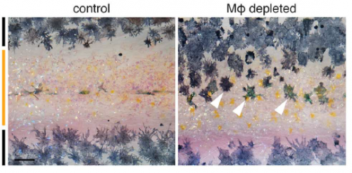
Macrophages are usually associated with immunity, but have increasingly appreciated functions in development and homeostasis. This week we meet the authors of a recent Science paper that identified a role for macrophages in zebrafish stripe patterning, revealing a remarkable ‘relay’ mechanism whereby macrophages help one type of cell signal to another via cytoplasmic extensions. Postdoc Dae Seok Eom and his supervisor David Parichy, recently appointed Pratt-Ivy Foundation Distinguished Professor of Morphogenesis at the University of Virginia, told us more.
David, can you give us your scientific biography and the questions your lab is interested in?
DP I started out in ecology and evolution as an undergrad at Reed College, studying maternal effects on tadpole growth and survivorship for four years with Bob Kaplan. With that experience I applied to E&E Ph.D. programs and ended up accepting an offer from Population Biology at UC Davis, with Brad Shaffer, a systematist and organismal biologist studying biogeography of salamander populations. But between when I applied and when I got there, I became more and more interested in developmental mechanisms and how they evolve. So I was really lucky that Brad has diverse interests and that Carol Erickson—a developmental biologist working on neural crest—was willing to serve as a co-advisor. For my dissertation I focused on the cellular bases for salamander pigment pattern development and evolution. Those are great animals, but the molecular biology and genetics were difficult at the time. So I switched to zebrafish and its relatives for my postdoc with Steve Johnson at Wash U Medical School in St. Louis. In Steve’s lab, we identified some of the mechanisms underlying the development of stripes and other patterns, and I carried this program into my independent career.
My lab has broad interests but an organizing theme has always been to understand the genes and cell behaviors underlying adult phenotypes, and how changes in developmental genetic mechanisms contribute to variation within and among species. We continue to work on pigment patterns, but we also want to know what regulates the stem cells that give rise to adult pigment cells and how “local” cellular mechanisms intersect with “global” endocrine control during development, homeostasis and regeneration. But our interests are wide-ranging so we’ve also worked on topics including skeletal development, zebrafish natural history, and the behavioural significance and cognitive processing of pigmentation. Right now we are doing a lot of work to understand scale morphogenesis and patterning in fish, and we’ve even started working on salamanders again, both pigment and regeneration.
The macrophage depletion phenotype, from Figure 1, Eom & Parichy, 2017.
And Dae Seok, how did you come to join David’s lab?
DSE Actually, when I was at PhD training at the University of Texas at Austin, Dave was a faculty of my department, and I was interested in his work, but by then he was then planning to move to the University of Washington. While I was finishing my PhD training, my wife started her PhD at UW, so I had to find a postdoc position in Seattle. I realised Dave was there, and it was obviously a great second chance to work with him – and he accepted.
Aside from aesthetics, what makes zebrafish stripe development an attractive developmental model?
DP When I was a grad student, I wanted to find a system that could be studied from many different angles—molecular through organismal. And this was why I ended up focusing on pigmentation. For anyone with broad interests it’s just a natural: there’s a deep literature on pigment cells and pattern going back to the turn of the last century, the cells are visible even in the living organism as the phenotype is developing, the patterns themselves often have profound ecological significance, and there’s tremendous diversity of pattern across even closely related species. Of course there are also a variety of pigmentary disorders, including melanoma, and pigment cells develop and regenerate from stem cells—so there is obvious biomedical utility in studying pigment cells, especially with a system like zebrafish in which the genetic and cellular mechanisms are so accessible.
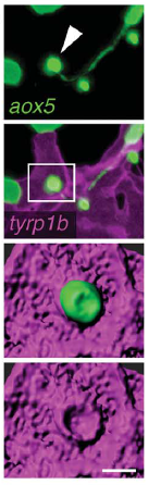
A macrophage drags an airineme to a melanocyte. Eom & Parichy, 2017.
Can you give us the key results of the paper in a paragraph?
DP We showed previously that consolidation of melanophores into adult stripes depends on interactions between these cells and xanthoblasts, the precursors of yellow xanthophores. During a specific stage of stripe development, xanthoblasts that happen to be present in future stripe regions extend very long, thin and meandering filopodia-like processes with membraneous vesicles at their tips. We called these projections “airinemes” after Iris—messenger of the gods—and Sir George Airy, who described limits on optical resolution, because the projections are such a pain to visualize. We saw airinemes “dock” with melanophores, and we found that blocking airineme extension prevented melanophores from consolidating properly into stripes, at least in part because of a defect in Delta-Notch signalling.
“Weird biology”
In Dae Seok’s new paper, we show that airineme extension and vesicle delivery depend on macrophages that are cruising around the local tissue environment. The macrophages recognize surface blebs on xanthoblasts and try to engulf them but continue to wander, pulling a membrane filament from the xanthoblast as they go. Eventually they wander across a melanophore and the vesicle and filament are deposited on the melanophore surface. If we get rid of macrophages, we prevent airineme extension and melanophores remain dispersed. Weird biology.
What convinced you to deplete macrophages in the first place? It might not seem like the most obvious place to look for patterning regulators…
DSE The idea initially came from the question of what would happen to unbound airineme vesicles. When xanthoblasts don’t meet the target melanophores, airineme vesicles detach from the airineme filaments and wander away. We knew the vesicles carry DeltaC and possibly other signalling molecules. Thus, these unbound vesicles should be somehow eliminated. The question then was what cell types can do that? One of the answers was the macrophage.
DP Plus we could constantly see macrophages wandering around in Dae Seok’s movies. How could we not try getting rid of them?
Airineme vesicle associated with melanophore membrane, from Fig. 4, Eom & Parichy, 2017.
Is there anything in the macrophage-depleted fish to suggest that other patterning or morphogenetic events may be affected, or is this just tissue specific?
DP We’re only starting to investigate roles for macrophages as well as airinemes in other tissues. Because macrophages wander all over, you could imagine a whole variety of possibilities for diffusion-like dissemination—or regulated attenuation—of signals in other contexts. The more people look, the more interesting things macrophages seem to do.
Can you hazard a guess as to whether you think the macrophages actively select signalling targets or just wander around randomly?
DSE We have no idea at this time but my guess is macrophages constantly probe the environment, and signals from the airineme vesicles instruct macrophages to detect the targets and drop the vesicles while they are randomly wandering around.
DP Yes, because Dae Seok showed that targeting is specific to a subset of melanophores, there has to be some sort of airineme vesicle–melanophore recognition system. It will be interesting to see whether this is somehow instructive to the macrophage itself or whether the macrophage is simply trying to eat the vesicle and can’t manage to do so, and therefore the vesicle gets displayed at the cell surface often enough that it can stick to a melanophore as its carrier macrophage wanders along.
When doing the research, was there a particularly exciting result or eureka moment that has stayed with you?
DSE I do not have good explanation for it, but I had very strong gut feeling that there are something going on between macrophages and airinemes. So of course, my most exciting moment was when I saw the macrophages interacting with extending airinemes.
Xanthoblast airinemes. Eom and Parichy, 2017.
And what about the flipside: any moments of frustration or despair?
DSE Our original plan was to add additional mechanisms of airineme vesicle-macrophage interactions beside phosphatidylserine, as we have several additional candidates. We ran into technical problems for this first paper but are now working on it again.
What next for you following this work?
DSE We have many questions about airineme-macrophage interactions, for example, what other signalling molecules carried in the airineme vesicles and how macrophages know what the targets are. I’m very excited about using our new super-resolution microscopes – they will open up many new possibilities.
“Our new super-resolution microscopes will open up many new possibilities.”
And where will this discovery take the Parichy lab?
DP Of course we’ll continue to pursue the mechanisms underlying this signalling relay in zebrafish pigment pattern formation. And we’d really like to know how these and other interactions have evolved to generate the very different patterns we see in Danio and beyond. But you know, a lot depends on the interests of the grads and postdocs who come to my lab. I just try to foster an intellectual environment and provide the resources to let people explore and go where the science leads. When we decided eight years ago to invest in live imaging we didn’t know what we’d find and we certainly didn’t expect this. So I’d be hesitant to make firm predictions.
Finally – what do you get up to outside of the lab when you are not playing with fish?
DSE One of my favourite things to do is visiting local breweries. My wife and I have a Saturday routine of having a lunch at a brewery, and then heading to the lab.
DP Legos. I like to spend as much time as I can with my four year old and my wife. So we do a lot of legos, trains and construction. Helicopters are big, too.
Our latest monthly trawl for developmental biology (and other cool) preprints. See June’s introductory post for background, and let us know if we missed anything
March was (yet) another bumper month for life sciences preprints. A glance at some of the names of last authors – Daniel St. Johnston, Denise Montell, Dennis Duboule, Roberto Mayor, Fiona Watt – shows that preprints are being embraced by established as well as early career scientists. The content itself covers every base in developmental biology, as well as a lot of exciting cell biology and modelling relevant for development, and an acronym-rich list of tools and resources.
The Molecular Link Between Auxin And ROS-Mediated Polar Growth. Silvina Mangano, Silvina Paola Denita-Juarez, Hee-Seung Choi, Eliana Marzol, Youra Hwang, Philippe Ranocha, Silvia M Velasquez, Jorge P Muschietti, Christophe Dunand, Hyung-Taeg Cho, Jose M Estevez
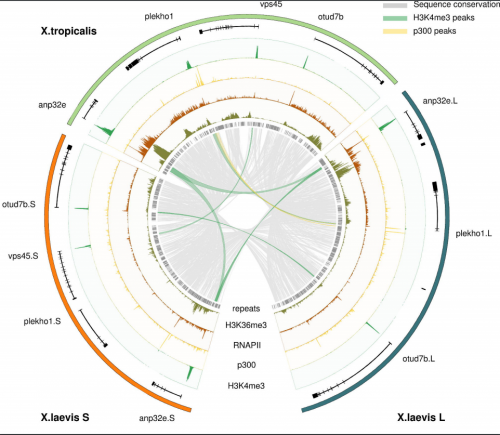
Alignment of X. tropicalis and the X. laevis L and S subgenomes, from Elurbe, et al.
Regulatory remodeling in the allo-tetraploid frog Xenopus laevis. Dei M. Elurbe, Sarita S. Paranjpe, Georgios Georgiou, Ila van Kruijsbergen, Ozren Bogdanovic, Romain Gibeaux, Rebecca Heald, Ryan Lister, Martijn A. Huynen, Simon J. van Heeringen, Gert Jan C. Veenstra
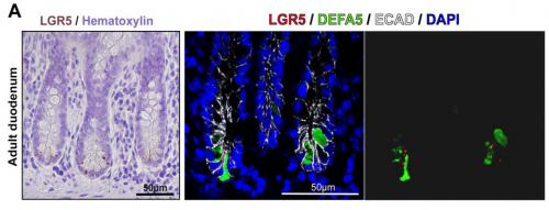
Looking for LGR5+ cells in the adult duodenum, from Dame, et al.
Identification, Isolation, and Characterization of Human LGR5-positive Colon Adenoma Cells. Michael K Dame, Durga Attili, Shannon D McClintock, Priya H Dedhia, Peter Ouilette, Olaf Hardt, Alana M Chin, Xiang Xue, Julie Laliberte, Erica L Katz, Gina M Newsome, David Hill, Alyssa Miller, David Agorku, Christopher H Altheim, Andreas Bosio, Becky Simon, Linda C Samuelson, Jay A Stoerker, Henry D Appelman, James Varani, Max S Wicha, Dean E Brenner, Yatrik M Shah, Jason R Spence, Justin A Colacino
An Algorithm for Cellular Reprogramming. Scott Ronquist, Geoff Patterson, Markus Brown, Haiming Chen, Anthony Bloch, Lindsey Muir, Roger Brockett, Indika Rajapakse
Rapid Sequential In Situ Multiplexing With DNA-Exchange-Imaging. Yu Wang, Johannes B Woehrstein, Noah Donoghue, Mingjie Dai, Maier S Avendano, Ron C.J. Schackmann, Shan Shan Wang, Paul W Tillberg, Demian Park, Sylvain W Lapan, Edward S Boyden, Joan S Brugge, Pascal S Kaeser, George M Church, Sarit S Agasti, Ralf Jungmann, Peng Yin
A toolbox of immunoprecipitation-grade monoclonal antibodies against human transcription factors. Anand Venkataraman, Kun Yang, Jose Irrizary, Mark Mackiewicz, Paolo Mita, Zheng Kuang, Lin Xue, Devlina Ghosh, Shuang Liu, Pedro Ramos, Shaohui Hu, Diane Bayron, Sarah Keegan, Richard Saul, Simona Colantonio, Hongyan Zhang, Florencia Pauli Behn, Guang Song, Edisa Albino, Lillyan Asencio, Leonardo Ramos, Luvir Lugo, Gloriner Morell, Javier Rivera, Kimberly Ruiz, Ruth Almodovar, Luis Nazario, Keven Murphy, Ivan Vargas, Zully Rivera-Pacheco, Christian Rosa, Moises Vargas, Jessica McDade, Brian S Clark, Sooyeon Yoo, Seva G Khambadkone, Jimmy de Melo, Milanka Stevanovic, Lizhi Jiang, Yana Li, Wendy Y Yap, Brittany Jones, Atul Tandon, Elliot Campbell, Stephen Anderson, Richard M Myers, Jef D Boeke, David Fenyo, Gordon Whiteley, Joel S Bader, Daniel J Eichinger, Ignacio Pino, Heng Zhu, Seth Blackshaw
Have you ever wondered what makes the shapes in the animal and plant kingdom so different? We take for granted the diversity of natural shapes that surround us, from a simple pine leaf to complex orchid flower. However, they pose one of the most beautiful scientific challenges. For centuries, scientists have been fascinated by how a certain shape is generated and what drives such diversity of shapes. I am no different! From my university days, I always wanted to study evolutionary development, to go into the depth of why and what lies behind the diversity of shapes in nature.
After my PhD days using genetics, molecular biology and comparative development as tools to study the evolution of shape in plants, I was keen to explore the upcoming computational modeling field as a tool to tackle complex development problems. This is where this article’s journey started, when I joined the lab of Enrico (Rico) Coen at the John Innes Centre to study the evolution and development of the complex 3D Snapdragon flower shape. I still remember my first contact with modeling. I was immediately hooked by the colorful shapes in the computer screen which looked so much like a snapdragon flower. However, nothing could have prepared me for the thousand lines of computational code that sustained the generation of the virtual Snapdragon model. At that moment I thought to myself, maybe I have bitten more than I can chew by wanting to do both the biology and the computational modeling.
The personal story that goes with the published article is a roller coaster of wrong hypothesis, failed and impossible experiments, and frustrating models but also glimpses of successes. You might be thinking, oh no this is going to be a tale of woe but let me disclose the end already – this is no sad story, this is the actual necessary evolution of thought, knowledge and personal growth that sustains any challenging scientific problem. For my feeling the story behind this paper feels more like a Tolkien novel with lots of downs, that you almost don’t expect to overcome, but also amazing ups.
Our approach at the start of my post-doc was to look into the working hypothesis generated by a previous in-house paper where the growth of the Snapdragon petals was analyzed and a virtual flower corolla was produced. The main assumptions underlying this model related to the spatiotemporal changes in the growth pattern due a local cell polarity inversion event combined with a local boost in growth rate.
To visualize the inversion of polarity in cells we decide to use an accepted marker of plant cell polarity that generally shows an asymmetric cellular localization – the PIN1 protein. For two long years, I struggled with PIN1 antibodies that didn’t work or produced slight promising subcellular signals that held us in the false hope of results. After changing the antibody company and spreading the risks by designing several different epitopes to the different PIN1s, I finally had a good working antibody that produced the expected pattern of cellular polarity in emerging organs. Armed with this tool, I initiated a marathon of immunolocalization sections across the petals at different stages of development to show the local inversion of cellular polarity. From the beginning it was obvious that something special was happening to the expression of the PIN1 in the predicted region at the predicted time by the model. However, after a year of thorough sectioning experiments and analysis, I was struggling to reconstruct the 3D PIN1 pattern from the 2D sections. At that point we wanted to start writing the paper to show the peculiar upregulation of PIN1 in the predicted region by the model but unfortunately the precise pattern of the PIN1 was still undetermined and led to a lot of team discussions and wild ideas on how to distil the result. Emotions were running high, as we were failing to grasp the final proof of the inversion of polarity. After we decided to put the paper on hold and re-think our approach, I got desperate! In other models system and exposed organs such as roots, the first solution would be to do whole-mount immunolocalization, where the whole organ is hybridized and the 3D patterns can be imaged. However, our tissue of interested was the inner surface of tiny petals, in buds as small as 500 microns. Desperate times require desperate measures, and after a bad night of sleep and probably a delirious morning, I started dissecting incredibly tiny petals and sticking them to a glass slide using prosthetic glue. Then, to make sure the antibody could penetrate the tissue, I boiled these fragile tissues cringing at every little water bubble brushing through the stuck petals. To check the cell/tissue integrity and in anticipation of no PIN1 signal, I also stained the petal tissues with a cellulose fluorescent stain called calcofluor. After three agonizing days of hoping that the tissue was not turned into a pulp, I descended the stairs that separate our lab from the confocal microscopy room with a heavy heart; this is our last crazy chance! If this doesn’t work, I am out of ideas. As I started up the confocal and focused on the first set of 5 petals, I couldn’t believe what I was seeing, a beautiful ladder of PIN1 signal in the regions of interest -I couldn’t contain my joy.
I picked up the phone and called some lab mates to be sure they could see what I was seeing (I couldn’t exclude delirious mirages at this point with my nerves on the fringe). After they confirmed what I was seeing and shared a happy dance with me, I went to get Rico from his office luring him with promises of something special without giving too much away. We still have photos of this moment that we shared with the four of us in a confocal room. There are moments that you don’t forget in your life and this one is engrained in my memory.
I finally could extract full epidermal patterns of PIN1 in wild-type and mutant petals at different stages of development, and guess what? I never saw an inversion or convergence of polarity in the region predicted by the model. I did see an upregulation of PIN1 at the predicted time and space but the pattern of PIN1 was mainly proximodistal (along one axis). This might feel like a bitter sweet result as it contradicted our model but actually this was quite an unexpected and exciting result. In my opinion, one of the beautiful givens in science is that it will surprise you. You make a hypothesis, your results prove it wrong and you need to re-evaluate your thinking. This was indeed what happened to us but as a result we realized that we didn’t need an inversion of polarity, as the key information required for growth lay in the orientation of the arrows (axiality), not in the direction in which they are pointing. So we came up with an alternative model that still involved a switch in growth orientations but without a polarity inversion (I can’t imagine I just summarized months of modeling with a sentence but there you have it!).
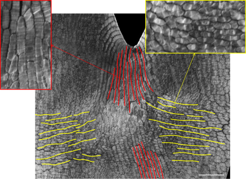
We then tested this model by correlating cell wall patterns with growth orientations and showing the switch in growth orientation at the predicted time and space.
Detail of Snapdragon petal stained with cell wall marker in order to visualize the orientation of cell division and growth (red lines indicate proximodistal growth while yellow lines indicate mediolateral growth with respective zoomed in areas). Adapted from Rebocho et al, 2017, eLife.
We were finally ready to write our results, publish our new model and make a contribution to the scientific understanding of 3D morphogenesis. However, one of our biggest challenges was still to surface – conveying the ideas developed over the years in words, while connecting the static 2D images of cell polarity and growth orientations with the 3D flower shape. When you read the paper now, the logic seems to fall into place, with the increasing complexity of the models and the experimental data driving a new model hypothesis. But the reality was a long winding road, with several versions of the paper; many rejected by ourselves, others by our critical colleagues, and others by peer-reviewed journals. This was a very frustrating time for the team and I am not really sure when we started to see the light, as this was an incremental and slow process, but at a certain point we felt that we were finally are on track. By deconstructing the model complexity (from square piece of tissue to wedge model to full corolla model and to mutant models) and conveying the idea of how differential growth behaviors can lead to tissue conflict resolutions, we were finally bringing the disjointed paper together. Our paper distilled three tissue conflict resolution behaviors leading to an out-of-plane deformation: surface conflict (differential growth/contraction rates between two surfaces), areal conflict (differential growth/contraction rates across a tissue) and directional conflict (differential orientation growth/contraction across a tissue). These three behaviors are the basis of many of the described morphogenetic events in plants and animals, and can be combined to produce the panoply of 3D shape complexity that surrounds us.
For me this journey made me a more complete scientist. I have explored new disciplines and learned to think in a different way. I have developed extra respect for the power of modeling and became humbler when it comes to tackling complex biological problems. Lastly, I realized that the essence of a piece of research lies in the evolution of thought of the researchers, their failures and frustrations, their stubbornness and their hunches, their commitment and their successes, and that is what makes science so amazing.
The Wellcome-Warwick Quantitative Biomedicine Programme Wellcome-Warwick Quantitative Biomedicine Programme was established to enhance the world-class interdisciplinary research environment at the University of Warwick by driving further development of our existing centres of excellence, including the Centre for Mechanochemical Cell Biology (www.mechanochemistry.org) and the Zeeman Institute for Systems Biology& Infectious Disease Epidemiology Research.
Warwick Medical School is seeking to appoint three outstanding early career scientists as Assistant Professors in Quantitative Biomedicine, aiming to expand our research on cell dynamics at the molecular, cellular and/or tissue scales. We are eager to recruit candidates using quantitative and/or interdisciplinary approaches and with an exemplary track record in cell biology, developmental biology, structural biology, computational biology or biophysics.
Successful candidates must have a strong track record with first class publications,together with the enthusiasm and expertise to contribute to our innovative undergraduate taught programme in Interdisciplinary Science. Evidence of being able to attract funding and/or fellowships would be a further advantage. You will also contribute to the Public Engagement interface of the QBP.
Successful candidates will receive a start-up package, laboratory space in the brand new extension to our mechanochemical cell biology building, access to state-of-the-art infrastructure, including light and electron microscopy, advanced proteomics, and support from our thriving and dynamic research community.
The posts will be in the Division of Biomedical Sciences, Warwick Medical School and the successful candidates will be expected to play an active role in advancing the mission of the QBP. The posts will be subject to a five year probation period and once successfully completed, promotion to Associate Professor will follow, subject to criteria set out by the University of Warwick being met.
Potential candidates are encouraged to make informal contact with the Directors of the Wellcome Warwick Quantitative Biomedicine Programme, Profs. Mohan Balasubramanian (M.K.Balasubramanian@warwick.ac.uk) and Andrew McAinsh (A.D.McAinsh@wawick.ac.uk).
To be considered, please fill out an online application, including a CV, names of three expert referees who are able to comment on your readiness to embark on an independent career, a one-page cover letter and a two-page research proposal describing an exciting research program in cell dynamics.
A postdoctoral position is available in the laboratory of Dr. Sophie Astrof to study roles of cell-extracellular matrix interactions in cardiovascular development and disease using cell biological approaches and mouse model system. The project will involve investigation of signaling by extracellular matrix in development and differentiation, utilizing state-of-the art imaging and genetic approaches. In our lab, we use genetics, conditional mutagenesis, and transgenic approaches to explore roles of tissue microenvironment during organogenesis and disease. Experience with genetic manipulation, embryology and cell biology is desirable. My laboratory is a part of the Center for Translational Medicine at Jefferson Medical College (http://www.tju.edu/jmc/medicine/translational_medicine/faculty/astrof.cfm?detail=0) located in the heart of Philadelphia. To apply, send a letter of interest, CV and names and contact information of three references to sophie.astrof@gmail.com…
 (No Ratings Yet)
 His Beddington Medal talk described the outcome of his most successful PhD project. About the background Erik explained: “The Drosophila segmentation cascade is a paradigmatic example of a developmental gene regulatory network, used to gain insight into transcriptional regulation in all animals… Using spatial information from graded domains of “gap” gene expression, seven “pair-rule” genes are expressed in periodic patterns of seven stripes each .. [which] then work in combination to specify precisely-phased 14 stripe patterns of “segment polarity” genes. These output patterns form the template for the segmental organisation of the insect body…[Although] the pair-rule genes have been studied in Drosophila for over 30 years,… there is still no good systems-level understanding of their regulatory interactions“.
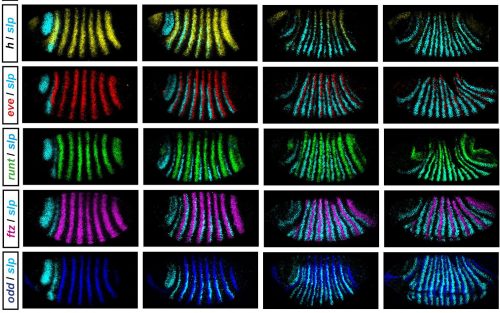
 (2 votes)
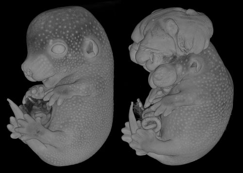


 Retinal degenerative diseases are the leading causes of blindness worldwide. In their
  Prostate organogenesis is a complex process that is regulated by androgens and subsequent mesenchyme-epithelial interactions. In their
  As every year, the