The laboratory of Zebrafish Neurogenetics, led by Dr. Laure Bally-Cuif at the Pasteur Institute in Paris, is seeking an outstanding and highly motivated Postdoctoral Research Associate to contribute to our ongoing research on adult neurogenesis in zebrafish. Our lab is interested in the molecular and cellular mechanisms underlying basic adult neural stem cell properties, including the control of their quiescence, their interactions with other neural stem cells of the germinal niche, and their recruitment towards neuron generation. The project will involve characterizing how the neurogenic activity of individual neural stem cells is controlled, and how neuronal identities are encoded and used to build a continuously growing but functional brain. Analyses will involve genetic approaches, whole-mount imaging and cell tracing in the adult animal, and will include tool development for conditional functional assays in adult neural stem cells.
Candidates must hold a Ph.D. in biology, and a strong interest and background in molecular and cellular neuroscience, in any model system. Previous experience with imaging and genomic techniques is preferred. Proficiency in English is required.
The position is funded by the European Research Council and Labex Revive.
The Bally-Cuif team is one of the 16 research groups of the “Developmental and Stem Cell Biology” Department of the Pasteur Institute, focusing on evolutionary, developmental and stem cell biology in various animal models. It is also co-affiliated with the Pasteur “Neuroscience” Department.
To apply, please submit your CV and the name of three references to:
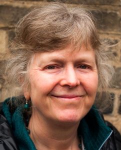
In 2016, the BSDB introduced the Cheryll Tickle Medal, which is being awarded annually to a mid-career, female scientist for her outstanding achievements in the field of Developmental Biology. The BSDB is proud to announce the 2017 awardee Jenny Nichols. The medal was presented at this year’s Spring Meeting where Jenny gave the Cheryll Tickle Award Lecture (available on YouTube). A post-award interview with Jenny was published in Development. For further awards at that meeting, see this post.
Jenny’s main research interests are the mechanisms that establish and maintain pluri-potency in the early embryo and during the formation of embryonic stem cells in mammals. She also uses animal models to understand defects which lead to type 1 diabetes. Jenny started her career at Oxford University where she worked as a research assistant to Prof. Richard Gardner (1981-90). In 1990 she moved to the University of Edinburgh to carry out her PhD project in the group of Prof. Austin Smith. She obtained her PhD in 1995 for her thesis entitled ‘A Study of the Expression and Function of Differentiation Inhibiting Activity and its Receptor in the Early Mouse Embryo‘. She stayed as a post-doctoral research fellow in the group of Austin Smith in Edinburgh, until she became a group leader at the Wellcome Trust-MRC Stem Cell Institute of the University of Cambridge, where she has stayed since then.
Jenny’s mentors: Richard Gardner (left) and Austin Smith (right)
Jenny has an impressive portfolio of current funding with 3 BBSRC, a Wellcome Trust and a Medical Research Council grant, she has published ~70 papers so far, supervised 11 PhD students, and has editorial responsibilities at three scientific journals (PLoS One, Biol Open, Dev Biol), in addition to a number of local administrative tasks. She is active in university teaching and has been the co-/organiser of a number of international stem cell workshops and engages in science communication with the public.
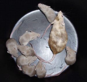
Germline chimaera generated in Jenny’s laboratory from ES cells derived in 2i medium
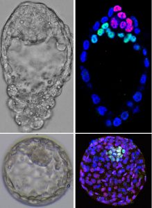
Top: E4.5 mouse embryo stained against Nanog (red) and Gata6 (green); bottom: D6 human blastocysts
Apart from the Cheryll Tickle Medal awarded this year, Jenny won the NC3Rs ‘3Rs’ prize (for research reducing refining or replacing the use of animals in biomedical research; 2009) and the Suffrage Science Award (2013), is an elected Fellow of the Royal Society of Biology (2010) and was an active member of the BSDB committee (2010-15).
The BSDB makes it a tradition to ask the Cheryll Tickle Medal awardees a number of questions concerning our field and its future. Please, read Jenny’s answers below.
What were the questions that inspired you to work in the field of Developmental Biology?
I was fascinated by the flexibility of the early mammalian embryo and curious as to how the lineages were specified and regulated. This is still my main research theme. The first questions were ‘when do the cells of the inner cell mass lose their ability to become troph-ectoderm?’ and ‘do they routinely supplement the developing troph-ectoderm?’ Most of all I just loved messing about with embryos. We had so few tools in those days and so many questions, but were quite restricted to using observation and grafting.
Why should young researchers continue to engage in Developmental Biology?
As someone spanning Developmental Biology and stem cell research I feel very strongly that studying Developmental Biology requires a rigorous and systematic approach that can often be by-passed by the stem cell biologists who work in vitro. Developmental Biology is necessarily a 3D system, so the questions of cell fate specification can be very tricky and exciting to tackle. One very satisfying thing about experimenting with embryos is that the final readout from any manipulation must measure up to the yardstick of normality.
Which were the key events or experiences in your life that influenced your career decisions and paved your path to success?
Firstly, having been in Richard Gardner’s lab surrounded by brilliant embryologists (Richard, Rosa Beddington, John West, Chris Graham) and having had the luxury of my own microscope and microinjection equipment and unlimited access to mice; secondly, joining Austin Smith’s lab and having the chance to work on embryonic stem cell derivation when it was such a mysterious process. Austin also gave me the chance to do a PhD and taught me how to think.
What advice do you give young researchers towards a successful career?
Go to a good, supportive lab and collaborate broadly.
The Waddington Medal, the only national award in Developmental Biology, is awarded for outstanding research performance as well as services to the subject community. The medal is awarded annually at the BSDB Spring Meeting, where the recipient presents the Waddington Medal Lecture (note that all awards of the 2017 Spring Meeting are listed here). The BSDB is delighted to announce the 2017 winner of the Waddington Medal: William Harris FRS FMedSci, Head of the Department of Physiology Development & Neuroscience at the University of Cambridge. Bill was awarded the medal for his pioneering contributions to the understanding of retinal development.
Bill is Canadian, but underwent his scientific education and early career in the U.S., where he did his B.A. in Biophysics (University of California, Berkeley; 1972), his Ph.D. on “Color vision in Drosophila” in the group of Seymour Benzer at the California Institute of Technology (Pasadena; 1972-76), carried out his postdoctoral research in the laboratory of David Hubel and Torsten Wiesel at the Dept. of Neurobiology, Harvard Medical School (1976-80), and joined the faculty of the Dept. of Biology, University of California (San Diego; 1980). He remained in San Diego until 1997, when he moved to the UK to take on a position as Professor of Anatomy at the University of Cambridge and, since 1999, Head of the Department of Anatomy (which became the Department of Physiology, Development and Neuroscience in 2006).
Bills achievements are best summarised in the nomination letter put forward by Sarah Bray, Michael Bate, Nancy Papalopulu, Daniel St Johnston and Steve Wilson:
“Through his passion for science, his leadership and his mentoring of people at all career stages, Bill has made outstanding contributions to Developmental Biology. Working at the interface of Developmental Biology and Neuroscience, he has championed the field within Cambridge, across the UK and throughout the world. His deep interest and scientific enthusiasm have led to major insights in the field of neuronal specification and wiring.
Bill has made many important contributions to our understanding of visual system development, which has been his focus throughout his career. Time and again, he has pioneered new fields of research. His early discoveries in the USA helped establish basic principles underlying axon guidance in brain wiring and revealed key mechanisms regulating cell differentiation in the retina. Starting with the identification in Drosophila of the sevenless gene (with Seymour Benzer), he went on to discover ways that axons in the brain are guided to the vicinity of their appropriate targets in the absence of neural activity, being one of the first to suggest there might be local chemotactic cues that guide retinal axons from a distance (see Figure 1; Dingwell et al., 2000, J Neurobiol 44, 246ff. – LINK). He used the developing Xenopus visual system to find the first vertebrate homologues of genes that influenced fate choice in Drosophila, including Notch, ASH, and ATH, leading to a new and fruitful research direction for Developmental Biologists.
Figure 1. Example of an elegant experiment where retinal ganglion cell growth cones that were severed from their axons, were video-images and shown to continue to navigate towards the tectum (thus demonstrating the navigational capacity residing in these structures; taken from Harris et al., 1987, Development 101, 123ff. – LINK).
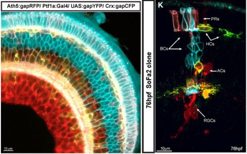
After moving to Cambridge in 1997, he built on these earlier observations. He established the retinal ciliary marginal zone as a powerful model to study not only retinal development per se, but also mechanisms controlling stem cells and the pathways regulating differentiation. Perpetually self-renewing and proliferative, this neuroepithelium at the perimeter of the retina in amphibians and fish gives rise to cells that are spatially ordered with respect to cellular development. Combining in vivo lipofection strategies in Xenopus retina with genetic approaches in zebrafish, Bill has uncovered roles for the cell cycle, metabolism and stochasticity in fate determination. A pioneer in the field of live imaging in developing systems, he made the very first time-lapse movies of axons growing in the brain. He has harnessed emerging technology to distinguish between different hypotheses. For example, by watching lineages evolve in vivo he ruled out the idea that their variance was due to random cell death. Through such cutting-edge studies he has shown how cell divisions, cell lineages and cell polarizations contribute to the process of retinal neuron specification (Fig. 2; Agathocleous & Harris, 2009, Annu Rev Cell Dev Biol 25, 45ff. – LINK). In doing so, he championed the establishment of the Cambridge Advanced Imaging Centre, whose main focus is on techniques that enable gentle deep imaging of cells in developing animals, and has forged strong links with physicists to develop powerful models.
Figure 2. The Spectrum of Fates approach can be used to assess various aspects of neural development, such as developmental waves of differentiation, neuropil development, lineage tracing and hierarchies of fates in the developing zebrafish retina (taken from Almeida et al., 2014, Development 141, 1971ff. – LINK)
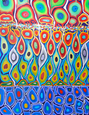
Bill has made important contributions to the community. He is an enormous presence in Cambridge, heading one of the major biology departments and taking a lead in the Cambridge Neuroscience initiative. Under his leadership and long-term vision, the Departments of Anatomy and Physiology were merged, establishing “PDN” where the letter D stands for Development. Bill is on the Advisory committee for the Gurdon Institute, and has also been instrumental in the establishment and renewal of the Wellcome Trust 4-year PhD Programme in Developmental Biology. As well as training first-rate researchers in his own lab (23 of whom have PI positions around the world), he has, as Department Head, nurtured many developmental biologists at different stages in their career, housing early researchers (e.g. Fiona Wardle), recruiting talented lecturers (e.g. Clare Baker, Benedicte Sanson, Kristian Franze) and supporting established leaders (e.g. Andrea Brand, Magda Zernicka-Goetz). Despite the burdens of being Department Head, Bill has retained a major teaching role throughout, with lectures and practicals introducing fundamentals of neural development to undergraduates at all stages. As organizer of a number of international conferences, he has included significant themes in Developmental Biology. He is also editor in chief of “Neural Development” and on the editorial boards for “PLoS Biology”, “Cell” and “Molecular Neuroscience”. Among his many other talents, Bill is an artist who also communicates his scientific vision through his paintings, as illustrated by his impression of a young zebrafish retina (see Figure 3).”
Figure 3. A painting by Bill Harris showing his impression of a young zebrafish retina.
To add to this, the high quality of Bill’s work is reflected in his many honours, fellowships and awards which include being a Fellow of the Academy of Medical Sciences (2007), Fellow of the Royal Society (2007) and Member of EMBO (2012). With respect to translational biology, he was founder of the drug discovery company DanioLabs (2002), which was successfully sold to VASTox (now Summit) plc, a leading UK biotechnology company [LINK]. Furthermore, for many of us, Bill has become an inspiring teacher, especially through his textbook Development of the Nervous System (Sanes et al., 3rd edition, 2011, Academic Press – LINK) which was a true eye opener: a highly entertaining read laying essential foundations for conceptual thought about the field and its many facets and directions.
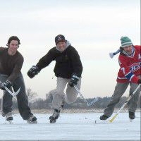
Finally, Bill’s passion for ice hockey deserves mentioning (Fig. 4). He decided to share it with the Cambridge community by founding the Cambridge Leisure and Ice Centre as a community-led initiative in 2001, which he still chairs today. The Cambridge Ice Arena will be the key result of the initiative and is scheduled to open late 2017.
The BSDB would like to congratulate Bill for his life achievements and for being the well-deserved awardee of the 2017 Waddington Medal. The Waddington Medal lecture is available on the BSDB YouTube channel and website, and an interview with Bill was published in Development.
In spite of our external appearance, our innards are asymmetric. For today’s interview, we feature a paper published recently in Development that provides a cellular and molecular investigation into symmetry breaking in a poorly understood organ, the stomach. We caught up with first authors Adam Davis and Nirav Amin, and their supervisor Nanette Nascone-Yoder, Associate Professor in North Carolina State University, Raleigh, NC.
Nanette, Adam and Nirav
Nanette, can you give us your scientific biography and the questions your lab is interested in?
NN-Y I have been interested in left-right asymmetry for a long time. I did my graduate work with Mark Mercola at Harvard Medical School, studying the induction and left-right asymmetric patterning of the heart. At NC State University, we’ve been investigating the morphogenesis and evolution of the gastrointestinal tract, specifically, how the embryonic gut tube forms the requisite three-dimensional shape necessary for proper physiological function, including appropriate length and left-right asymmetric curvature.
And Nirav and Adam, how did you come to join Nanette’s lab?
NA During my career, my research interests have focused on the transcriptional control of cell fate specification and organ development. I did my PhD work in the lab of Dr. Jun “Kelly” Liu at Cornell University, where I focused on the transcription factors, and their targets, that controlled muscle and non-muscle fates in the C. elegans mesoderm. From there, I wanted to apply the tools and techniques I used in C. elegans to the classic vertebrate model for developmental biology, Xenopus. I did my postdoctoral work in the lab of Dr. Frank Conlon at the University of North Carolina at Chapel Hill, where my research focused on a transcription factor, Casz1, and its role in cardiac development. As part of my project, I also participated in some RNA-seq and ChIP-seq studies to identify targets of Casz1 in the heart. Eventually, I began exploring methods to determine the functional roles of genes in cardiac development by editing frog genomes via mutagenesis and transgenesis.
Towards the completion of my postdoctoral work, I met Nanette and she told me about a research project in her lab in which she was using a non-model organism, the Budgett’s frog, to investigate how organs establish left-right asymmetry. These frogs have large embryos (three times larger than the Xenopus embryo!), and she was using this to her advantage in some transcriptomic studies. My experience with this type of data and functional studies in the frog made me a perfect fit for the project. I was excited by the possibility of developing this frog as a new model for developmental biology research and I joined the lab three years ago.
AD I’ve always been interested in animals, particularly in how they are shaped. I’ve also always been interested in how our DNA shapes our organs and organ systems. I performed my PhD work in the lab of Dr. Ed Stellwag at East Carolina University. I studied how Hox genes are regulated in the developing brain and pharyngeal arches. I used the Japanese medaka (Oryzias latipes) as a model to study the development and evolution of morphology. I enjoyed learning how transcription factors and their respective cis-regulatory elements regulate gene expression during development. I first met Nanette when I presented a poster of my research at the Southeast regional conference meeting for the Society of Developmental Biology at UNC, Chapel Hill in 2007. I was amazed by her talk and the research her lab was performing at NC State, particularly in how genes influence cellular morphogenesis. Once I finished my Ph.D., I was lucky enough to obtain a postdoc position in her lab under a NIEHS training grant. I was excited to work with Pitx2, as it is a homeodomain-encoding transcription factor. I was also excited to understand how organs are shaped at the cellular level.
Schematic of alternate models for stomach curvature, from Figure 1, Davis, Amin, et al. 2017
You reference some papers from the 1960s hypothesising as to how the stomach gets its curvature. Why do you think it has taken until 2017 for the first experimental investigation into its cellular and molecular basis?
NN-Y The rotation theory was actually put forth in the late 19th century, based on retrospective observations of human embryos. Rotation of the organ nicely explained the final anatomical location of left and right nerves along the front and back of the stomach; hence, the idea of early organ rotation became widely accepted and propagated such that it became dogma in all the textbooks. We were certainly not the first to question this model. Dissenting theories have been proposed by other scientists before now, but these studies were also based largely on retrospective studies in human embryos, and conducted prior to modern molecular developmental biology.
It is important to clarify that the stomach probably does undergo rotation events during later development, in order to refine the final contour and position of the organ as it descends into the abdominal cavity. However, our results indicate that wholesale rotation is not the mechanism of establishing the initial LR asymmetric curvature, as has been assumed for over a century.
What led you to address this question using both mice and frogs?
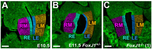
NA One day Nanette pulled me into her office to show me some pictures publicly available on emouseatlas.org. It was clear that a lot of the observations she and Adam had made in Xenopus held true in the mouse stomach. We were fortunate that the Ghashghaei lab here at North Carolina State University had the Foxj1 mouse mutant, enabling us to directly test how curvature is affected when left-right asymmetry is randomized, and to show that the origins of stomach curvature are conserved in mammalian development.
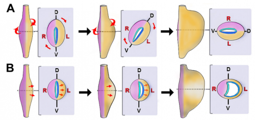
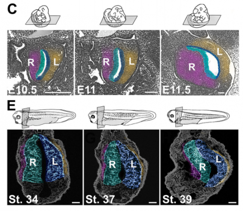
Leftward expansion in the early stomach of mice and frogs, from Figure 1, Davis, Amin, et al. 2017
Can you give us the key results of the paper in a paragraph?
NA The J-shaped stomach is characterized by a longer “greater” curvature and a smaller “lesser” curvature. Historically, this shape has been thought (and taught) to be the result of a rotation of the dorsal surface of the gut tube to the left. However, others have suggested that this curvature is not the result of rotation, but an intrinsic asymmetric growth of the stomach to give rise to the greater curvature. In this paper, we use frog and mouse embryos to show that the latter hypothesis is true –differential lengthening of the left wall drives the curvature of the stomach. This lengthening occurs due to radial rearrangement which cause the left stomach wall to thin. Importantly, this process is dependent on Foxj1/Nodal/Pitx2c, key determinants of left-right asymmetries in multiple organisms.
Do you have any idea how Pitx2 is directing stomach morphogenesis?
NA Our working hypothesis for how Pitx2 is directing stomach morphogenesis is through the regulation of cellular effectors that drive radial intercalation. Our current research focus is in the identification of such factors within the left stomach.
Control and mutant mouse stomachs, from Fig. 2, Davis, Amin, et al. 2017
Are all asymmetric internal organs made asymmetric in different ways? How does the stomach relate to the rest of the digestive system, for instance?
NN-Y Different types of morphological asymmetries form in different organs. For example, some organs form an acute curvature like the stomach or early heart tube. Other organs adopt left right asymmetric positions, or undergo asymmetric regression or remodeling, such as the spleen and vasculature. In others, grossly different morphologies develop out of their left versus right halves/ counterparts; examples include the cardiac atrial chambers or the contralateral lobes of the liver and lungs. Unfortunately, for the majority of organs, we know surprisingly little about the cellular and molecular events that break symmetry during organogenesis, so it is unclear whether these varied types of morphological asymmetries may form in different ways.
The recent work in intestinal rotation does provide one point of comparison. The appearance of cellular differences between the left and right sides of the dorsal mesentery breaks the symmetry of the structure that suspends the gut tube from the body wall, leading to a leftward shift in gut position, ultimately biasing the direction of intestinal rotation. Interestingly, no developmental asymmetries are thought to exist in the intestine itself. In contrast, we find that the left and right sides of the foregut tube itself undergo distinct morphogenetic processes to drive stomach curvature. At the cellular level, left side cells are more polarized /organized than right side in both stomach and mesentery; however, at the tissue level, the left stomach wall thins and expands, while left mesentery condenses. Only the mesoderm layer of the gut is involved in the mesentery, while both mesoderm and endoderm become asymmetrical in the stomach. So there are both similarities and differences in the development of asymmetry in each context. In the future, we will need to look at a variety of organs in order to determine the degree of universality or divergence in organ-level symmetry breaking.
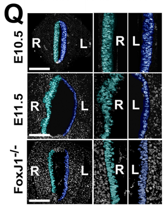
Nuclear staining reveals cell numbers in the two walls of the mouse stomach, from Fig. 2, Davis, Amin, et al. 2017
When doing the research, was there a particularly exciting result or eureka moment that has stayed with you?
NA For me, I was excited by the success of the CRISPR reagents I generated to look at Pitx2c function in the stomach. Though it wasn’t the most crucial data pertaining to this paper, it was ground-breaking for me in that I could use CRISPR/Cas9 to systematically interrogate gene function in the F0 generation for Pitx2 and other genes I identify to be involved in asymmetry. This has proven to be a very time- and money-saving finding.
AD Absolutely! It was when I was examining immunochemically-stained transverse sections of wild-type Xenopus stomachs at several developmental stages. I noticed that several morphometric factors (E-cadherin, aPKC, and γ-tubulin) showed much more robust apical localization in the left endodermal cells of the developing stomach than the right. This was observed in developmental stages prior to gut curvature at the gross anatomical level.
And what about the flipside: any moments of frustration or despair?
NA Having worked with worms and frogs, I have gotten used to being able to follow embryogenesis in real time and in large numbers of animals. When we embarked on the mouse side of this project, the frustration for me came in not being able to 1) get more than 2-3 mutant animals per litter and 2) precisely control the timing of when we get embryos. On the bright side, it made me appreciate the advantages of Xenopus even more!
AD There were a lot more of these than the eureka moments, including antibodies not working properly, needles clogging when trying to inject embryos, frogs not yielding viable eggs, me wanting to pull my hair out, etc., etc.
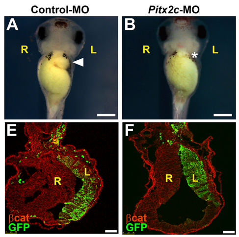
Frog embryos injected with control or Pitx2c morpholinos, from Fig. 4, Davis, Amin, et al. 2017.
What next for you following this work?
NA I am very excited to continue this work – we have used transcriptome profiling in the Budgett’s frog to identify some interesting genes that function together with Pitx2 in regulating stomach curvature. Hopefully soon, you’ll be reading more about these!
AD Thanks to my experience with my PhD, and as a postdoc in Nanette’s lab, I am currently a tenure-track assistant professor at Gordon State College. I enjoy teaching Developmental Biology and Human Anatomy and Physiology. I learned a wealth of information regarding molecular and developmental genetics, cellular and gross morphogenesis, anatomy, histology, and microscopy. I used this information to build my developmental biology course. I also enjoy training undergraduate students in research in developmental biology.
And finally – what do you get up to when you are not in the lab?
NA The majority of my non-lab life is dedicated to my wife, 4 year-old daughter, and 2 year-old son. They keep me balanced and make me feel young (and old at the same time!). Now that the weather is getting nicer here in North Carolina, we are constantly outside staying active – we have started our garden and will be camping, biking, and more in the coming months.
AD My non-academic life is dedicated to my wife, Rebecca, and our 7 year-old son, Finn. Like Nirav, they give me balance with my work life. We’re near the Appalachian Mountains, so we love hiking and camping. Finn and I enjoy searching for and photographing salamanders and other wildlife when we hike. Also, last summer, we made our first insect collection together.
Back to you Nanette – where will this paper take your lab?
NN-Y We are currently identifying the cellular morphogenetic events that underlie symmetry-breaking in two other organs. As Nirav mentioned, we have also devised a novel strategy for identifying the molecules involved, using a novel model organism (the Budgett’s frog). The hope is that these genes could be candidates for human organ defects. Variation in these processes may also be involved in generating novel organ anatomy and morphology during evolution.
Last Sunday evening found me sitting in the BBC Cambridge radio studio, headphones on and mic in front of me, talking about developmental and stem cell biology with Dr. Chris Smith, better known as the naked scientist. Fortunately, both of us were fully clothed. For those of you who aren’t familiar with The Naked Scientists, it’s an award-winning radio show and podcast that discusses the latest scientific research, answers questions on diverse scientific topics from listeners, and generally aims to make science more accessible to the general public. I’d met Chris a few weeks earlier to talk about a program he was planning that would link developmental and stem cell biology to regenerative medicine, and he asked whether I’d be willing to contribute to the show – providing an introduction and commentary to the interviews he was conducting. Hence the headphones and mic.
Having never been in a radio studio before, let alone appeared on live radio, I found the experience fascinating and daunting in equal measure. How did the whole thing work? What if I said something stupid? Fortunately, Chris and Tom (Crawford – the producer working on this show) were great at putting me at ease and guiding me through the program. And I think (hope!) the end result makes for an interesting listen. In the show, you’ll hear Roger Barker talking about his plans for a clinical trial for Parkinson’s Disease using embryonic stem cell-derived dopaminergic neurons, Hans Clevers discussing how gut organoids can be used for personalised drug testing, and Don Ingber on his amazing organ-on-a-chip technology. Plus Chris and me trying to pull these threads together and provide a perspective on where the field is going.
You can find the full show here; our discussion starts around 25 minutes in, but I’d actually encourage you to listen to the whole thing – which covers topics as diverse as heroin addiction, why airplane travel is likely to get more turbulent, and what determines whether something will ‘go viral’.
Regeneration, the ability to restore lost parts of the body, is a widespread phenomenon in animals. Whilst this ability is somehow limited in classical developmental model organisms, a variety of animals are able to regenerate complex structures, such as limbs or important parts of their body. Regeneration is often based on the presence of populations of stem cells which are either pluripotent and able to regenerate all tissues, or multipotent with a much more restricted potential. Regeneration can also rely on local cell dedifferentiation processes by which differentiated cells are reprogramed into proliferating progenitor or stem cells.
In the our team (Stem Cells, Development and Evolution team), we use the emerging developmental biology annelid model Platynereis dumerilii to investigate the evolution of stem cells and regeneration. Platynereis worms are able to grow and regenerate their posterior (caudal) part following posterior amputation during most of their life. These two abilities end when the worms become sexually mature and are dependent on the presence of a brain hormone (methylfarnesoate) that blocks sexual maturation. In the frame of a collaborative ANR-FWF funded project with the team of Florian Raible (Origin and Diversification of Hormone Systems, MFPL Vienna, Austria), the postdoctoral fellow will study, using cellular and molecular approaches, how the brain hormone controls the growth and regeneration abilities of Platynereis worms, and participate to the molecular and cellular characterization of the regeneration process.
The Vervoort team belongs to the Institut Jacques Monod (IJM) in Paris (France). The IJM is a leading French biological research institute, comprising about 30 interactive research groups and high-quality technological facilities, including a cutting-edge imaging platform. The working language at the IJM is English, and knowledge of French is therefore not a prerequisite for this position. Successful candidates will collaborate with a dynamic team of developmental, cellular and evolutionary biologists at both the IJM and MFPL.
Starting date is flexible but should be around September 2017 onwards and is funded initially for 14 months. The successful applicant must have, or be in the process of completing, a PhD thesis in a relevant research area and not more than two years of postdoctoral experience. Desirable qualifications include expertise in molecular biology, immunohistochemistry, qPCR, and DNA sequence analysis. Potential candidates should send their application by e-mail to michel.vervoort@ijm.fr with a statement of interests and expertise, a Curriculum Vitae and contact information from two referees. The position will remain open until filled; however, applications received by May 30th will be given priority. Please contact Michel Vervoort for more information.
We are seeking an enthusiastic and motivated Postdoctoral Research Scholar to join research projects investigating the molecular basis of neurodevelopmental disorders in the laboratory of Kristen L. Kroll at Washington University School of Medicine. Working in collaboration with Washington University’s Intellectual and Developmental Disabilities Research Center (http://iddrc.wustl.edu/About/MissionOverview), we use directed differentiation of human pluripotent stem cells (embryonic stem cells and patient-derived induced pluripotent stem cells), mouse models, and a wide range of cellular, molecular, biochemical, and genomic approaches, to define gene regulatory networks that control neural cell fate acquisition and the specification and differentiation of specific neuronal cell types, such as cortical interneurons. We are defining roles for epigenetic regulation in controlling these networks and identifying mechanisms by which their dysregulation alters neural development and function and contributes to neurodevelopmental disorders, including inherited epilepsies, autism spectrum disorder, and neural tube defects.
For additional information about our ongoing work and research interests, please see: http://krolllab.wustl.edu/
Setting/Salary/Benefits:
Our laboratory is in an academic setting in the Department of Developmental Biology at Washington University School of Medicine (St. Louis), an internationally recognized research institution with a dynamic research environment and extensive infrastructural and core facility support. Postdoctoral appointees at Washington University receive a starting salary based on the NIH NRSA guidelines and a generous benefit package. Complete information on the benefit package is located on the WUSM Human Resources Benefits Website (http://medschoolhr.wustl.edu). The St. Louis area combines the attractions of a major city with family-friendly and affordable lifestyle opportunities (https://explorestlouis.com/)
Qualifications:
Candidates should hold a recent PhD with less than 2 years of prior post-doctoral experience. Preference will be given to applicants with a strong interest in and research training relevant to the areas of neural development, stem cell biology, and transcriptional or epigenetic regulation. US citizens are preferred. Interested candidates should send a CV/names of references by email to kkroll@wustl.edu or by regular mail to Kristen L. Kroll, Washington University School of Medicine, Campus Box 8103, 660 S. Euclid Ave, St. Louis, MO 63110.
A postdoctoral position is available in the laboratory of Dr. Martin Basch to study the regenerative potential of the stria vascularis in cases of congenital deafness. Highly motivated, creative and enthusiastic individuals are particularly invited to apply.
Qualifications include a PhD in developmental biology, neuroscience, cell biology or related field. Priority will be given to candidates with experience in cell culture and basic molecular biology. Mouse genetics and/or neonate mouse surgery skills are desired but not required.
Our laboratory is part of the Hearing Research Program, at Case Western Reserve University School of Medicine. Currently the program involves five Research Faculty PIs and Physician Scientists with key areas of research, which include molecular otology, otitis media, congenital/acquired hearing loss, inner ear development, and hair cell biology. The strength of our program is enhanced by an excellent interdisciplinary and collaborative intellectual environment at Case.
Interested candidates should submit their CV and a letter of application (including a brief description of previous research experience and a statement of interests) to: Martin Basch Ph.D. at mlb202@case.edu.
For more information on our laboratory, please visit our website at http://www.baschlab.org
In employment, as in education, Case Western Reserve University is committed to Equal Opportunity and Diversity. Women, veterans, members of underrepresented minority groups, and individuals with disabilities are encouraged to apply.
Case Western Reserve University provides reasonable accommodations to applicants with disabilities. Applicants requiring a reasonable accommodation for any part of the application and hiring process should contact the Office of Inclusion, Diversity and Equal Opportunity at 216-368-8877 to request a reasonable accommodation. Determinations as to granting reasonable accommodations for any applicant will be made on a case-by-case basis
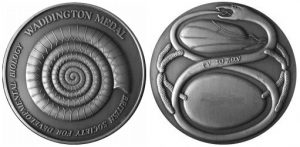
The Beddington Medal is the BSDB’s major commendation to promising young biologists, awarded for the best PhD thesis in Developmental Biology defended in the year previous to the award. Rosa Beddington was one of the greatest talents and inspirational leaders in the field of developmental biology. Rosa made an enormous contribution to the field in general and to the BSDB in particular, so it seemed entirely appropriate that the Society should establish a lasting memorial to her. The design of the medal, mice on a stylised DNA helix, is from artwork by Rosa herself. For further medal and award winners at the 2017 Spring Meeting see here.
The BSDB congratulates the 2017 Beddington Medal winner Erik Clark. Erik did his BA in Biological Sciences at the University of Oxford (1st in his year group) and his MSc in Bioinformatics & Theoretical Systems Biology at Imperial College London where he worked on the project entitled “Evolution of Mutation Rate in Fluctuating Environments”. He then moved on to do his PhD within the BBSRC Genes to Organisms Program supervised by Michael Akam at the Department of Zoology, University of Cambridge, where he worked on his project entitled “The Drosophila Pair-Rule System” and where he continues to work now. Erik won an impressive number of prizes, fellowships and grant awards, including the Gibbs Prize in Animal Biology (Univ. Oxford, 2011), an Isaac Newton Trust Research Grant (2016-17), a Junior Research Fellowship (Trinity College, Cambridge) and he is co-investigator on a BBSRC research grant (2017-20).
His Beddington Medal talk described the outcome of his most successful PhD project. About the background Erik explained: “The Drosophila segmentation cascade is a paradigmatic example of a developmental gene regulatory network, used to gain insight into transcriptional regulation in all animals… Using spatial information from graded domains of “gap” gene expression, seven “pair-rule” genes are expressed in periodic patterns of seven stripes each .. [which] then work in combination to specify precisely-phased 14 stripe patterns of “segment polarity” genes. These output patterns form the template for the segmental organisation of the insect body…[Although] the pair-rule genes have been studied in Drosophila for over 30 years,… there is still no good systems-level understanding of their regulatory interactions“.
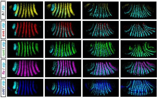
In his thesis, Erik used a combination of modelling and experiment to reverse-engineer the structure of the Drosophila pair-rule network and understand how it generates expression dynamics that lead to the patterning of segmental boundaries. He set out to collect a complete time-resolved dataset of relative expression for all pairwise combinations of the 7 pair-rule genes in wild type embryos (see Figure), and a partial dataset for a number of mutant genotypes. Using these data he defined how the regulatory interactions of pair-rule genes change at the mid-cellularisation stage, and identified odd-paired as a temporally regulated factor responsible for these network changes. As an outcome of his work, Erik proposes that that spatial resolution emerges from temporal dynamics, rather than static positional information.
A further part of his thesis proposes that the standard model for how parasegment boundaries are specified, by the interpretation of local gradients of Even-skipped protein, may not be correct. He suggests instead that the shifting of even-skipped stripes across the field of cells in the blastoderm, driven by dynamic gap gene expression, coupled with the temporal control of network interactions, may generate the key offsets in downstream gene expression, and this is an entirely novel idea.
To illustrate Erik’s path to success, Michael Akam writes: “Unlike any other student I have had, Erik spent pretty much the whole of his first year reading. He worked through the entire literature on Drosophila segmentation (spanning 30 years and hundreds of papers), assessing the claims made on the basis of the data presented, and with the advantage of hindsight that the original authors lacked. I suspect he has a more detailed and critical knowledge of this literature than any other researcher.” Michael concludes his support letter with the words: “Erik’s work is strikingly original, and represents a major innovation in thinking about Drosophila segmentation.”
Erik’s publications so far:
Clark, E. (2017) ‘Dynamic patterning by the Drosophila pair-rule network reconciles long-germ and short-germ segmentation’. bioRxiv: 1101/099671
Clark E & Akam M (2016). Odd-paired controls frequency doubling in Drosophila segmentation by altering the pair-rule gene regulatory network. eLife: 7554/eLife.18215
Clark E. & Akam M. Drosophila pair-rule gene double FISH Data (data from Clark & Akam 2016). org:10.5061/dryad.cg35k 2016
BertaVerd, ErikClark, Karl R.Wotton, HildeJanssens, EvaJimenez-Guri, AntonCrombach, JohannesJaeger (2017). A damped oscillator imposes temporal order on posterior gap gene expression in Drosophila. bioRxiv: 10.1101/068072
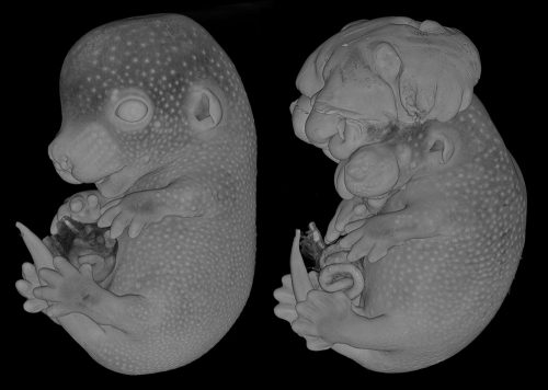
A large-scale study of DMDD data has shown that inactivating the same gene in mouse embryos that are virtually genetically identical can result in a wide range and severity of physical abnormalities. This suggests that the relationship between gene mutation and embryo development is more complex than previously thought.
A comparison of two embryos that are both missing the embryonic lethal gene Coro1c. The embryo on the right has abnormal viscerocranium (facial skeleton) morphology, while the embryo on the left does not.
The study considered 220 mouse embryos, each with one of 42 different genes inactivated. These genes are part of a set known as ‘embryonic lethal’, because they are so crucial to development that an embryo missing any one of them can’t survive to birth. Studying these genes can help us understand how embryos develop, why some miscarry and why some mutations can lead to abnormalities.
 (No Ratings Yet)
 Jenny’s main research interests are the mechanisms that establish and maintain pluri-potency in the early embryo and during the formation of embryonic stem cells in mammals. She also uses animal models to understand defects which lead to type 1 diabetes. Jenny started her career at Oxford University where she worked as a research assistant to Prof. Richard Gardner (1981-90). In 1990 she moved to the University of Edinburgh to carry out her PhD project in the group of Prof. Austin Smith. She obtained her PhD in 1995 for her thesis entitled ‘A Study of the Expression and Function of Differentiation Inhibiting Activity and its Receptor in the Early Mouse Embryo‘. She stayed as a post-doctoral research fellow in the group of Austin Smith in Edinburgh, until she became a group leader at the Wellcome Trust-MRC Stem Cell Institute of the University of Cambridge, where she has stayed since then.
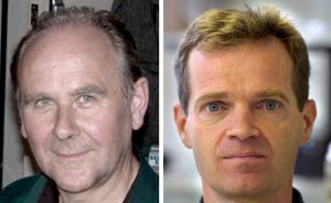
 Bill is Canadian, but underwent his scientific education and early career in the U.S., where he did his B.A. in Biophysics (University of California, Berkeley; 1972), his Ph.D. on “Color vision in Drosophila” in the group of Seymour Benzer at the California Institute of Technology (Pasadena; 1972-76), carried out his postdoctoral research in the laboratory of David Hubel and Torsten Wiesel at the Dept. of Neurobiology, Harvard Medical School (1976-80), and joined the faculty of the Dept. of Biology, University of California (San Diego; 1980). He remained in San Diego until 1997, when he moved to the UK to take on a position as Professor of Anatomy at the University of Cambridge and, since 1999, Head of the Department of Anatomy (which became the Department of Physiology, Development and Neuroscience in 2006).
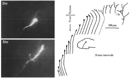
 (6 votes)
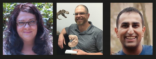


 His Beddington Medal talk described the outcome of his most successful PhD project. About the background Erik explained: “The Drosophila segmentation cascade is a paradigmatic example of a developmental gene regulatory network, used to gain insight into transcriptional regulation in all animals… Using spatial information from graded domains of “gap” gene expression, seven “pair-rule” genes are expressed in periodic patterns of seven stripes each .. [which] then work in combination to specify precisely-phased 14 stripe patterns of “segment polarity” genes. These output patterns form the template for the segmental organisation of the insect body…[Although] the pair-rule genes have been studied in Drosophila for over 30 years,… there is still no good systems-level understanding of their regulatory interactions“.