Stockholm University, Sweden, invites applications for one postdoctoral position in the laboratory of Professor Mattias Mannervik at the Department of Molecular Biosciences, The Wenner-Gren Institute (http://www.su.se/mbw). The position is scheduled to start as soon as possible.
Transcriptional coregulators are proteins that facilitate communication between transcription factors and the basal transcription apparatus, in part by affecting chromatin through post-translational modification of histones. As such, they contribute to generation of cell-type specific gene regulatory networks and epigenetic control of animal development (see Mannervik et al. Science, 284, 606-609, Mannervik Exp Cell Res. 321(1):47-57). This laboratory is using molecular, genetic, and transgenic approaches in Drosophila melanogaster to elucidate the molecular mechanisms of transcriptional co-regulator and chromatin regulator function during development. To investigate if histone modifications can be instructive in regulating gene expression this project uses two approaches. A histone replacement system is used to examine the effects of amino acid substitutions in the histones on organismal development, and a modified CRISPR/Cas9 system is employed to target chromatin modifying enzymes to endogenous loci.
The position is available immediately and requires a recent Ph.D. as well as extensive experience in molecular biology techniques. The successful applicant should have a high-quality publication record, and motivation to study underlying mechanisms of gene regulation in development. The position will be funded with a fellowship, and includes health insurance.
Stockholm University is one of the largest and most prominent universities in Sweden, located in the nation’s capital city, beautifully surrounded by the first national city park in the world. For further information, see http://www.su.se/english/ and http://www.academicstockholm.se/
Application: Applications marked with reference number SU FV-2.1.9-2187-16 should be submitted electronically as a single PDF file to mattias.mannervik@su.se and to registrator@su.se. The application deadline is September 1, 2016
Applications should comprise the following:
1) a personal statement describing research interests (1-2 paragraphs), research experience (1–2 paragraphs) and career goals (1-2 paragraphs)
2) curriculum vitae
3) bibliography
4) names, e-mail adresses, and phone numbers of three references
On the 23rd of June the United Kingdom held a referendum on whether to remain a member of the European Union or to leave. Prior to the vote, Nature reported that 83% of nearly 2000 polled scientists favoured remaining, and letters from Royal Society members and Nobel Prize winners urged the public to vote to remain in the interests of British research. The result was called early on Friday morning: the UK had voted to leave the EU, by 51.9% to 48.1% (around 17 million to 16 million voters). We’re now faced with the question of what this might mean for UK science, and, given how interconnected science is, also how it might influence science in the EU and the rest of the world.
So we’ve got two Questions of the Month for June, one personal and one more global:
What does the Referendum result mean for you, scientifically and career-wise?
Are there practical steps we as a community can do to ensure a bright future for UK and EU science?
We’re hoping to hear from as broad a selection of people as possible: UK and EU nationals working here in the UK, elsewhere in the EU or in the wider world; students, postdocs, PIs, and people who have left the lab bench!
We recognise what an emotive subject this is, and would like to try to keep this discussion to the referendum’s impact on science and careers. Let us know what you think in the Comment boxes below, or via social media (we’ll also be collating these answers via Storify).
Last month saw the return of the Young Embryologist Network annual meeting held this year at the UCL Institute of Child Health. To settle into the long weekend, a number of us from the Briscoe Lab at the Crick Mill Hill site headed on down to central London to spend the day being inspired by talks from PhD students, Postdocs and several invited guest speakers. We were met with an impressive array of speakers, highly interactive poster sessions and an atmosphere buzzing with young embryologists demonstrating their clear passion for cell and developmental biology. With such excellent organisation and enthusiasm, we are happy to write about our thoughts of the meeting, and greatly anticipate the next event.
Poster session (photo provided by Ruben Perez).
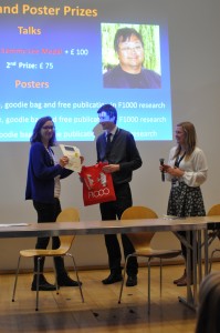
To kick off the day, our newest lab member, Teresa Rayon, opened the “Cell Fate Determination” session. Presenting her PhD project, Teresa demonstrated the importance of enhancer usage and developmental pathways that direct tissue-specific expression of Cdx2 in the early mouse blastocyst. Following this tour de force, the opening session only continued to satisfy our appetite for developmental biology, concluding with the two prize-winning talks of the day. Sarah Bowling (Rodriguez Lab), presented an elegant story on the role of mTOR signalling, a main component involved in cell competition in early embryos. Her studies showed the link between developmental signalling, metabolism and cell survival, for which she received the Sammy Lee Memorial Medal. This award recognises an early stage career embryologist, who not only carries out inspiring work but also has the ability to communicate their science in a personable manner, a skill that Sammy Lee was well known for. Following this, the session concluded with Sarah Harrison (Zernicka-Goetz lab), who was awarded a second place prize for her work on organoids, and how asymmetry occurs in the early embryo. At this point we were left with a clear feeling of excitement that in vitro systems such as stem cells have afforded developmental biologists, as more and more becomes possible at the tissue and organ level….
The second session of the day concentrated on polarity and asymmetry with the best place to focus being the zebrafish brain. The talks up until this point all had one thing in common: stunning imagery, which gave a natural build up into the first external speaker of the day, Dale Moulding from UCL Institute of Child Health. His talk covered new and emerging technologies in microscopy, covering a range of approaches from micro-CT, to OPT, SPIM and light-sheet imaging – a veritable repertoire of optical imaging solutions, which are continually pushing the boundaries of image resolution and depth of field.
After the lunch break the second guest session was filled with inspiring presentations on early embryonic development from Kathy Niakan (Francis Crick Institute) and Shankar Srinivas (University of Oxford). Kathy discussed mouse and human blastocyst development, focusing on the key transcriptional regulators involved in early lineage commitment. Shankar then followed with an examination of the cellular mechanisms involved in driving early AP patterning in the mouse, and in establishing a beating heart. Echoing earlier sentiments of the day, he demonstrated how light-sheet microscopy can be exploited to reveal impressive cellular-level detail in the context of whole organisms as they develop in real time. The generation of this type of live imaging data now poses new challenges to the developmental biology field, in how best to reconstruct individual cells and extrapolate the intricate details of developing organs. Such a feat will undoubtedly rely on the continued collaboration between biologists and physicists alike to develop the best possible data analysis solutions.
The final session, “Morphogenesis, Repair and Regeneration” had talks on cell matrix adhesions during contact inhibition of locomotion as well as cell movement in distal neuropore closure. To complete the session, Tapan Papilia (Hughes lab) gave an insight into Pax7 stem cells in muscles and their roles in regeneration and repair.
YEN awards ceremony (photo provided by Ruben Perez).
Closing the talks for the day, we welcomed Professor Paul Martin (University of Bristol), invited to give The Sammy Lee Memorial Lecture. Capturing the essence of YEN, Paul’s talk reminded us that it’s not only important to work hard, but to enjoy the work that you’re doing – an important aspect that can drive discovery. His captivating presentation highlighted that no matter what area of development you may find yourself in, the basic underpinnings of biology can have far reaching implications. Some of these are being realised in the field of tumour biology, where studies that began in fly have transitioned to vertebrate models of wound healing that are informing cancer treatments. Never knowing exactly where one will end up is definitely an attraction for many developmental biologists, and a motivation that drives us to work long (and sometimes tedious) hours at the bench or desk.
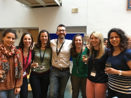
YEN meeting participants. From left to right: Lorena Garcia-Perez, Vicki Metzis, Marta Moris Sanz, Ruben Perez, Teresa Rayon, Katherine Exelby, Neda Mousavy (photo provided by Teresa Rayon).
From cells to embryos, the YEN meeting makes a compelling case that the UK harbours many talented young scientists. Importantly, these individuals come from across the globe and are fuelled by a potent combination of excellent research leaders, high quality facilities and resources – a tradition that we are proud to be a part of now and into the future. An important conclusion that we take away from this meeting is that a successful “young embryologist” is in our view not only a creative, open-minded, cross-disciplinary individual, but also an excellent communicator, both to the public and to scientists alike. Events such as these are important to promote these aspirations. For this, we would like to thank the organising committee and sponsors who enabled this excellent meeting. We look forward to continuing our participation in future events organised by the young embryologists network and strongly encourage you to do the same.
Here is some developmental biology related content from other journals published by The Company of Biologists.
Cartilage development downstream of Notch
Notch signalling regulates various aspects of vertebrate cartilage development, and Hilton and colleagues now investigate the role of the Notch effectors HES1 and HES5. These transcription factors suppress chondrogenesis and promote chondrocyte hypertrophy, with some overlapping and some distinct functions.
Scribble promotes proliferation
The scaffolding protein Scribble is critical in establishing apical-basal polarity during epithelial development. Muthuswamy and coworkers now show that during pregnancy, Scribble has an unexpected role in promoting cell proliferation during alveologenesis, potentially via keeping the prolactin receptor at the cell surface.
A molecular pathway to haptotaxis
Haptotaxis is directional cell migration in response to a gradient of substrate-bound cues. Bear and colleagues investigate the cellular and molecular basis of haptotaxis using microfluidic chambers, and show that differential actin and lamellipodial dynamics, regulated by Arp2/3 and its upstream regulators, contribute to the process.
A new way to generate photoreceptor-like cells
De novo generation of photoreceptor cells is therapeutically promising for patients with retinal degenerative diseases. Seko and colleagues describe efforts to directly reprogram blood cells into photoreceptor-like cells using the CRX transcription factor. This method may provide a cost-effective alternative to induced pluripotent stem cells for personalised drug screening and disease modelling [OA].
Polyamines in pigmentation
Zebrafish pigmentation is an established model system for developmental patterning. Irion and colleagues now identify a new player in pigmentation: the polyamine spermidine. Mutations in the spermidine synthase gene leads to loss or interruption of the dark stripes of the flanks and fins [OA].
Picking the right model
In his Editorial, Leonard Zon explores how complementing his lab’s primary model organism – the zebrafish – with other model systems helped in the translation of research to the clinic , and the importance of collaboration and infrastructure [OA].
Distinct cellular contributions to muscle repair
Hughes and colleagues identify stem cell diversity in the wound healing response of zebrafish muscle, and propose that pax7a-expressing cells initiate de novo fibre formation, while pax7b-expressing cells promote fibre growth [OA].
Linking neural crest cell migration to craniofacial disorders
Pera and co-workers useXenopus to model Musculocontractural Ehlers–Danlos syndrome, which is caused by mutations in dermatan sulfate enzymes. The craniofacial abnormalities associated with the disorder may arise from defective migration, rather than specification, of neural crest cells [OA].
Optogenetics has been widely used in the study of neural activity and behavior. By using genetic tools to target light-sensitive proteins to one or populations of neurons in specific region of brain, we can active those neurons by optical stimulation.
The foundation of neuron interaction bases on the ion transmission through the neuron membrane. The positively charged ions and negatively charged ions flow across the membrane via the transmission proteins and the balance of these ions in both inside and outside of the neurons contribute to the neurons trigger action potential. The action potentials, as known as spikes, are the key points in neural communication. Therefore, if we control the transmission proteins by genetic methods, then we can control the transmission of ions flow, which means that we can control the neurons communication.
Recently, the conventional way to achieve optogenetics is introducing light via optical fibers. However, light delivered by optical fibers is not at high spatial resolution in the brain because of light absorption and scattering. Therefore, Robert et al., show a novel optogenetic probe that achieves cell-type-specific perturbation precisely at high spatial resolution. The probe presented in the paper integrates micro-LEDs so that the light source is brought into the deep brain which allows high-spatial stimulation. Also the dimension of probe is small enough to minimize insertion damage. Unlike other similar approaches, the micro-LEDs are fabricated on silicon substrate, not on sapphire substrate. Sapphire-based LEDs cannot be thinned beyond 100 mm but silicon substrate can be thinned to a proper size that suitable for the probe. Therefore, the probe is able to reach the deep region of brain without unnecessary damage. I think this novel probe offers a good opportunity to study the neuron communication.
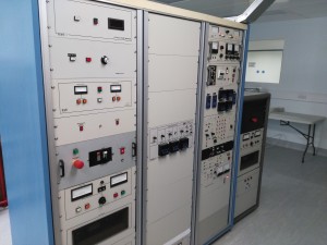
Hello guys, I am a PhD student from University of Strathclyde, UK. My PhD career has two parts: microfabrication and neural recordings. With the help of novel semiconductor fabrication techniques, I can make micro-level devices for neuroscience applications such as neural recordings and optogenetics.
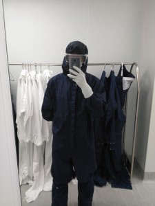
The whole fabrication process is done in the cleanroom which can block any tiny particles in the air and make all the fabrication steps are done in micro- or even nano-scale precisely. In order to keep the good fabrication environment of cleanroom, everyone who wants to enter into the cleanroom will be required to wear this specific suit.
It will cover your whole body from head to feet; even your eyes are protected by goggles. Thanks to my poor eyesight, I have to wear two glasses! (PS: It really reminds me the nightmare of watching IMAX 3D movie. Every time I go to cinema to watch IMAX 3D movie, the 3D glasses is always unsuitable to my own glasses! T_T)
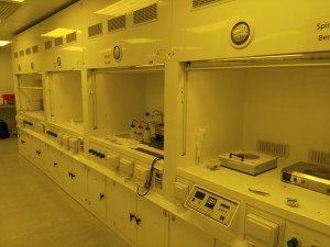
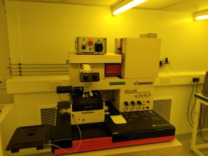
Well, let’s go back to the story. The device I design for neural recording has multi-layer structure. So Mask Aligner can help me to transfer the designed patterns to the sample layer by layer. I also need to work on these benches to do some chemical works which I call “Magic”! Haha!
Wet benches
Mask Aligner
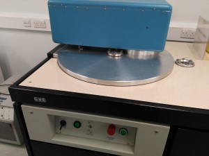
When the work is done here, some big guys are waiting for me in the white room. Neurons communicate with each other via electrical signals which are called Spikes and the amplitude of spikes is about microvolt. Therefore, metal with low resistance is required.
Sputter (process chamber)
Sputter (control system)
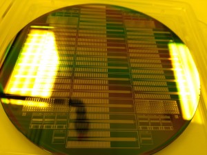
Yes! Gold! It is you! See the big guy there? His name is Sputter and he will deposit a uniform thin layer of gold on the sample. Then RIE (reactive ion etching) will draw a picture on the gold layer to form patterns that I want to have.
Devices on 4” wafer
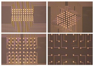
Closed-up image of microelectrode array
See! Great job!! Well done!!!
The device is used to record signals in vitro. The tissues will be cultured on the top of the array and the spikes will be recorded by these electrodes and transmitted to the outer electronic system. The fabrication work is a little bit tricky because it requires time to practise and optimize. Sometimes it is annoying to be honest but I have already drawn into it.
Post doctoral position available to study the genetic and epigenetic control of stem cell attributes and pluripotency, focusing on the neural crest gene regulatory network (NC-GRN). Neural crest cells are stem cell-like progenitors that migrate extensively and whose genesis was central to the evolution of vertebrates. Misregulation of components of the NC-GRN underlies numerous human diseases and congenital disorders. Studies involve post-translational regulation of known network components, and use of proteomics and next generation sequencing to identify novel components.
Highly motivated applicants with a PhD and strong background in cell and molecular biology and/or developmental biology are encourage to apply. Please send a CV, brief description of research interests, and the names of three references to:
Carole LaBonne, PhD (clabonne@northwestern.edu)
Department of Molecular Biosciences
Northwestern University, Evanston, IL 602028
The Fight for Sight charity have provided a three year studentship to Stephen Wilson and Gaia Gestri to support a student to work on the role of the Yap/Taz pathway in the morphogenesis of the eye. The project will use zebrafish as a model and will involve a combination of imaging and molecular genetic approaches to resolve the cell behaviours that regulate eye formation in health and disease.
We are looking for a talented, motivated and enthusiastic student to work in a team of scientists studying various aspects of brain and eye development (www.ucl.ac.uk/zebrafish-group/). Candidates will have suitable MSc and/or BSC qualifications and ideally experience and expertise in imaging and image analysis and in working with zebrafish as a model system.
We are seeking a highly motivated and collaborative postdoc in the area of human embryology and stem cell biology to join Dr. Kathy Niakan’s laboratory.
We have identified several transcription factors and components of key signaling pathways that are highly expressed in pluripotent epiblast cells of the developing human embryo. The pluripotent epiblast has the unique potential to give rise to the entire fetus in vivo and can self-renew indefinitely as embryonic stem cells (hESCs) in vitro. Understanding the molecular basis of lineage specification in the early human embryo is of fundamental biological importance and has significant clinical implications for infertility treatment as well as the use of hESCs to treat various diseases. Importantly, the genes we identified as enriched in human embryos are not expressed in mouse embryos at the equivalent developmental stage, further suggesting differences in pluripotency mechanisms between these species.
The aim of the project is to characterise putative regulators of human pluripotency and embryogenesis using currently the most efficient and precise genome editing technique (CRISPR/Cas9) in human embryos and stem cells. This will provide fundamental insights into human biology and facilitate the development of conditions for the establishment of novel human stem cells. We also seek to establish novel human embryonic stem cells by modulating signaling pathways that we have identified as specific enriched and functional in the development of the pluripotent human epiblast.
The successful candidate is likely to be an energetic, focused, and productive individual with a desire to work in a congenial, dynamic, and collaborative research environment. Good organisational, analytical, and communication skills are essential.
ORGANISATION
Dr Niakan’s laboratory focuses on understanding the mechanisms of lineage specification in human embryos and the derivation of novel human stem cells. Details of research projects currently being undertaken can be seen at: http://www.crick.ac.uk/kathy-niakan
Research techniques used in the laboratory include: molecular biology, advanced microscopy and image quantification, human and mouse preimplantation embryo culture and micromanipulation, genome modification, genome-wide techniques including single-cell RNA-sequencing, human embryonic and induced pluripotent stem cell derivation.
OBJECTIVES
In this project, some of the specific objectives could include, but not be limited to:
Stem cell derivation from embryos
Reprogramming using induced pluripotent stem cell approaches
Genome editing using CRISPR-Cas9
Genomic profiling of early human embryos and microdissected cells
Ensuring the design and implementation of the project
Liaising with collaborators within the Crick, the UK and abroad
Writing and contributing to the preparation of scientific manuscripts, reports, presentations and records of experimental plans and results
Working closely with the Group Leader and other team members to report on the results via publications
Supervising and providing technical advice to more junior members of the team when appropriate
ABOUT US
The Francis Crick Institute has a distinctive vision of how biomedical research is conducted. It is one of the most significant projects in UK biomedical science for a generation. The institute’s labs have an international reputation for cutting edge research into basic biology and are committed to training the next generation of research scientists.
On 1 April 2015, staff from the London Research Institute (CRUK) and National Institute for Medical Research (MRC) transferred to the Crick to form a fully functional research institute on four sites. In 2016, the Crick will move to a single new, purpose built research centre in St. Pancras which will house some 1,500 staff.
PERSON SPECIFICATION
The post holder should embody and demonstrate our core Crick values: Bold, Imaginative, Open, Dynamic and Collegial, in addition to the following:
Essential
PhD in the areas of Developmental Biology, Stem Cells, Molecular Biology or similar (or in the final stages of PhD submission)
Good knowledge and experience in molecular biology and microscopy
Technical expertise in embryo and/or cell culture
Proven track record of research (i.e. publication record)
Excellent communication skills required – both oral and written presentation
Ability to communicate ideas and results effectively and interact fluidly with computational biologists
Ability to work independently and organise own workload
Ability to design experiments, report on research progress and outcomes openly and review methodologies in response to feedback
Highly motivated, organized and analytical
Ability to update knowledge in the specialist area and implement relevant technologies to advance the project
Desirable
Experience in preimplantation mouse or human embryo culture
Experience in human and mouse pluripotent stem cell culture
Experience in preparing samples for advanced sequencing
Experience in genome editing using CRISPR-Cas9 technology
Experience in lentivirus production and transduction
Postdoctoral Training Fellows are expected to lead their own projects, contribute to other projects on a collaborative basis (both in the lab and with external collaborators) and guide PhD students in their research. The ability to work in a team is essential.
If you are interested in applying for this role please apply through our online system:
 (No Ratings Yet)
 and letters from
  (1 votes)
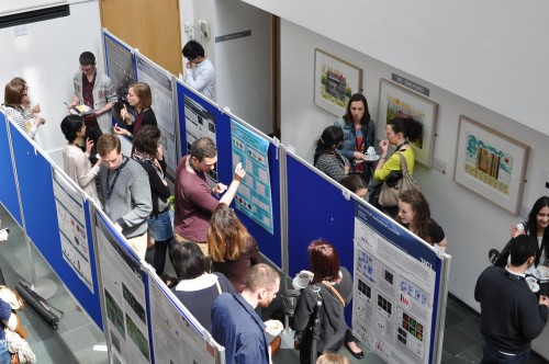
 (7 votes)
 Notch signalling regulates various aspects of vertebrate cartilage development, and Hilton and colleagues now
  protein Scribble is critical in establishing apical-basal polarity during epithelial development. Muthuswamy and coworkers now
 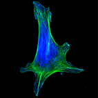 Haptotaxis is directional cell migration in response to a gradient of substrate-bound cues. Bear and colleagues
 De novo generation of photoreceptor cells is therapeutically promising for patients with retinal degenerative diseases. Seko and colleagues
  pigmentation is an established model system for developmental patterning. Irion and colleagues
 identify
 Pera and co-workers