Postdoctoral position to study pancreas development and plasticity
Pancreatic acinar cells are highly specialized entities dedicated to produce and secrete digestive enzymes. Acinar cells are also very plastic and they undergo striking morphologic and functional changes upon injury or under the effects of oncogenes. A POSTDOCTORAL POSITION is available immediately in the Sosa-Pineda laboratory to study how the acinar differentiation program is established during development, maintained in adult cells, and modified during regeneration or neoplastic transformation. The candidate will use available mouse models, 3D acinar cultures, organoid cultures and biochemical and bioinformatics approaches to investigate the former processes.
Highly motivated candidates who recently obtained a PhD or MD degree and have expertise in developmental biology studies, transcription regulation, mouse husbandry, and cell culture and molecular biology techniques are encouraged to apply. For more information on our research please visit our site at: http://labs.feinberg.northwestern.edu/sosa-pineda/index.html
Interested individuals should send their curriculum vitae, a brief description of their research interests, and the names of three references to:
Beatriz Sosa-Pineda, PhD
Associate Professor
Department of Medicine
Northwestern Feinberg School of Medicine, Chicago
beatriz.sosa-pineda@northwestern.edu
Northwestern University is an Equal Opportunity, Affirmative Action Employer of all protected classes, including veterans and individuals with disabilities. Women and minorities are encouraged to apply. Hiring is contingent upon eligibility to work in the United States.
The Society for Developmental Biology (SDB) Professional Development and Education Committee (PDEC) created the John Doctor Education Prize to highlight great developmental biology education research. In previous years, the committee awarded a prize for the best education poster at the SDB annual meeting. This year, the PDEC has reinvented the award as a best education video competition.
The challenge is for SDB members to produce short videos (5 min max) demonstrating a creative and engaging method for teaching difficult-to-learn topics in developmental biology to an undergraduate, graduate, or lay public audience. This year’s developmental biology concept is induction. Videos with broad appeal and those demonstrating active learning exercises or hands-on activities are preferred. The creator(s) of the best video will be awarded a certificate and a check for $1000 at the 75th SDB Annual Meeting awards banquet in Boston.
For more information see links to the guidelines for submission and online form below. The submission deadline is Monday, June 6, 2016 (11:59 pm EDT).
It is our greatest pleasure to welcome you to the 2016 BSDB autumn meeting that will be held from the 28-30 August in Edinburgh. This conference is our only theme driven meeting for 2016 and provides a unique forum to network and socialise with a wide cross-section of the developmental biology and stem cell community.
In this meeting we will explore the importance that chimeric analysis has had on our understanding of developmental mechanisms. Our speakers have made seminal contributions to the field using a diversity of organisms, including slime moulds, plants, chick, mouse and human. Our plenary speakers, Profs Nicole le Douarin and Richard Gardner have been at the forefront of research in the area, and their work has laid the foundations to our understanding of cell fate and plasticity in vertebrates. The sessions of the meeting will cover lineage tracing and potency, signalling mechanisms, regenerative therapy and human, disease models and gene function, and therefore will provide a broad perspective on how chimeras have shaped our understanding of developmental and stem cell biology.
Additionally, we have been fortunate enough to be able to organise this meeting at the end of the Edinburgh International Fringe Festival, providing the attendees with a unique opportunity to enjoy not only an excellent scientific meeting, but also the cultural offerings of this wonderful city.
We look forward to seeing you in Edinburgh!
organisers: Jenny Nichols and Tristan Rodriguez; Warwick
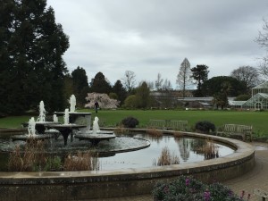
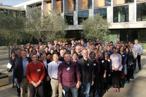
Set in the idyllic location of the Sainsbury Laboratory Cambridge University (SLCU) building adjacent to the Cambridge University Botanic Garden, the inaugural Sainsbury Lab Symposium (#SLS16 on Twitter) attracted over 100 researchers from across the world. This three-day symposium focused on the topic of Induced Plant Development and featured 18 speakers, 10 poster flash talks and 36 posters, covering a variety of research topics. Several themes emerged from this meeting that we highlight in this report.
Cambridge University Botanic Garden
The symposium kicked off with a Keynote Address by Sofie Goormachtig (VIB Department of Plant Systems Biology – Ghent University), discussing her elegant work on the role of Strigolactones in controlling root architecture. The importance of Strigolactone signalling was further supported by, Thomas Greb (COS – Heidelberg University) presenting his results on the role of Strigolactones in primary energy metabolism and growth regulation. The implication of hormones in induced plant development was repeatedly discussed during the symposium, including several posters emphasizing the role of Auxin, Cytokinins and Abscisic Acid signalling in the regulation of cell elongation, regeneration and differentiation.
In addition to hormones, the importance of abiotic environmental signals in controlling plant development was highlighted. The environmental cues discussed included light, water, calcium, sugar and mechanical induction. Seisuke Kimura (Kyoto Sangyo University) presented his exciting results on phenotypic plasticity and heterophylly in the herbaceous semiaquatic plant Rorippa aquatica. Discussing his latest results, Seisuke showed that leaf form in R. aquatica is also affected by light and temperature. Further, Elena Baena-González (IGC – Portugal) underlined the importance of monitoring plant carbon status by SNF1-related Protein Kinase1 (SnRK1) in the restoration of post-stress homeostasis.
Biotic regulation of plant development was another central feature of the symposium. This included the discussion of parasitic plants, insects and fungi. Of particular interest was work from Melissa Mitchum(University of Missouri) on the signalling mechanisms of cyst nematode parasitism. In the classic co-option of host mechanisms, the cyst nematode delivers CLE-like effector proteins to mimic the plant CLE peptides. The subsequent signalling through the CLE pathway is essential for the establishment of nematode feeding sites. In considering parasitic plants, Ken Shirasu (RIKEN) reported the draft genome sequence of Striga asiatica andrevealed a recent whole genome duplication as a potential driving force for the adaptation to host plants. In parallel, he presented a new model for forward and reverse genetics in parasitic plants, Phtheirospermum japonicum. Recent results from his lab provide evidence for the cooption of a lateral root developmental program for haustorium (penetration structure required for host invasion) formation.
Each of these areas of induced plant development revealed the inherent problems with studying environmental influences on plant development and the need for improved technology. Several researchers presented their emerging technologies for improving plant phenotyping and environmental control. For example, Olivier Loudet(INRA) discussed his development of a high throughput phenotyping robot called the Phenoscope. This impetus for this design was the importance of controlling water content per pot and the growth room position, which are vital for his drought response studies. Along these same lines, Christian Fankhauser (University of Lausanne) also developed a phenotyping platform to analyse leaf growth and positioning with high spatial and temporal resolution. Least we forget about the belowground portion of plants, Malcolm Bennett (University of Nottingham) updated us on the progress of the Hounsfield Facility for 3D x-ray imaging of root systems. With a particular interest in how plants find and respond to water, Malcolm discussed his latest work on the molecular control of hydropatterning.
The link between fundamental sciences and agronomics has never been so tight as it was during this symposium. Many speakers have emphasized how their research can be applied to agronomics and industry. Kerry Franklin (University of Bristol) described a nice industrial partnership with a company interested in UV-B treatment of their plants undergoing shade avoidance and thermomorphogenesis problems. Christian Fankhauser (University of Lausanne) also revealed industrial interest in his research on light-induced morphogenesis and accessibility to sunlight.
It is noteworthy to mention the extreme diversity of plant species used in the topics discussed during the symposium, reflecting the vitality of the plant induced development field. From Arabidopsis thaliana to Rorippa aquatica, to the parasitic plant Striga asiatica (aka witchweed) to a variety of crops, not less than a dozen different species were represented, pushing the boundaries of plant development to new and exciting topics.
All together the inaugural Sainsbury Laboratory Symposium was a rousing success. In our humble opinion, this symposium highlighted the reason many of us attend meetings – there was stimulating discussions, new ideas, cutting edge research and a great diversity of topics. Congratulations to Dana MacGregor, postdoctoral scientist in Steve Penfield’s lab (John Innes Centre), awarded best poster of the Symposium for her work on natural variation and environmental regulators of seed dormance. The success of this symposium is due in large part to the organizer, Sebastian Schornack, who did a wonderful job and has our gratitude for the substantial effort required in organizing such an event. This symposium will definitely be included on the “must attend” list for future years.
Germ cells are unique among all metazoan cells in their ability to persist from one generation to the next. In seeking to understand how germ cells acquire and maintain immortality, a logical place to start with is the distinctive cell biological properties these cells display. For example, in many organisms, developing germ cells socially interact just before and during meiosis. In Drosophila, a founder germ cell called a cystoblast synchronously divides four times without undergoing cytokinesis to produce sixteen interconnected cells known as a germline cyst. Interconnected germ cells within ovarian cysts transport cytoplasm, organelles and RNAs to one germ cell that will survive and develop as oocyte. The rest of the fifteen sister germ cells that transfer materials, undergo apoptosis and are called nurse cells (de Cuevas et al., 1997).
Whether mammals also nurse developing oocytes has remained unclear. In fetal mammalian ovaries, germ cells appear morphologically similar, rather than as recognizable oocytes and nurse cells. After migrating to the fetal ovary, primordial germ cells (PGCs) proliferate and the resulting cells either differentiate into primary oocytes or undergo apoptosis. In humans, PGCs proliferate into about 7 million germ cells at 20 weeks of gestation and these give rise to 1 million primary oocytes at birth (Baker, 1963). In mice, PGCs generate about 20,000 germ cells at embryonic day 14.5 (E14.5) that differentiate into about 4,000 primary oocytes in the postnatal day 4 (P4) ovary (Lei and Spradling, 2013). These germ cell losses could be explained if the distinction between nurse cell and oocyte was not difficult to recognize in mammals. In that case, the cells undergoing apoptosis would be nurse cells that had finished transferring important materials to the oocytes.
If mammalian fetal ovaries do contain nurse cells as well as differentiating oocytes, they should be interconnected in germline cysts, like in Drosophila and many other species. Efforts to characterize mouse germline cysts began in our group in 1995 when Dr. Melissa Pepling joined the laboratory. Pepling systematically studied intercellular bridges and nest development within the fetal ovary. She found evidence that cysts form based on an increasing number of intercellular bridges, and observed multiple bridges per cell, bridges containing microtubules and with luminal mitochondria apparently moving between sister germ cells. Strikingly, she documented that synchronous germ cell mitotic divisions occur at this time, initially in groups corresponding to powers of two. These observations strengthened the case that germline cysts form between E10.5 when PGCs reach the gonad, and E14.5, when germ cells cease mitosis and enter meiosis (Pepling and Spradling, 1998). A few years later, Pepling demonstrated that the breakdown of mouse cysts by apoptosis correlated with the formation of primary oocytes and primordial follicles (Pepling and Spradling, 2001). However, it was not possible to precisely describe the structure of the starting cysts or how they broke down into oocytes. How many oocytes derived from an initial cyst remained unknown.
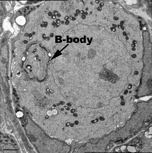
Oocytes from diverse species contain a visible mass of organelles known as the Balbiani body or mitochondrial cloud at the time they are first enclosed within a follicle. Specific mRNAs that later localize within the oocyte, including in the germ plasm, associate with the Xenopus Balbiani body (Kloc and Etkin, 1995). Up until this time, mouse oocytes were reported to be symmetrically organized and to lack a Balbiani body. However, studies in Drosophila showed that the Balbiani body is built mostly from organelles that are transferred from nurse cells into the oocyte around the time of follicle formation (Cox and Spradling, 2003, 2006). This prompted Pepling et al. (2007) to reinvestigate and show that a Balbiani body does form in mouse oocytes around the time of birth (Figure 1). The existence of a mouse Balbiani body raised the question of whether it forms like in Drosophila, by the transfer of its organelles and other components from connected nurse cells.
Figure 1. A primary oocyte in a P4 primordial follicle. Arrow indicates a Balbiani body (B-body) .
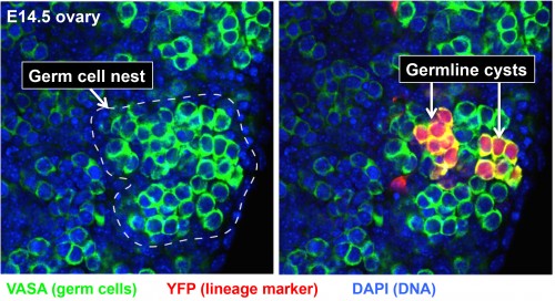
This was the situation when Lei Lei, a new postdoc with extensive previous experience in studying the mouse ovary, joined the lab in 2009. Clearly, the major roadblock to analyzing the behavior and function of cysts in the mouse ovary was simply the difficulty in recognizing individual cysts and their component germ cells. Since cyst cells share a common lineage, lineage-tracing methods offered a solution. Lei began to experiment with labeling only about one PGC per ovary at E10.5 when individual PGCs arrive at the fetal gonad using a general CreER-loxP system and low doses of Tamoxifen that rarely remove a blocker segment and activate yellow fluorescent protein (YFP) production. This approach, dubbed “single-cell lineage labeling,” would render the cells making up at least one cyst unambiguous in each ovary (Figure 2). Of course, using a low dose of Tamoxifen could not actually guarantee that exactly one PGC would be labeled. Some ovaries would have zero and a few would have two. However, even when two were labeled, they would usually be widely separated in the ovary, ensuring that their progeny remained separate. Lei would come to realize that this simpler paradigm could act as a key to throw open the treasure chest of mammalian oogenesis.
Figure 2. Two germline cysts (red) derived from one PGC that are revealed by the single PGC lineage tracing.
By analyzing the development of 164 individual marked E10.5 PGCs in this manner (about 500 are present per ovary), many longstanding questions were resolved (Lei and Spradling, 2013). All the PGCs follow the same developmental program, undergoing a similar number of divisions and producing a similar number of primary oocytes. PGCs start by generating germline cysts, since the initial number of labeled cells produced is always a power of two, reflecting the synchronous divisions first seen by Pepling. No PGCs from the fetal ovary remained undifferentiated, generated any other cell types or gave rise to oogonial stem cells, as previously postulated by some. Overall, between E10.5 and P4, 80% of PGC daughter cells underwent apoptosis, while the surviving 20% became primary oocytes. On average, each PGC produced an initial cyst that fragmented into about 5 derived cysts by E14.5 when mitotic division ceased. All germ cells then entered meiosis and generated about 6 primary oocytes by P4. The close agreement between the number of final derived cysts and the number of oocytes suggested that after an initial period of fragmentation, each remaining derived cyst generates one oocyte.
These experiments finally laid the groundwork for Lei to directly address the question of whether mouse cysts function like Drosophila cysts to nurse the oocyte and build the Balbiani body. First, she mapped the pattern of cellular interconnections within large, unfragmented cysts. In Drosophila cysts the oocyte always has 4 ring canals (intercellular bridges). Mouse germ cells with 3 or 4 ring canals were scattered throughout the early cysts, and this pattern may somehow presage how the initial cyst breaks into its derivatives. Lei went on to investigate whether organelles move between the interconnected cells within mouse cysts and accumulate in cells that become oocytes. She found that the Balbiani body could be used to distinguish cells transferring organelles (which lack a Balbiani body), from cells receiving organelles (which have a forming Balbiani body). By following centrosomes, mitochondria, Golgi elements and total cytoplasmic volume from E14.5 until all the cells have separated and either formed oocytes or died, she was able to prove that both organelles and cytoplasm are transferred non-randomly from the 80% of cells that later undergo apoptosis into the 20% that accumulate these materials, form a Balbiani body and become primary oocytes (Lei and Spradling, 2016). When cytoplasmic transport was blocked by inhibitors of microtubule polymerization or dynein-mediated movement, oocyte production and Balbiani body formation were greatly reduced. Concomitantly, apoptosis also declined, arguing that transfer is necessary to induce apoptosis. Thus, it is finally clear that fetal mammalian germ cells are divided into nurse cells and oocytes despite the fact that both enter meiosis and cannot easily be distinguished throughout much of fetal development by chromosomal or synaptonemal complex morphology.
During her final Spring and Summer in Baltimore, as this work was nearing completion, problems with the “Drosophila model” of transport through the ring canals became apparent. Mouse ring canals seemed to be important only during the early cyst stages. They shrink after E14.5, detach from the membrane joining adjacent germ cells, and disappear entirely after E19.5, well before oocyte differentiation is complete. The membranes separating germ cells develop large gaps at this same time, connecting the cells directly. The number of organelles and amount of cytoplasm moving into the oocytes is so large, it seemed more likely that they moved directly through membrane discontinuities rather than through the lumens of these small bridges. Moreover, confocal microscopy and EM studies began to make it clear that the germ cells undergoing apoptosis frequently have lost most of their cytoplasm, with little more than the nucleus being excluded from transfer. This suggested that germ cell apoptosis at these stages mostly destroyed nuclei, rather than entire cells. This realization should probably not have seemed so surprising. In Drosophila, the nurse cells in full-grown follicles synchronously transfer their cytoplasm into the oocyte just prior to entering apoptosis, a process known as “nurse cell dumping.” The nurse cell nuclei are excluded and show signs of apoptotic degeneration. Thus, the process of mouse oocyte formation by cytoplasmic dumping appears to be a variation on germ cell behaviors that are widespread among oocytes across many phyla.
These new insights require a revision of the traditional concept that mammalian oogenesis is highly inefficient. The largest single phase of germ cell loss, that occurring during oocyte differentiation, can now been seen as resulting from nurse cell apoptosis following the transfer of their cytoplasm and organelles to oocytes. Thus, “germ cell apoptosis” does not represent a waste of material, but rather a mechanism to start increasing the cytoplasmic/nuclear ratio of oocytes that will grow to be the largest cells in the mammalian body. The transfer of RNAs from nurse cells, including small RNAs, may also constitute an important part of the genome re-programming that takes place in the oocyte about this time.
The realization that mammalian oocytes develop in cysts and are supported by nurse cells provides new evidence that mammalian oocyte development is fundamentally similar to oogenesis in other animal groups. Indeed, even plant gametophytes transfer RNAs and other components between the somatic endosperm and the oocyte, in part to aid in the control of transposable elements (Creasey and Martienssen, 2010). Our work encourages the view that many of the unique properties of germ cells in all organisms, arise from fundamentally similar mechanisms that have been extensively conserved during evolution.
References:
Baker T.G. (1963). A quantitative and cytological study of germ cells in human ovaries. Proc. R. Soc. B.150, 417-433.
Cox R.T. and Spradling A.C. (2003). A Balbiani body and the fusome mediate mitochondrial inheritance during Drosophila oogenesis. Development. 130,1579-1590.
Cox R.T. and Spradling A.C. (2006). Milton controls the early acquisition of mitochondria by Drosophila oocytes. Development. 133,3371-3377.
Creasey K.M. and Martienssen R.A. (2010). Germline reprogramming of heterochromatin in plants. Cold Spring Harb Symp Quant Biol. 75,269-274.
de Cuevas M., Lilly M.A. and Spradling A.C. (1997). Germline cyst formation in Drosophila. Annu. Rev. Genet.31,405-28.
Kloc M. and Etkin L.D. (1995). Two distinct pathways for the localization of RNAs at the vegetal cortex in Xenopus oocytes. Development. 121,287-297.
Lei L. and Spradling A.C. (2013). Mouse primordial germ cells produce cysts that partially fragment prior to meiosis. Development. 140, 2075-2081.
Lei L. and Spradling A.C. (2016). Mouse oocytes differentiate through organelle enrichment from sister cyst germ cells. Science. 352,95-99.
Pepling M.E. and Spradling A.C. (1998). Female mouse germ cells form synchronously dividing cysts. Development. 125,3323-3328.
Pepling M.E. and Spradling A.C. (2001). Mouse ovarian germ cell cysts undergo programmed breakdown to from primordial follicles. Dev. Biol.234, 339-351.
Pepling M. E., Wilhelm J.E. O’Hara A.L. Gephardt G.W. and Spradling A.C. (2007). Mouse oocytes within germ cell cysts and primordial follicles contain a Balbiani body. Proc Natl Acad Sci U S A.104,187-192.
I had the chance to get involved for the second time and I can attest it is unbelievably fun and rewarding!! Individual research labs and the Science Outreach team from Rockefeller University set up over 25 booths where they present scientific concepts and methods through different activities to engage kids and (importantly!) their families. Children get to learn about DNA structure, brain function, model organisms and – of course – ice cream making (because… ice cream) through games, simple experiments and handcrafts. As usual, you can catch up with the action on twitter.
Such a large event is a lot of work and requires resources, an organizing team like the university’s excellent Science Outreach program (led by the amazing Jeanne Garbarino) and the coordinated effort of tens of volunteers (from PIs to high school students), from both Rockefeller and the neighboring institutions. However, science outreach can be done at any level. Do it next time a relative asks you ‘what do you do in the lab all day?’. Do it when you’re at a party and someone asks ‘what do you exactly work on?’. Prepare a big-picture, short explanation, practice and deliver it. Bring a friend to the lab and show them around! The goal is always the same: engage non-scientists, show them that they too can understand science and make them aware of the way science actually works (and how it often doesn’t!). To boot, it will make you understand your own work better and will help you put it into context! Let’s create a more scientifically literate society that demands more and better science. Get involved!
Department/Location: Department Molecular Biology, UT Southwestern Medical Center, Dallas Texas 75390
Closing date: June 30, 2016
Position: The funds for this post are available for up to 4 years, however postdoc will be encouraged to seek independent fellowship funding.
General area of research includes regenerative medicine, developmental biology, molecular biology, stem cells, and tissue engineering. We are interested in how the vasculature develops coordinately with organs, including the kidney and pancreas. We will investigate basic mechanisms of blood vessel patterning, cell-cell interactions and tissue morphogenesis and function. In particular, we want to engineer replacement nephrons, within the framework of the NIH-NIDDK consortium (https://www.rebuildingakidney.org/collaborate.html).
For this position experience in molecular biological and biochemical techniques, basic mammalian cell and tissue culture, mouse husbandry, histology and tissue engineering is desirable. The position is based in the Cleaver laboratory and is available immediately.
The closing date for applications is June 30 2016, but the position is available immediately.
Please provide your Curriculum Vitae (CV) including at least 3 referent contact information, as well as a cover letter in initial inquiries.
UTSW values diversity and is committed to equality of opportunity. The University has a responsibility to ensure that all employees are eligible to live and work in the USA.
The Department of Human Evolutionary Biology at Harvard University invites applications for a Postdoctoral Fellow in the Evolutionary and Developmental Genetics Laboratory of Dr. Terence D. Capellini. The focus of the position will be to study the genetic and developmental basis of human-specific adaptations and disease.
While the lab primarily concentrates on skeletal adaptations in humans and primates, an important research focus is to identify causal genetic variants that mediate human phenotypes resulting from more recent selection. The lab uses a variety of experimental and computational tools, such as functional genomics (e.g., ATAC-seq), developmental genetics in the mouse (e.g., CRISPR-Cas9), and in vitro methods on human cells, to explore the consequences of genetic variants on human biology.
The research will take place in the Evolutionary and Developmental Genetics Laboratory directed by Dr. Capellini and located in the Peabody Museum on Harvard University’s Cambridge, Massachusetts campus.
A doctoral degree is required for this position. Desired qualifications include research in developmental genetics, functional genomics, evolutionary developmental biology, mouse developmental biology and/or human evolutionary genetics.
Please submit a letter of interest, an updated CV, and the names and email addresses of three references to Terence Capellini at tcapellini@fas.harvard.edu. Evaluation will begin at the time this advertisement is posted and will continue until the position is filled.
Harvard University is an equal opportunity employer and all qualified applicants will receive consideration for employment without regard to race, color, religion, sex, sexual orientation, sexual identity, national origin, disability status, protected veteran status, or any other characteristic protected by law.
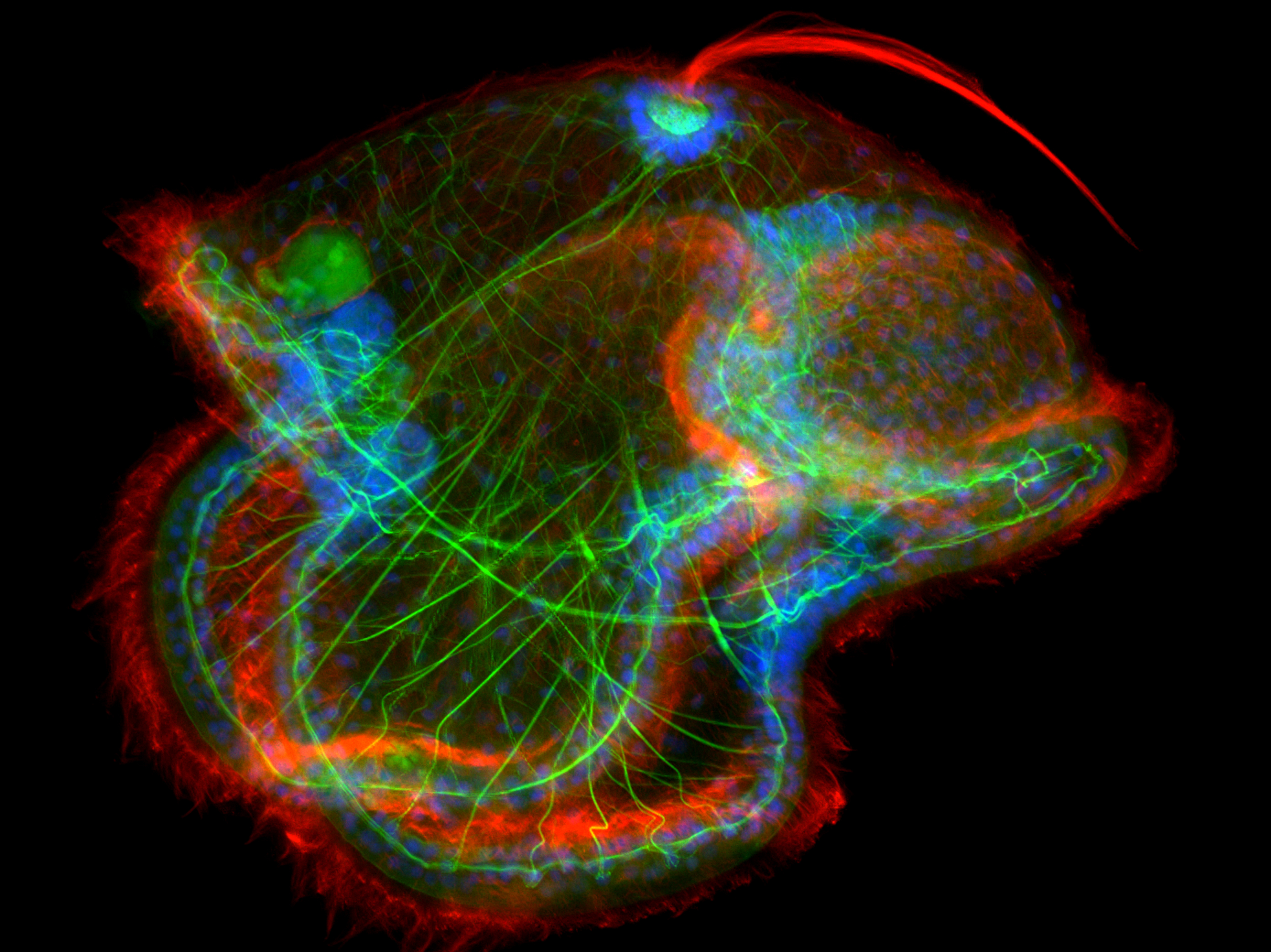
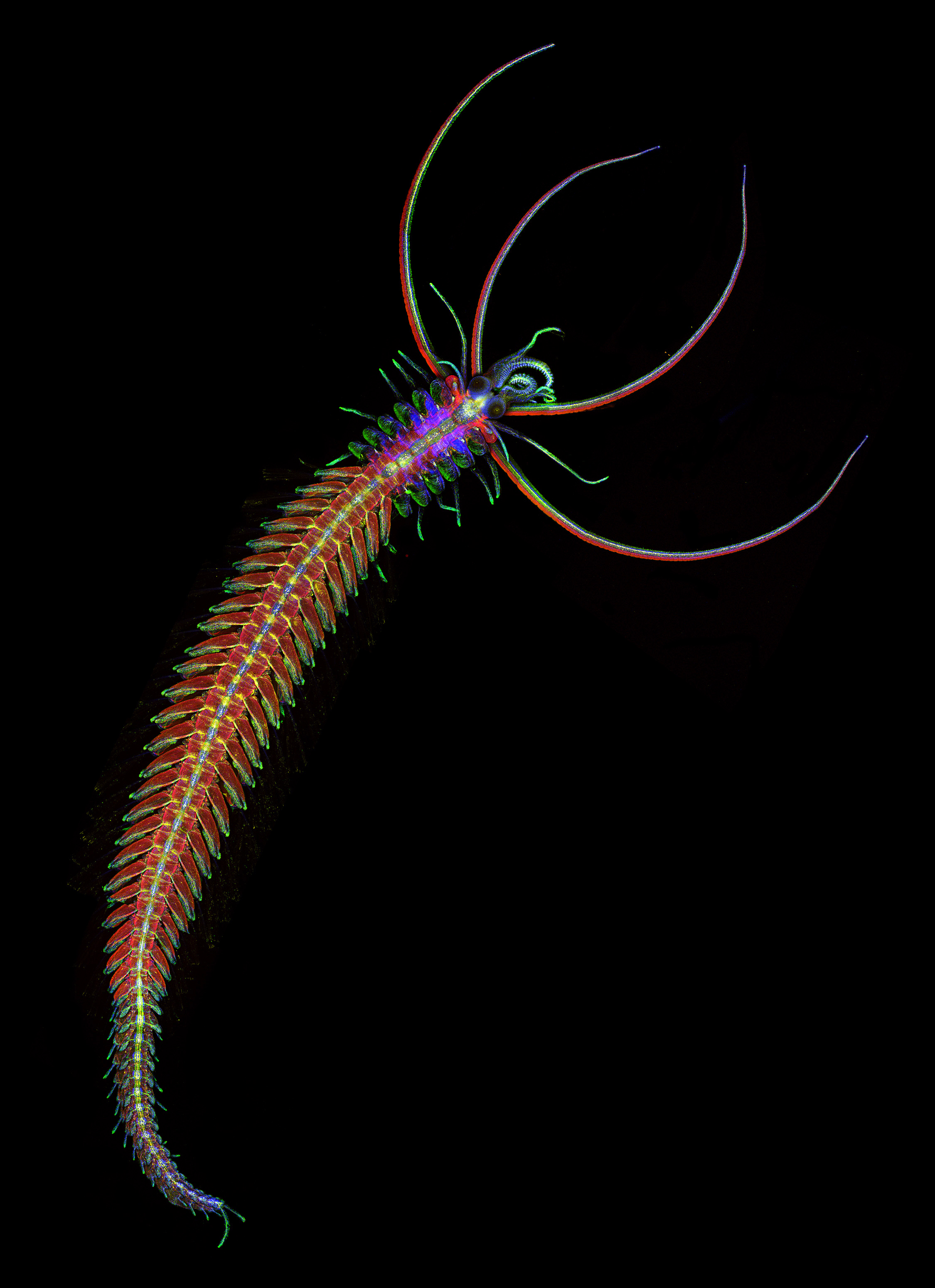
As you may have noticed, we have a series of new Node banners (alongside some of our old favourites). As there isn’t really space to give credit to the authors on the banner itself, we thought we would write this short post to give credit where credit is due!
Below is the list of the authors of the beautiful images decorating the Node banner:
Joseph Campanale, Aracely Lutes, and Stephanie Majkut took this awesome image of pilidium larvae of the Nemertean ribbon worm at the 2011 Woods Hole Embryology course.
Also at Woods Hole, but during the 2012 Embryology course, Eduardo Zattara (University of Maryland, College Park) took this image of the male stolon of an annelid.
The human embryo images were taken by Juan Carlos Izpisua Belmonte (The Salk Institute for Biological Studies).
The last banner image featured on the cover of Development:
This Arabidopsisthaliana stem section was taken by Truernit et al (paper here).
Thank you to all the authors for giving us permission to use their images on the Node!
I was kindly asked to shortly summarize my experience at the Career Workshop at the BSDB/BSCB Meeting at the University of Warwick.
My name is Hamze Beati and I am currently a postdoctoral researcher in the laboratory of Arno Müller in the Department of Cell and Developmental Biology in the School of Lifesciences at the University of Dundee. I am about to finish my postdoc after doing my PhD in the lab of Andreas Wodarz in Göttingen, Germany (now in Cologne). Later this year I will start my own junior research group “Nachwuchsgruppe” at the University of Kassel in Germany, which of course made the Career Workshop an interesting opportunity to learn about individual careers/career paths.
The first session was led by James Wakefield from the University of Exeter. The discussion was very interesting for me as he had chosen an academic career path, establishing his own group following up his own research interests. We have learnt that he changed the places he lived and worked frequently, also including times when he had to commute extensively. It was particularly nice to see that he managed to balance his work/life balance, also having a family with children at home. From our discussion I have learnt that a very important factor for an academic career path is to work together with PIs where one can follow up own ideas and interests to a particular extend. That covers my own experience so far as I was always able to develop my own ideas and thoughts about particular questions in Cell and Developmental contexts.
The second session I have attended was of great interest to me and was hosted by Claudia Barros, as she is working with the same model organism (Drosophila melanogaster) as I do. She was trying to establish the most important things for a successful academic career by looking back at her own career path. Similarly to James Wakefield we learnt that she had to change the places she lived and had to go through a hard time working in the US while her partner still lived in Europe. Things she pointed out were that winning awards are important for a successful career, including winning poster prizes, travel grants, etc.. These are all factors for a good CV. Both sessions agreed that the publication record is the most important determinant for a successful career, which was not surprising to me. Also, both sessions pointed out that during an academic career work in the laboratory will decrease, while work in the office is increasing drastically (University duties, paper and grant writing, etc.).
The last session I have attended was led by Anne Wiblin from Abcam. This discussion was also of big interest to me as she is working for a company and had left an academic career path. I learned that it was not very easy for her to find a job in industry coming from Lifesciences, something I was aware of before talking to many young researchers who decided to leave academia. Anne had to apply to many companies to finally get a position. We were able to ask her about the kind of work which is done once one decides to leave Lifescience, starting work in a company. She told us that the scientists in her company are quite busy testing new reagents, antibodies, etc. for their specificity, etc., which is quite nice as many people leaving Lifesciences would prefer to continue doing “benchwork”.
I really liked the Career Workshop and would highly recommend people to try and attend in the future. It also helped to network with the hosts, which sometimes is not very easy elsewhere at the conference.
 (No Ratings Yet)
 (1 votes)
 It is our greatest pleasure to welcome you to the 2016 BSDB autumn meeting that will be held from the 28-30 August in Edinburgh. This conference is our only theme driven meeting for 2016 and provides a unique forum to network and socialise with a wide cross-section of the developmental biology and stem cell community.