I was kindly asked to shortly summarize my experience at the Career Workshop at the BSDB/BSCB Meeting at the University of Warwick.
My name is Hamze Beati and I am currently a postdoctoral researcher in the laboratory of Arno Müller in the Department of Cell and Developmental Biology in the School of Lifesciences at the University of Dundee. I am about to finish my postdoc after doing my PhD in the lab of Andreas Wodarz in Göttingen, Germany (now in Cologne). Later this year I will start my own junior research group “Nachwuchsgruppe” at the University of Kassel in Germany, which of course made the Career Workshop an interesting opportunity to learn about individual careers/career paths.
The first session was led by James Wakefield from the University of Exeter. The discussion was very interesting for me as he had chosen an academic career path, establishing his own group following up his own research interests. We have learnt that he changed the places he lived and worked frequently, also including times when he had to commute extensively. It was particularly nice to see that he managed to balance his work/life balance, also having a family with children at home. From our discussion I have learnt that a very important factor for an academic career path is to work together with PIs where one can follow up own ideas and interests to a particular extend. That covers my own experience so far as I was always able to develop my own ideas and thoughts about particular questions in Cell and Developmental contexts.
The second session I have attended was of great interest to me and was hosted by Claudia Barros, as she is working with the same model organism (Drosophila melanogaster) as I do. She was trying to establish the most important things for a successful academic career by looking back at her own career path. Similarly to James Wakefield we learnt that she had to change the places she lived and had to go through a hard time working in the US while her partner still lived in Europe. Things she pointed out were that winning awards are important for a successful career, including winning poster prizes, travel grants, etc.. These are all factors for a good CV. Both sessions agreed that the publication record is the most important determinant for a successful career, which was not surprising to me. Also, both sessions pointed out that during an academic career work in the laboratory will decrease, while work in the office is increasing drastically (University duties, paper and grant writing, etc.).
The last session I have attended was led by Anne Wiblin from Abcam. This discussion was also of big interest to me as she is working for a company and had left an academic career path. I learned that it was not very easy for her to find a job in industry coming from Lifesciences, something I was aware of before talking to many young researchers who decided to leave academia. Anne had to apply to many companies to finally get a position. We were able to ask her about the kind of work which is done once one decides to leave Lifescience, starting work in a company. She told us that the scientists in her company are quite busy testing new reagents, antibodies, etc. for their specificity, etc., which is quite nice as many people leaving Lifesciences would prefer to continue doing “benchwork”.
I really liked the Career Workshop and would highly recommend people to try and attend in the future. It also helped to network with the hosts, which sometimes is not very easy elsewhere at the conference.
Today is my last day on the Node, so this is my chance to say goodbye! It has been 3 very busy years on the Node, but I have really enjoyed myself! When I first started on the Node I was fresh out of my PhD (actually, I was still writing my thesis) and my background was really cell biology. But I quickly discovered how wonderful the developmental biology community is, and you were all very welcoming. Thank you! The definite highlight of this job has been meeting all of you, either online or in person. I had the opportunity to chat to you about science, give career advice, discuss online communication and even, on one notable occasion, a heated but friendly argument on whether anarchy was a viable social system! So it wasn’t all just work, and I had fun as well.
During the last three years I have been involved in a lot of projects on the Node, which I hope you have enjoyed. The most obvious change was the new look of the site and its new logo, which I hope you approve. This was also when we celebrated the Node’ s 5th anniversary, a very respectable age, and had a great time filming some of you to mark the occasion! One of the series that I launched during my time on the Node was the ‘A day in the life‘ series, on model organisms in developmental biology. This was actually an idea that I suggested at my job interview, so it was great to see it come to fruition. This year we even turned a selection of the posts into a small booklet that you can collect at conferences. Your feedback on this series has always been really positive, so this is definitely a highlight! Other highlights included designing our Node postcards, kicking off the new forgotten classics series and interviewing people at conferences. Ok, ok, and I must admit that the conference locations were one of the pluses of the job as well!
My very first conference location (ISDB in Cancun). It was a tough job, but someone had to do it!
When I started this job I often still had to explain what the Node was, and ensuring that there was a constant flow of content was one of my priorities. Three years on the Node is a household name within the community, averaging more than 1 new post a day, and boasting almost 8,000 visitors a month. This is only possible because you have embraced the Node as your community site, and have read, posted and commented, and shared the word with your colleagues. Thank you for making my job so easy! And this is also a good occasion to ask for your help again. My replacement will only start in mid June, which means that for a few weeks the Node will be without a community manager to keep things ticking at the usual pace. I hope that you will help the Node team by continuing to post and comment during this period!
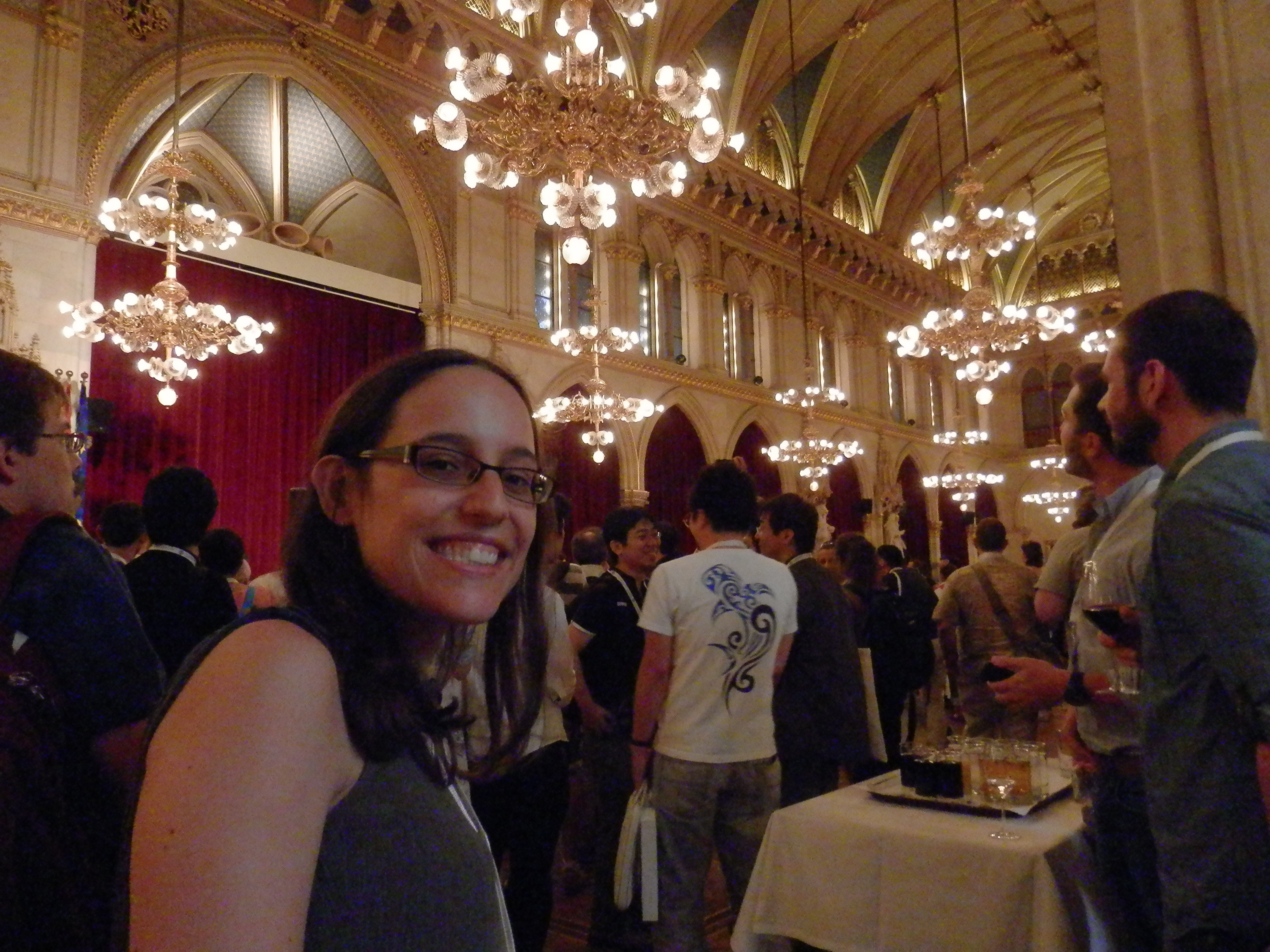
At the Vienna city hall (European Evo Devo meeting)
I hope that this post is not a goodbye, but only a see you later! My new job is based at the University of Oxford, where I will be involved in academic online communications, although not specifically about science. While I will not be travelling to exciting conferences any more, I hope I will come across some of you in the future. If you would like to keep in touch, my twitter account is @catcvicente. Thank you and see you around!
As a neglectful member of this parish over the last few months/years (insert standard academic administration/teaching workload complaints here), I have the great pleasure to come out of my slumber to drum up interest in one of the best things about being (a developmental biologist) in London:
If you have been lots before, you have probably forgotten when it is (I had): this month (27th). If you have never been before, you should come because it is interesting, friendly, and all the lovely things a good conference should be. If you are a PhD student looking for a nice place to give your first talk, YEN is absolutely brilliant. A medium sized, very supportive, and very friendly audience. If you are a desperate postdoc looking to engage in much needed shameless self-promotion, it is good for that too, but you’ll need to get a move on: the abstract deadline is 5TH MAY.
See you there for science and beer (probably in that order).
This post highlights the approach and findings of a new research article published in Disease Models & Mechanisms: ‘Stem cell-specific endocytic degradation defects lead to intestinal dysplasia in Drosophila’. This feature was written by Elan Strange as part of a graduate level seminar at The University of Alabama (taught by DMM Editorial Board member, Prof. Guy Caldwell) on current topics related to use of animal and cellular model systems in studies of human disease. The course is designed to expose students to recent research in a variety of diseases, and for this assignment, students were asked to read and provide a scholarly summary of an assigned research article ‘in press’ at DMM. Elan’s summary was selected by the editorial team for publication at the Node. The text has been edited and shortened by DMM in conjunction with the author.
A useful approach to investigating the mechanisms underlying complex diseases such as cancer involves exploring common genetic mutations. Understanding the phenotypic impact of such mutations can help to identify risk, estimate prognosis and guide treatment for specific forms of cancer. For example, screening for BRCA1 and BRCA2 mutations has been shown to be effective in determining risk of developing breast and ovarian cancer (Mavaddat et al., 2013). Similarly, Marisa et al. (2013) showed that grouping colon cancer patients into subtypes based on genetic mutations can provide a better indication of prognosis. Researching genetic mutations that correlate with oncogenesis has proven to be an invaluable means of learning more about the causes of cancer and guiding the development of new chemotherapeutics.
UVRAG, the metazoan homolog of yeast Vps38, is well characterized as a regulator of autophagy (Liang et al., 2006), a conserved mechanism by which cells digest and recycle dispensable or dysfunctional organelles and cellular components. Although loss-of-function mutations in UVRAG are known to correlate with tumorigenesis (Ionov et al., 2004) and overexpression of the protein has been shown to reduce cell proliferation (Liang et al., 2006), the precise mechanisms by which UVRAG acts as a tumor suppressor have not yet been elucidated. Given that autophagy has been shown to be involved in several types of cancer (Bento et al., 2016), the most intuitive hypothesis for the role of UVRAG in tumorigenesis implicates its autophagy-regulating function. However, this hypothesis was explored by Knævelsrud et al. (2010), who determined using qualitative and quantitative readouts for autophagy that the tumorigenicity of UVRAG mutations in colorectal cancer cell lines is independent of its role in regulating autophagy. Additionally, UVRAG has functions in DNA repair, maintenance of centrosome stability, and endocytosis (Zhao et al., 2012), all of which are implicated in cancer and could explain the role of UVRAG as a tumor suppressor. In a new study published in DMM, Nagy et al. sought to investigate the role of UVRAG as a tumor suppressor, using the fruit fly Drosophila melanogaster. Drosophila represents a powerful model for exploring the pathology and molecular mechanisms of human intestinal disorders due to the highly similar histological and cellular stress response mechanisms (specifically those involving cell proliferation and renewal) in the guts of mammals and flies.
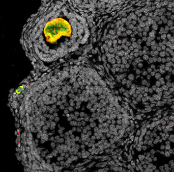
The authors began by using RNAi silencing to study the effects of adult-onset loss of Uvrag in Drosophila intestinal stem cells (ISCs). They report that the Notch ligand Delta and Wnt ligand Wingless (Wg), two biomolecules known to be trafficked via endosomes, accumulate in intracellular compartments of ISCs lacking UVRAG, thus indicating that these cells are deficient in endosomal trafficking. They validated their RNAi silencing experiments by showing that cells expressing null alleles of Uvrag show similar patterns of Delta and Wg accumulation.
The team then analyzed the effects of Uvrag silencing on ISC proliferation and morphology. The most noteworthy observations made were an increase in ISC number and concomitant thickening of the intestinal wall, both of which are characteristics of intestinal dysplasia (a precancerous lesion, see related DMM Review). To analyze the effects of silencing Uvrag on gut function, the authors fed the flies food containing a blue dye that enabled movement through the gut to be tracked. The feces of flies with UVRAG-deficient ISCs contained less dye, indicating
that these animals retain food more efficiently. Consistent with a previous finding that proper gut function is essential for normal lifespan (Biteau et al., 2010), Nagy et al. found that the fly mutants had significantly reduced lifespan. To look at how flies with UVRAG-deficient guts respond to environmental stress, the authors treated the flies with the toxin dextran sodium sulfate (DSS), and, in a separate experiment, infected them with the pathogen Pseudomonas aeruginosa. Under both treatments, UVRAG-deficient flies were killed faster than control flies. Overall, these experiments show that UVRAG deficiency induces gut dysplasia and sensitizes the gut to external stressors.
ISCs maintain integrity of the gut by proliferating and differentiating via a process dependent on Notch, which induces differentiation by activating the well-studied kinase, target of rapamycin (TOR) (Kapuria et al., 2012). This process involves individual ISCs undergoing asymmetric cell division to produce a new ISC and an enteroblast, the latter of which can then differentiate into an enterocyte (90% of the time) or an enteroendocrine cell (Zeng et al., 2011). The authors wanted to see if Notch signaling can regulate this process in the absence of UVRAG.They report that despite the presence of Notch activity in UVRAG-deficient cells, there is a significant lack of differentiation and active TOR. Interestingly, Uvrag silencing resulted in larger and selective impairment of enteroblast differentiation into enterocytes (but not into enteroendocrine cells).
Based on a previous finding showing that JAK/STAT regulates ISC proliferation in Drosophila (Jiang et al., 2009), the authors sought to determine the activity of key proliferation/differentiation signaling pathways in UVRAG-deficient intestines. They found that while AKT and Ras-MAPK pathways were not involved, JNK activity was misregulated in UVRAG-deficient ISCs. Subsequent knockdown of the JNK homolog Basket or STAT homolog Stat92E suppressed the hyperproliferation seen in UVRAG-deficient ISCs.
The authors then looked at how Notch signaling, which has been implicated in regulating ISC differentiation (Micchelli and Perrimon, 2006), is affected by Uvrag silencing. Silencing of both Uvrag and Notch suppressed ISC differentiation, while Notch overexpression rescued the impaired differentiation phenotype induced by Uvrag silencing. To address the question of whether or not the effects of Uvrag silencing are a consequence of autophagic defects in ISCs, the authors measured levels of the trafficked nucleoporin p62 homolog Ref(2)P. No differences in the endogenous levels of Ref(2)P in UVRAG-deficient and wild-type ISCs were detected, suggesting that autophagy is not impaired in ISCs in the absence of UVRAG.
In perhaps the most revealing experiment presented in the paper, the authors expressed a dominant-negative form of the dynamin homolog (shibire) and silenced Rab7 using RNAi (both in ISCs) to inhibit early and late endocytosis, respectively. Expression of dominant-negative shibire was lethal in young flies; however, silencing of Rab7 induced gut dysplasia in a manner that mimicked Uvrag silencing. This exciting result provides compelling evidence that intestinal dysplasia induced by knocking out Uvrag expression is a result of impaired endocytic trafficking.
The goal of this study was to learn more about the pathogenic role of loss-of-function mutation of UVRAG often observed in human colorectal cancer. The authors determined that deregulation of endocytic trafficking in ISCs, driven by loss of UVRAG, leads to intestinal dysplasia in Drosophila. Given that intestinal dysplasia is a common precancerous lesion in the human gastrointestinal tract, this finding provides important insight into the potential role of UVRAG in colorectal cancer.
References
Bento, C. F., Renna, M., Ghislat, G., Puri, C., Ashkenazi, A., Vicinanza, M., Menzies, F. M. and Rubinsztein, D. C. (2016). Mammalian Autophagy: How Does It Work?Annu. Rev. Biochem.85, annurev–biochem–060815–014556.
Department/Location: Wellcome Trust – Medical Research Council Cambridge Stem Cell Institute, University of Cambridge
Salary: £28,982-£29,847
Reference: PS08402
Closing date: 22 May 2016
Fixed-term: The funds for this post are available for 2 years in the first instance.
The Wellcome Trust – Medical Research Council Stem Cell Institute at the University of Cambridge provides outstanding scientists with the opportunity and resources to undertake ground-breaking research into the fundamental properties of mammalian stem cells (http://www.stemcells.cam.ac.uk/).
Transcriptional control of lineage decisions in embryonic stem cells.
Applications are invited for a postdoctoral position to investigate the molecular control of embryonic stem cell lineage commitment and differentiation. The successful applicant will be part of an interdisciplinary collaboration between The Cambridge Stem Cell Institute and Microsoft Research to understand how information is processed by individual stem cells to bring about cell fate decisions.
For this position demonstrated experience in the analysis of transcriptional mechanisms will be required. The candidate is expected to have considerable expertise in molecular biological and biochemical techniques, basic mammalian cell culture, and to be familiar with basic programming and computational methods. Previous experience in higher-level programming, mammalian stem cell biology, and/or chromatin biochemistry is highly desired. The position will be based in the Hendrich laboratory and is available immediately.
You should have been awarded a PhD degree or equivalent and have several years laboratory experience.
To apply online for this vacancy and to view further information about the role, please visit: http://www.jobs.cam.ac.uk/job/9561. This will take you to the role on the University’s Job Opportunities pages. There you will need to click on the ‘Apply online’ button and register an account with the University’s Web Recruitment System (if you have not already) and log in before completing the online application form.
The closing date for all applications is the Sunday 22 May 2016.
Please upload your Curriculum Vitae (CV) and a covering letter in the Upload section of the online application to supplement your application. If you upload any additional documents which have not been requested, we will not be able to consider these as part of your application.
Here are the highlights from the current issue of Development:
Making inroads into spermatogonial differentiation
Differentiation of spermatogonial cells is a crucial part of spermatogenesis. Many of the key signalling pathways and molecules that are involved in spermatogonial differentiation have been identified, but their precise function at the cellular level as well as their downstream targets are not well understood. In this issue (p. 1502), Ming-Han Tong and colleagues address this with an in-depth look at the role of retinoic acid (RA) in spermatogonial differentiation. The authors specifically block retinoid signalling by introducing a dominant-negative mutant of RA receptor alpha (RARα) targeted to the spermatogonia of the transgenic mice. With this model, they show how a lack of RA signalling completely blocks spermatogonial differentiation in homozygous mice, which is due to the arrest of the undifferentiated cells in the G1/S phase. The authors then use RNA-Seq to probe for possible downstream targets of RA signalling in this context, and identify a role for replication-dependent core histone genes in promoting spermatogonia differentiation. These data make a significant contribution to our understanding of the mechanisms underlying spermatogonial differentiation, and the creation of a novel mouse mutant will be a valuable tool for the field.
Surprising role for CP110 in cilia biogenesis
Primary cilia are antenna-like cellular organelles that act as sensory receptors and also play an important role in signal transduction. Formation of these structures occurs as cells exit the cell cycle, whereupon centrioles migrate to the apical domain and become the basal bodies that anchor the new cilia as it forms. Centrosomal protein CP110 is a crucial regulator of centriolar division during the cell cycle and is thought to act as a key suppressor of ciliogenesis, based on in vitro studies. In this issue (p.1491), Anand Swaroop and colleagues add a new twist to this theory and show that, in vivo, the absence of CP110 results in a failure to make cilia in a Cp110−/− mouse model. The authors show that ablation ofCp110 causes lethality shortly after birth due to organogenesis defects that are similar to those observed in ciliopathies. Using serum-starved embryonic fibroblasts derived from Cp110−/− mice, they further demonstrate a failure of basal body docking to membranes during cilia formation. These data challenge the prevailing view and demonstrate a more complex role of CP110 in the ciliogenic pathway, and highlight the importance of in vivo studies for our understanding of ciliogenesis in a physiologically relevant setting.
New model for organ growth termination
Robust growth termination is essential to ensure that organs reach their correct size and grow no further. The precise mechanism of growth termination and the relative contributions of reduced cell proliferation and increased cell differentiation are elusive, and it is not known to what extent these mechanisms may be conserved in different evolutionary contexts. In this issue (p. 1482), Dagmar Iber, Fernando Casares and colleagues combine quantitative three-dimensional measurements with mathematical modelling to investigate growth dynamics in the Drosophila eye disc. The authors show that, much as in other organs and species, the growth rate declines continuously in the eye disc. Moreover, they computationally evaluate how well different candidate growth laws fit with the observed kinetics of organ growth and differentiation, and find that both an exponential and an area-dependent decline in the growth rate fit the data, although the latter offers the most parsimonious explanation. By testing this model prediction in a Drosophila strain with smaller eyes, they confirm experimentally that the area growth rate declines inversely proportional to the total eye disc area, even when the growth rates and relative areas are very different. The area-dependent growth mechanism proposed by the authors is an alternative model to explain the still unresolved issue of how organs know when to stop, and to stop consistently.
Improved protocol for purification of differentiated hepatocytes
Directed differentiation of pluripotent stem cells (PSCs) into hepatocyte-like cells (HLCs) shows great promise for disease modelling as well as regenerative medicine. Unfortunately, current differentiation protocols result in heterogeneity in differentiation efficiency as well as the production of immature and undesirable cell types. In this issue (p. 1475), Chad Cowan and colleagues report an in-depth transcriptional and functional analysis of mature HLCs purified using the membrane marker asialoglycoprotein receptor 1 (ASGR1) from amidst the heterogeneous population of differentiating cells. The authors perform microarray profiling as well as functional assays for albumin and urea secretion and cytochrome activity, and find that the ASGR1+ cells exhibit a gene profile and functional characteristics similar to primary human hepatocytes, as compared with the HLCs negative for ASGR1. Although the cells isolated by this method are not perfect mimics of primary adult hepatocytes, the observed increase in homogeneity represents a substantial improvement in the differentiation of HLCs. This approach might therefore serve as a means to overcome the variation in the efficiency of HLC differentiation when starting from different PSC lines.
PLUS…
Heartbreak hotel: a convergence in cardiac regeneration
In February 2016, The Company of Biologists hosted an intimate gathering of leading international researchers at the forefront of experimental cardiovascular regeneration, with its emphasis on ‘Transdifferentiation and Tissue Plasticity in Cardiovascular Rejuvenation’. As discussed by Michael Schneider, participants at the workshop revealed how understanding cardiac growth and lineage decisions at their most fundamental level has transformed the strategies in hand that presently energize the prospects for human heart repair. See the Meeting Review on p. 1435
Plant regeneration: cellular origins and molecular mechanisms
Compared with animals, plants generally possess a high degree of developmental plasticity and display various types of tissue or organ regeneration. Here, Keiko Sugimoto and colleagues summarise how plants control these various types of regeneration and how developmental and environmental constraints can influence plant regenerative regulatory mechanisms. See the Review on p. 1442
Here are some of the posts we featured on the Node in the last month!
Research
– Chen-Hui wrote about skinbow, a new system to study cell dynamics during epithelial regeneration in zebrafish.
– Our latest evo devo post was by Andrew, who discusses his recent Development paper highlighting the similarities between Shh in the gill arches and the tetrapod limb.
– Icha shared a recent video protocol on how to use light sheet microscopy to image zebrafish eye development.
– Mark described how high pressure freezing can be used to study the Drosophila trachea.
– Last month we were at the Spring Meeting of the British Society for Developmental Biology, where we interviewed the winner of the Beddington Medal for best PhD thesis Elena Scarpa, whose thesis work focused on contact inhibition in the neural crest. We also featured a new instalment of our ongoing poster interview chain, with SDB poster winner Valeria Yartseva interviewing BSDB poster winner Mathew Tata. If you weren’t at the meeting you can check out the full list of award winners here, and several of the talks, including the Waddington Medal Lecture by Enrico Coen and the Cheryll Tickle Medal lecture by Abigail Tucker are now available on YouTube!
– How do you mend a broken heart? A recent Company of Biologists workshop brought together experts in the field of heart development, regeneration and tissue engineering to discuss this topic, and Juliane wrote for the Node about it!
Interviews
– Last month we featured two connected interviews. The first one was with limb developmental biologist Cheryll Tickle, who told us about how the field changed during her long career as a developmental biologist. Our second interview was with Abigail Tucker, the winner of the first Cheryll Tickle medal. Abigail told us about her work on craniofacial research, the challenges and rewards of working with funky critters and the importance of science outreach.
– We also featured an interview with Drosophila genetics pioneer Gerry Rubin, originally published in Disease Models & Mechanisms.
Also on the Node
– Plagiarism, data manipulation, author disputes… what are the biggest ethical issues in life science publishing at the moment? And how can we prevent them? Share your thoughts!
– And Bento lab is a low-cost, portable DNA laboratory that aims to bring genetic analysis to everyone. Read more about this interesting project!
Here is some developmental biology related content from other journals published by The Company of Biologists.
New neural crest EMT reporter
Stewart and colleagues describe a novel neural crest EMT reporter for rapid in vivo drug screening in zebrafish. They use to identify a small-molecule EMT inhibitor that blocks this process by activating retinoic acid signaling. Read the paper here [OPEN ACCESS].
Untangling developmentally programmed obesity
5-hydroxytryptamine (5-HT) is a trophic factor whose synthesis is nutritionally regulated. Martin-Gronert and colleagues show that maternal protein restriction increases fetal brain 5-HT and might contribute to changes in production and function of hypothalamic 5-HT2C and 5-HT2A receptors in the offspring later in life. Read the paper here [OPEN ACCESS] and find out more about this work on this Node post.
A role for LRP2 in cardiac development
DeRuiter and colleagues examine the role of the second heart field and neural crest cells in outflow tract formation in the mouse embryo. They show that depletion of the LPR2 results in a disturbed contribution pattern and subsequent common arterial trunk. Read the paper here [OPEN ACCESS].
CPEB1 and DAZL cooperate in oocytes
Conti and co-workers devise a new strategy to quantify the ongoing translation of specific mRNAs by measuring the extent of their co-immunoprecipitation with tagged ribosomes. Using this method, they show that the RNA-binding proteins DAZL and CPEB1 cooperate to regulate mRNA translation and protein synthesis during the meiotic cell cycle in mouse oocytes. Read the paper here.
Septate junction formation in Drosophila midgut
Izumi, Furuse and colleagues show that a tetraspanin family protein, Tsp2A, is an essential component of septate junctions in the Drosophila endodermal epithelia and is involved in intestinal barrier function. Read the paper here.
Surviving hypoxia
Rundle and co-workers examine whether the timing of cardio-respiratory development in the marine gastropod Littorina obtusata is important for determining whether embryos survive hypoxia. They show that individuals that develop their adult cardiovascular system early survive low oxygen conditions. Read the paper here.
Famished bee larvae cope better with starvation in later life
Two papers examined whether honeybees can capitalise on the experience of food shortages as larvae to prepare for times of scarcity when adults. They show that bees that experienced deprivation during development were better prepared to survive starvation in later life. Read the papers here and here.
Planar Cell Polarity
Lawrence and colleagues show that Drosophila utilises the Dachsous/Fat system differently as it develops. They also show that the localised expression of four-jointed in the tendon cells may help polarise all rows of denticles in late larval stages. Read the paper here [OPEN ACCESS].
In the last few years, the life sciences have been plagued by cases of scientific misconduct which led to corrections, retractions and, to some extent, in a lack of trust on the scientific record. This has encompassed a variety of issues, from manipulation to fabrication of data, from inappropriate use of statistics (unintentional or otherwise) to the inability to reproduce results, from authorship disputes to plagiarism. Some of these practices are clearly misconduct, while others may have become almost common practice under the current publishing and funding pressures. Which of these do you think is most widespread? Which do the most damage? And what can we do to prevent them? This month we are asking:
What do you think are the biggest ethical issues in life science publishing at the moment?
Share your thoughts by leaving a comment below! You can comment anonymously if you prefer. We are also collating answers on social media via this Storify. And if you have any ideas for future questions please drop us an email!
 (1 votes)
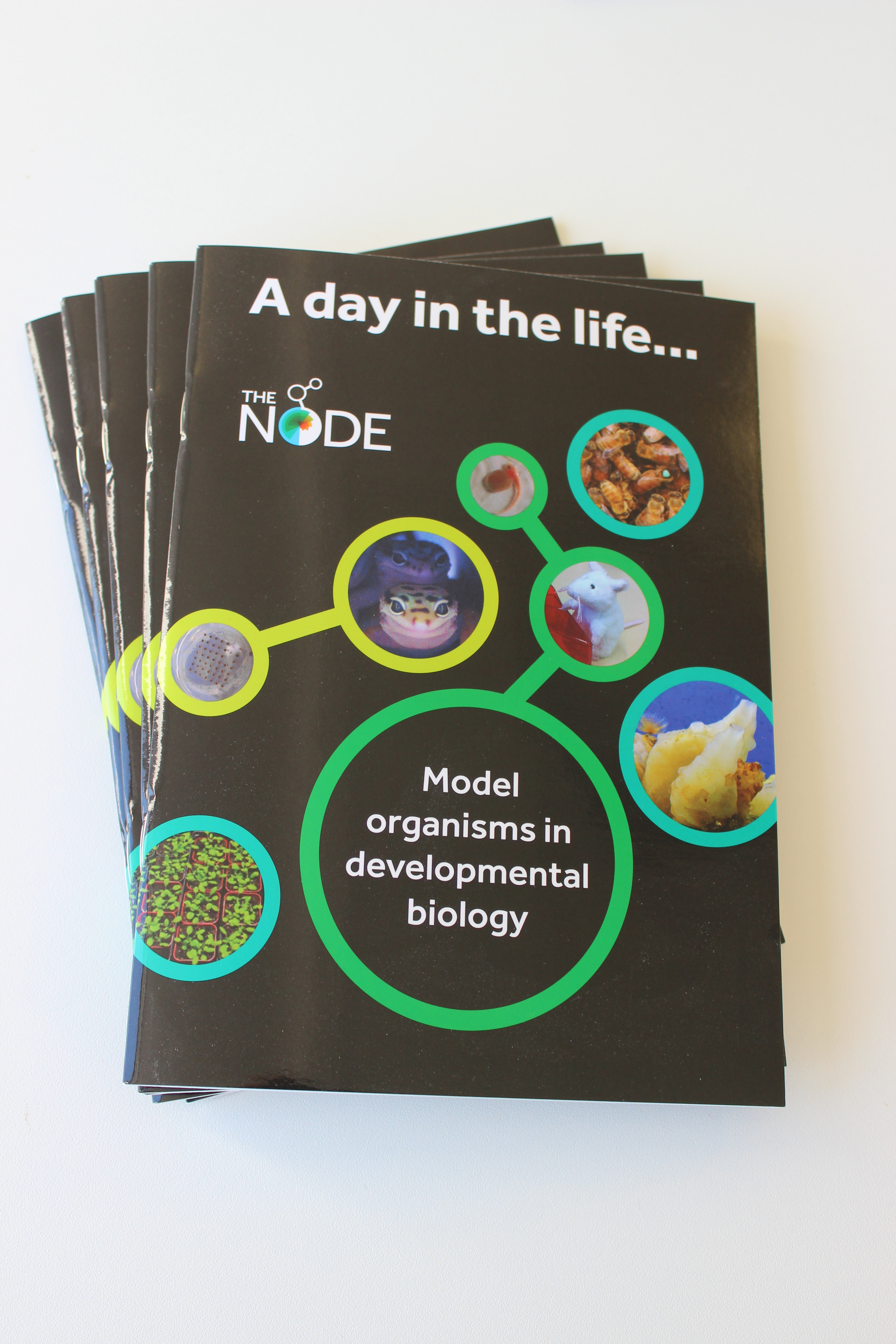 During the last three years I have been involved in a lot of projects on the Node, which I hope you have enjoyed. The most obvious change was the
 (No Ratings Yet)


 In February 2016, The Company of Biologists hosted an intimate gathering of leading international researchers at the forefront of experimental cardiovascular regeneration, with its emphasis on ‘Transdifferentiation and Tissue Plasticity in Cardiovascular Rejuvenation’. As discussed by Michael Schneider, participants at the workshop revealed how understanding cardiac growth and lineage decisions at their most fundamental level has transformed the strategies in hand that presently energize the prospects for human heart repair. See the Meeting Review on p.
  Compared with animals, plants generally possess a high degree of developmental plasticity and display various types of tissue or organ regeneration. Here, Keiko Sugimoto and colleagues summarise how plants control these various types of regeneration and how developmental and environmental constraints can influence plant regenerative regulatory mechanisms. See the Review on p.
 – Last month we were at the Spring Meeting of the British Society for Developmental Biology, where we interviewed the winner of the Beddington Medal for best PhD thesis
 – Plagiarism, data manipulation, author disputes… what are the
 Stewart and colleagues describe a novel neural crest EMT reporter for rapid in vivo drug screening in zebrafish. They use to identify a small-molecule EMT inhibitor that blocks this process by activating retinoic acid signaling. Read the paper
 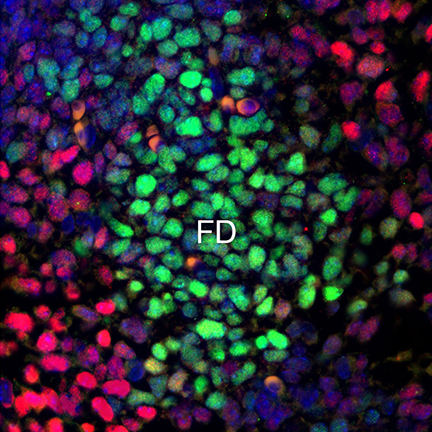 A role for LRP2 in cardiac development
 Conti and co-workers devise a new strategy to quantify the ongoing translation of specific mRNAs by measuring the extent of their co-immunoprecipitation with tagged ribosomes. Using this method, they show that the RNA-binding proteins DAZL and CPEB1 cooperate to regulate mRNA translation and protein synthesis during the meiotic cell cycle in mouse oocytes. Read the paper
  Septate junction formation in Drosophila midgut
 Rundle and co-workers examine whether the timing of cardio-respiratory development in the marine gastropod Littorina obtusata is important for determining whether embryos survive hypoxia. They show that individuals that develop their adult cardiovascular system early survive low oxygen conditions. Read the paper
 Planar Cell Polarity