The community site for and by
developmental and stem cell biologists

Light sheet microscopy 101: Get started with a short video protocol

Posted by , on 13 April 2016

Here you can find out more about our video protocol on using light sheet microscopy to image zebrafish eye development.


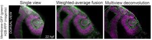
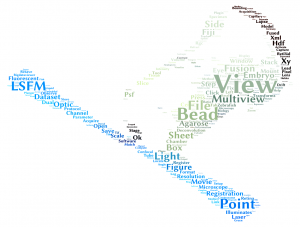
Light sheet fluorescence microscopy has quickly become a popular technique in developmental biology. This method is very gentle to the samples, with fast acquisition speed and allows capturing the samples from any angle or from multiple angles at the same time (so called multi view imaging) (Stelzer, 2015). Such multi view imaging overcomes the degradation of the signal in Z axis and allows imaging of large specimens with high and almost isotropic resolution. The fact that light sheet microscopy is trending was confirmed when it was voted the method of the year 2014 by Nature Methods.

A comparison of image quality among single view data and data processed by two different strategies of multiview fusion
A comparison of quality among single view images and data processed by two different multiview fusion algorithms. The image shows developing zebrafish eye and neural tube.


The major obstacle to mainstream adoption of this method was until recently the technical complexity for many biologists without a background in optics and computer science (Reynaud et al., 2015). The good news is that things have started to change and performing successful light sheet experiments is getting easier. For some time now, there are commercially available light sheet microscopes, which are easy to operate and the software solutions are not lagging behind, with plugins to process the resulting datasets as clickable GUIs (Amat et al., 2015; Preibisch et al., 2014; Preibisch et al., 2010) or Zeiss own solution in ZEN software. One word of warning, your hardware still has to be prepared to handle large volumes of data.

We recently contributed a video protocol (Icha, Schmied et al., 2016) to document a versatile light sheet microscopy experimental pipeline using a commercial microscope and an open source software solution for data processing. Specifically, we used the Lightsheet Z.1 microscope from Zeiss and the Multiview reconstruction application in Fiji to process the data. We demonstrated our approach by imaging several stages of retinal development in zebrafish. The general application for our pipeline would be long-term time-lapse imaging of morphogenetic processes during development using single or multi view acquisition. The protocol will take you through the essential steps of a light sheet microscopy experiment from mounting the sample to processing the data. The most complicated step is not taking the images, but the subsequent combination of image information from multiple views together. The solution we use is embedding fluorescent beads around the sample to register the different views onto each other and thereby to reconstruct the imaged volume of the sample.

A) a snapshot from BigDataViewer with overlapping views shown in different colors B) a magnified area with two fluorescent beads
A) Snapshot from BigDataViewer with overlapping registered views shown in different colors. B) Magnified view showing two fluorescent beads and the overlap of the point spread functions from the different views.


In the associated text, you will find a step-by-step protocol including all the experimental details plus troubleshooting in the discussion section. This should be especially useful for the newcomers to the light sheet fluorescence microscopy field.

This video protocol was created as a collaboration between the Norden lab (twitter @NordenLab) and the Tomancak lab (twitter @PavelTomancak) at MPI-CBG in Dresden. Enjoy and don’t hesitate to contact us in case you have questions. The full video is also available on the Norden lab website.

wordle_final

Other useful links:

EMBO course in Light sheet microscopy 2016

SPIM – Light Sheet Microscopy Literature Database

Open source hardware: DIY light sheet microscopes

Open SPIM wiki page

Open SPIN microscopy

Nature Methods method of the year 2014 thematic issue


References:

Amat, F., Höckendorf, B., Wan, Y., Lemon, W. C., McDole, K. and Keller, P. J. (2015). Efficient processing and analysis of large-scale light-sheet microscopy data. Nat Protoc 10, 1679–1696.

Icha, J., Schmied, C., Sidhaye, J., Tomancak, P., Preibisch, S., Norden, C. (2016). Using Light Sheet Fluorescence Microscopy to Image Zebrafish Eye Development. J Vis Exp 110, e53966.

Preibisch, S., Amat, F., Stamataki, E., Sarov, M., Singer, R. H., Myers, E. and Tomancak, P. (2014). Efficient Bayesian-based multiview deconvolution. Nat Meth 11, 645–648.

Preibisch, S., Saalfeld, S., Schindelin, J. and Tomancak, P. (2010). Software for bead-based registration of selective plane illumination microscopy data. Nat Meth 7, 418–419.

Reynaud, E. G., Peychl, J., Huisken, J. and Tomancak, P. (2015). Guide to light-sheet microscopy for adventurous biologists. Nat Meth 12, 30–34.

Stelzer, E. H. K. (2015). Light-sheet fluorescence microscopy for quantitative biology. Nat Meth 12, 23–26.

Thumbs up (4 votes)
Loading...

Tags: , , , ,
Categories: Research, Resources, Video

Navigate the archive

Use our Advanced Search tool to search and filter posts by date, category, tags and authors.

BSCB/BSDB Spring Meeting 2016: BSDB medals & all other awards

Posted by , on 13 April 2016

BSDBlogoThe joint BSDB/BSCB spring meeting has yet again been a great and most successful event. As every year most of our Awards are announced on this meeting and the BSDB would like to congratulate all prize winners and awardees.

Quick AccessWaddington | C Tickle | Beddington | Summary

Enrico Coen: winner of the 2016 BSDB Waddington Medal

EnricoCoen2The BSDB is delighted to announce Enrico Coen CBE FRS (John Innes Centre, Norwich) as the 2016 winner of the Waddington Medal. Professor Coen was awarded the medal for his pioneering contributions to understanding patterning and morphogenesis in plants, particularly snap dragon flowers. His work elegantly combines molecular genetics, diverse imaging techniques and computational modeling (see the Coen lab site). He is also well known for his popular science books ‘The art of genes’ (1999) and ‘Cells to civilisations’ (2012), and his painting, which has appeared on the cover of Cell and the walls of the Royal Society.

The medal talk was a pleasure to watch and is now available on YouTube. It was a scholarly masterpiece of conceptual brilliance, presented with inspiring enthusiasm, enriched with beautiful images, illustrated with enlightening and entertaining movies of pottery (!!!) and computer models, and even spiced up with live experiments.It will soon be available on the BSDB’s YouTube channel. An interview performed by Cat Vicente during the Spring meeting is scheduled to be published in Development.

The Cheryll Tickle Medal revealed

AbigailTucker As reported previously, the BSDB has introduced the Cheryll Tickle Medal, awarded annually to a mid-career, female scientist for her outstanding achievements in the field of Developmental Biology. The BSDB is proud to announce the inaugural awardee Abigail Saffron Tucker who gave her outstanding and memorable Cheryll Tickle Award Lecture on the evolution of shape which can be watched on YouTube. To read more about Abigail, please download the BSDB Newletter 2015 or go to the post on The Node.

CherrylTickleMedal-2On this occasion, for the first time the actual medal was revealed, which was designed by Andreas Prokop and Megan Davey in discussion with Cheryll Tickle. It shows the famous digit aberrations that occur upon transplantation/manipulation of the zone of polarizing activity (ZPA) or implantation of beads soaked in retinoic acid or sonic hedgehog (for details see Towers & Tickle, 2009, Int J Dev Biol 53, 805ff.). Cheryll performed much of this work in chick as illustrated by the feather on the medal’s flip side, which also shows the typical tool set required for experimental operations and the BSDB logo depicting in ovo development from egg to embryo.

Elena Scarpa: the BSDB Beddington Medal winner

ElenaScarpaThe BSDB is proud to announce the 2016 Beddington Medal winner Elena Scarpa. Elena studied in Turin (Italy), went for her Wellcome Trust fellowship-funded PhD project to the laboratory of Roberto Mayor (UCL, London), and works now in the group of Benedicte Sanson (Univ. Cambridge) on the role of mechanical tension in orienting cell divisions in the Drosophila embryo. Her Beddington Medal talk described the outcome of her successful PhD project (submitted in April 2015) and was entitled Cadherin switch during EMT in neural crest cells leads to contact inhibition of locomotion via repolarisation of forces”.Elena introduced her project with the following words: “Contact Inhibition of Locomotion (CIL) was discovered by Abercrombie and colleagues over 60 years ago as the process through which migrating cells move away from each other after cell-cell contact. More recently, it has been shown to play important roles in vivo during morphogenesis and cancer invasion, but its molecular mechanisms have not been elucidated. In all systems where it has been investigated, the CIL response seems to rely on cell-cell contact dependent signalling. In particular, Eph-Ephrin signalling has been found to be responsible for CIL in cancer cells and in neurons, while in neural crest Wnt-PCP and N-Cadherin dependent cell-cell adhesion are required for CIL. However, it remained unclear why certain cells display an efficient CIL response while many other cell types do not exhibit CIL and instead remain in contact after cell collision, thus forming a stable cell-cell adhesion. During my PhD, I undertook a comparative approach to pursue this mechanism and ask why some cells exhibit CIL, while others, like epithelial cells, remain in contact and form stable junctions.”

The details of this research are described in her 2015 publication entitled “Cadherin Switch during EMT in Neural Crest Cells Leads to Contact Inhibition of Locomotion via Repolarization of Forces” (Dev Cell 34, 421-34), and an interview with Elena has been published here on The Node.

Summary of all BSCB/BSDB awards

Medal Awards

  • BSDB Waddington Award winner: Enrico Coen CBE FRS (John Innes Centre, Norwich) who gave a talk about his pioneering contributions to understanding patterning and morphogenesis in plants (available on YouTube), and an interview will be published soon in Development.
  • BSCB Hooke Award winner: Thomas Surrey (Crick) who presented the lecture “Microtubule cytoskeleton dynamics: mechanistic insight from reverse engineering” that is available on YouTube. See more information about Thomas here.
  • BSCB WICB Award winner: Lidia Vasilieva (Dept Biochem., Univ. Oxford, Oxford) who presented the talk “Towards understanding mechanisms of gene expression” that is available on YouTube. Read more information about Lidia here.
  • The BSDB Cheryll Tickle Medal winner: Abigail Saffron Tucker who gave her Cheryll Tickle Award Lecture about the evolution of shape available on YouTube. To read more about Abigail, please download the BSDB Newletter 2015 or go to the post on The Node.
  • BSDB Beddington Award winner: Elena Scarpa (now Univ. Cambridge in the group of Benedicte Sanson) for her work entitled “Cadherin switch during EMT in neural crest cells leads to contact inhibition of locomotion via repolarisation of forces” which was performed in the laboratory of Roberto Mayor (UCL).

PhD Poster Prizes

  • 1st BSDB PhD Poster Prize winner (visit to 2016 SDB-ISD meeting, Boston): Mathew Tata (University College London, group of Christiana Ruhrberg) – P117 “Regulation of embryonic neurogenesisi by germinal zone vasculature” – read an interview in The Node.
  • 1st BSCB PhD Poster Prize winner (visit to 2016 ASCB meeting, San Francisco): Emma Stewart (University of York) –  P65 “RNA-dependent localisation of the nuclear matrix protein CIZ1 to the inactive X chromosome
  • 2nd BSDB PhD Poster Prize (£75 cash prize): Laura Martin-Coll (DanStem, University of Copenhagen) – P87 “A single-cell analysis of progenitor heterogeneity at the onset of pancreas formation
  • 2nd BSCB PhD Poster Prize (£75 cash prize): Alex Pool (Barts Cancer Institute, Queen Mary University) – P56 “DDR1 localisation to adherens junctions prevents efficient clustering of supernumerary centrosomes
  • 3rd BSDB PhD Poster Prize (£50 cash prize): Leila Thuma (University of Bristol) – P159 “Modelling immune cell diapedesis from vessels to wounds in the Drosophila pupal wing veins
  • 3rd BSCB PhD Poster Prize (£50 cash prize): Saroj Saurya (Oxford University) – P74 “Drosophila Ana1 stabilises centrioles and also promotes centriole elongation in a dose-dependent manner

Postdoc Poster Prizes

  • Joint 1st BSDB Prize (£150 cash prize): Guilherme Costa (University of Manchester) – P144 “Cellular localisation of mRNA during angiogenesis”
  • Joint 1st BSDB Prize (£150 cash prize): Sophie Gilbert (University of Oxford) – P29 “How the worm completes its skin
  • 1st BSCB Prize (£300 cash prize sponsored by MDPI): Dimitra Aravani (University of Leicester) – P104 “HHIPL1: a new gene that promotes atherosclerosis
  • 2nd BSDB Prize (£125 cash prize): Filip Wymeersch (MRC Centre for Regenerative Medicine, University of Edinburgh) – P134  “Transcriptionally dynamic neuromesodermal progenitors coexist alongside a stable niche during axis elongation
  • 2nd BSCB Prize (£75 cash prize): Kyojiro Ikeda (Sir Dunn School of Pathology, University of Oxford) – P141  “PTEN regulates CDC42-dependent morphogenesis through beta-arrestin1/ARHGAP10 signalling
  • 3rd BSCB Prize (£50 cash prize): Amy Barker (Queen Mary University of London) – P18 “Characterising intracellular trafficking of Junctional Adhesion Molecule C (JAM-C)

Others

  • The BSCB Science writing Prize winner (chosen by Barbara Melville; @keyeri) is Girisaran Gangnatharan (PhD student, Montpellier) for an essay about zebra fish models of regeneration entitled “Heart disease: fishing for a cure
  • The BSCB Image Award winners are:
    • Anna Franz (School Biochem, Univ Bristol)
    • Ronan Mellin (IGMM, Human Genetics Unit, Edinburgh)
    • Helen Weavers (School Biochem, Univ Bristol)

Quick AccessWaddington | C Tickle | Beddington | Summary

Thumbs up (1 votes)
Loading...

Tags:
Categories: Events, News, Societies

Three Group Leader Positions at DanStem: Stem Cell Biology, Pancreatic Cancer and Bioengineering (Fixed-term, 6 years)

Posted by , on 13 April 2016

Closing Date: 15 March 2021

The Novo Nordisk Foundation Section for Basic Stem Cell Biology, Danish Stem Cell Center, Faculty of Health and Medical Sciences University of Copenhagen

The University seeks to appoint three Group Leaders in Pancreatic Cancer, Stem Cell Biology and Bioengineering to The Novo Nordisk Foundation Section for Basic Stem Cell Biology (BasicStem) at the Danish Stem Cell Center (DanStem) to commence as soon as possible. The positions are for six years with possible extensions depending on the outcome of a peer reviews.


Background

The Danish Stem Cell Center (DanStem) is an international research center at the University of Copenhagen. The overall scientific goal is to develop new stem cell-based therapeutic approaches, currently in the area of diabetes and cancer addressing basic questions in stem cell and developmental biology and seeking to identify the factors that govern the development of different cell types in the body. Read about DanStem at www.danstem.ku.dk/.


Group Leader in Pancreatic Cancer

Particular interest in basic and disease-oriented pancreatic cancer biology

Group Leader in Stem Cell Biology

Particular interest in addressing fundamental questions in stem cell biology by using single cell behaviour analysis

Group Leader in Bioengineering

Particular interest in addressing fundamental questions in stem cell biology using bioengineering approaches. Experience with materials science and/or devices (e.g. microfluids) would be an advantage.

The Group Leaders duties will primarily consist of:

  • Developing a strong research program.
  • The Group Leaders must be willing to synergize with other DanStem scientists and contribute to common activities at DanStem such as seminars and PhD courses.
  • The Group Leaders are expected to take full responsibility for training and supervision of young researchers, for management of each of their own group, and for publication/dissemination of research results.
  • The Group Leaders are expected to actively contribute to teaching activities and education activities
  • Academic assessments

The closing date for applications is 23.59 pm, May 1st, 2016

Apply online: http://employment.ku.dk/all-vacancies/?show=795295

Only online applications will be accepted.

Thumbs up (No Ratings Yet)
Loading...

Categories: Jobs

Enrico Coen: the 2016 BSDB Waddington medal winner

Posted by , on 11 April 2016

BSDBlogoEnricoCoen2The BSDB is delighted to announce Enrico Coen CBE FRS (John Innes Centre, Norwich) as the 2016 winner of theWaddington Medal. Professor Coen was awarded the medal for his pioneering contributions to understanding patterning and morphogenesis in plants, particularly snap dragon flowers. His work elegantly combines molecular genetics, diverse imaging techniques and computational modeling (see the Coen lab site). He is also well known for his popular science books ‘The art of genes’ (1999) and ‘Cells to civilisations’ (2012), and his painting, which has appeared on the cover of Cell and the walls of the Royal Society.

The medal talk was a pleasure to watch and is now available on YouTube. It was a scholarly masterpiece of conceptual brilliance, presented with inspiring enthusiasm, enriched with beautiful images, illustrated with enlightening and entertaining movies of pottery (!!!) and computer models, and even spiced up with live experiments.It will soon be available on the BSDB’s YouTube channel. An interview performed by Cat Vicente during the Spring meeting is scheduled to be published in Development.

Thumbs up (2 votes)
Loading...

Tags: ,
Categories: News

‘Rising Stars’ fellowships (based at Welsh institutions)

Posted by , on 8 April 2016

Closing Date: 15 March 2021

‘Rising Star’ fellowships will be very prestigious and highly competitive positions, designed to attract the very best ‘rising stars’ of academic research. ‘Rising star’ packages will be funded at approximately £0.2m per annum and can involve collaboration with relevant commercial or third sector organisations

‘Rising Star’ applications can be submitted at any time.

Eligibility Criteria

Rising Star Fellowships applicants should meet the eligibility criteria set out below:

Applicants should have over 7 years of experience since completion of PhD (or equivalent degree) and scientific track record showing great promise
Applicants should have an excellent research proposal
Applicants can be of any nationality
Applicants must submit a completed application form and associated documents (supervisor form, ethics form, and CV)
Applications must comply with the fundamental ethic principles as detailed in the ethics section
Applicants must have the support of their chosen host institution

Link


Thumbs up (1 votes)
Loading...

Categories: Jobs

POSTDOCTORAL POSITION IN CELL and DEVELOPMENTAL BIOLOGY

Posted by , on 7 April 2016

Closing Date: 15 March 2021

Post doctoral position available to study the genetic and epigenetic control of stem cell attributes and pluripotency, focusing on the neural crest gene regulatory network (NC-GRN). Neural crest cells are stem cell-like progenitors that migrate extensively and whose genesis was central to the evolution of vertebrates. Misregulation of components of the NC-GRN underlies numerous human diseases and congenital disorders. Studies involve post-translational regulation of known network components, and use of proteomics and next generation sequencing to identify novel components. 

Highly motivated applicants with a PhD and strong background in cell and molecular biology and/or developmental biology are encourage to apply. Please send a CV, brief description of research interests, and the names of three references to:

Carole LaBonne, PhD (clabonne@northwestern.edu)
Department of Molecular Biosciences
Northwestern University, Evanston, IL 602028


Thumbs up (No Ratings Yet)
Loading...

Categories: Jobs

The Node at the BSDB Spring meeting (with new freebies!)

Posted by , on 7 April 2016

booklet coverThe 2016 Spring meeting of the British Society for Development Biology (held jointly with the British Society for Cell Biology) will start this Sunday at the University of Warwick and the Node will be there!

Pop by The Company of Biologists’ stand to chat with Cat, our community manager, and collect our new freebies! If you are a fan of our ongoing series on model organisms in developmental biology you may like to take a copy of our brand new booklet, which includes a selection of some of the earlier posts in the series. Thank you to all the authors of the posts who gave us permission to use their text and images and helped us put this booklet together!


IMG_0909


We also have a new set of postcards featuring beautiful images from the Woods Hole Embryology course. Make sure to come to the stand to collect yours!

IMG_0876

IMG_0887


This meeting is also a great opportunity to chat with other people at The Company of Biologists. Development’s executive editor Katherine Brown, and reviews editor Seema Grewal will also be at the meeting, and Nicky Le Blond, who runs our travelling fellowships and grant program and organises our fabulous workshops, will be at the stand on Tuesday. Cat and Katherine will also be leading discussion tables on social media and publishing respectively at the Careers Workshop on Sunday afternoon, so plenty of chances to say hello. We look forward to meeting you then! If you can’t make it to the meeting you can always follow us on Twitter. The Node will be tweeting using the hashtag #cbdb16


Thumbs up (1 votes)
Loading...

Tags:
Categories: News

From London to Paris – Travelling through Cell Science

Posted by , on 7 April 2016

Thanks to the Cell Science Travel Fellowship of the Company of Biologists, I was able to work for 2 months at the Institut National de la Recherché Agronomique (INRA) in Jouy-en-Josas, a small town close to Paris, in France.


I just started the second year of my PhD in the group of Dr. Mostowy at Imperial College London. The Mostowy group is well known to study host cell biology, focusing on the eukaryotic cytoskeleton (actin, microtubules, intermediate filaments and septins). The discovery that prokaryotes exhibit counterparts of the major cytoskeletal components (e.g. MreB, FtsZ, CreS) radically changed the context how bacteria are studied and helped to inspire the field of bacterial cell biology. To travel from eukaryotic to prokaryotic cell biology, I went to the lab of Dr. Rut Carballido-López, who is well recognised for her pioneering work on the actin-like MreB cytoskeleton in Bacillus subtilis.


New techniques to study the cytoskeleton include genetic modifications to follow the spatiotemporal location of cytoskeletal proteins (e.g. fluorescently tagging) and to analyse their function (e.g. gene inactivation). Both approaches are applied in eukaryotes and prokaryotes, however the precise methods are completely different between these kingdoms (e.g. siRNA versus knockouts). In eukaryotes as well as in prokaryotes, cytoskeletal proteins are key structural determinants that assemble into filaments and their genes are essential for viability, which makes their genetic manipulation even more challenging. Luckily, I was working with Arnaud Chastanet, a highly experienced Research Scientist (CR1) with great visualisation and explanation skills, and Charlène Cornilleau, an Ingénieur d’Etudes with ‘magic cloning hands’, who supported me greatly with my clonings.


mreb

Picture: The actin-like MreB cytoskeleton (green) in Bacillus subtilis (red).


It was a great experience to work in the lab of Dr. Rut Carballido-López. Everyone in the working group was really friendly and helpful and I had many interesting discussions – science-related and beyond. My visit allowed me to expand my knowledge in microbial genetics. Now back in London, I can share my knowledge and tools, and allow other scientists to benefit from my stay at INRA. Working in France provided me with the unique opportunity to experience science internationally, and to network with people for my future career in science. I am deeply grateful to Dr Rut Carballido-López for enabling my collaborative visit and to The Company of Biologists for awarding me with a Cell Science Travel Fellowship.

Thumbs up (3 votes)
Loading...

Tags:
Categories: Funding

High-pressure tubes

Posted by , on 6 April 2016

Notes on “Intracellular lumen formation in Drosophila proceeds via a novel subcellular compartment” by Linda S. Nikolova and Mark M. Metzstein. Development 2015 142: 3964-3973; doi: 10.1242/dev.127902

In this post, I provide additional details to a paper which we published last year in Development. In particular, I expand on our description on the method of high pressure freezing/freeze substitution, as well as why we developed this technique to examine Drosophila tracheal terminal cells, a part of the insect breathing system.

Like all terrestrial animals, insects require the ability to take oxygen from the atmosphere and deliver it to internal tissues. In vertebrates, this function is carried out by two independent systems: the lungs, used to take air into the body, and the vasculature, used to distribute oxygen throughout the body. Insects and other invertebrates use a different strategy, in which gas intake is directly coupled to the distribution system. This organization is accomplished by a single network of epithelial tubes, called trachea by analogy with vertebrate breathing systems. Openings on the surface of the insect, called spiracles, connect to large, multicellular tracheal tubes. In turn, smaller, unicellular tubes branch off the multicellular tubes and extend toward different regions of the animal. Finally, located at the ends of unicellular tubes, a number of specialized cells, known as terminal cells, are responsible for distribution of gas to all individual cells and tissues. Insect respiration is thought to be driven entirely by passive diffusion of air through the tracheal network, a method of respiration that obviously works well for this class of animals given their huge numbers and species diversity. However, it is also likely this passive diffusion of air sets the limits to organismal size, thus explaining why giant ants do not regularly rampage over the countryside.

We are particularly interested in the cell biological mechanisms of terminal cell morphogenesis. To perform their function, terminal cells undergo fascinating cell shape changes, with each individual cell undergoing an iterative process of plasma membrane outgrowth and bifurcation during larval stages. Eventually, each cell produces 20-100 thin (typically less than 1µm in diameter) subcellular branches (labeled with a cytoplasmic GFP in Fig. 1A). In terms of complexity, terminal cells rival the elaborate axonal and dendritic arbors found in neurons. However, unlike neurons, terminal cells have to undergo an additional form of morphogenesis: tubulogenesis.  For gas to flow efficiently, each of the thin branches terminal cell branches develops into a tube, with a membrane-bound, gas-filled open space, called a lumen, running through it.


Terminal Cell fig


In our recent paper, we focus on the cellular and molecular mechanisms by which the subcellular lumen forms in terminal branches. The lumen of terminal cell branches is extremely thin, smaller in diameter than the width of a bacteria such as E. coli. At this scale, the formation of the lumen is akin to the mechanisms required to form subcellular organelles, such as mitochondria or lysosomes, albeit the lumen is different as it is a continuous structure extending through all the branches of the terminal cell. Despite recent amazing advances in light microscopy, the only high resolution technique available to examine the membranes that form the lumen is transmission electron microscopy (TEM; EM on left; schematized on right in Fig. 1B).

One important consideration in using EM is that the technique does not visualize biological structures directly, as the electron beam immediately vaporizes essentially all biological material. Instead, tissues must be immobilized (“fixed”) and treated with stains, typically heavy metals, that can be visualized directly in the EM. By far the most common method of fixation has been the use of chemical cross-linkers, usually aldehydes. This approach has been successfully applied to numerous samples and has produced many high quality studies. However, chemical cross-linking has some significant disadvantages. First, different biological polymers, such as membranes, proteins, or nucleic acids, differ significantly in how well they are preserved by any particular cross-linking reagent, and it is hard to find conditions in which all are simultaneously well preserved. Second, chemical cross-linking takes time, during which tissues can undergo deformation. Third, it is necessary to get the fixatives rapidly into the cells. While this is relatively easy for cells in culture and for dissected tissues, it is a significant problem for an intact organism, such as a Drosophila larvae.

To avoid the problems of chemical fixation, we turned to an alternative, very different method of fixation: high pressure freezing/freeze substitution (HPF/FS). The basic principle of HPF fixation is straightforward: cells are preserved by rapid freezing. However, as is well known, water has the unusual property of expanding upon freezing. Since most tissues are composed mainly of water, expansion from water ice crystals then causes disruption of cellular structures. However, as was proposed some 40 years ago, a procedure to avoid ice crystal damage is to rapidly freeze samples while subjecting them to high pressure. Under this regime, ice crystals cannot form. Instead the water forms an amorphic arrangement, similar to a glass. Amorphic ice has essentially the same density as liquid water, thus occupies the same volume, so damage from ice crystal expansion or tissue shrinkage does not occur. Importantly, amorphic ice maintains its structure when pressure is released, as long as the sample is kept cold. This allows the second step of the HPF/FS –freeze substitution– to proceed. During this step, water ice in the sample is replaced (substituted) with solvents that do not expand upon freezing, such as ethanol or acetone. Metal stains can be included in this substitution “cocktail” to label internal cell components. After the substitution step is completed, the samples can be returned to ambient temperature. This is followed by standard procedures of embedding in a plastic resin, sectioning, and observation by an electron microscope. Fortunately, many of the steps of HPF/FS are automated with commercial instruments available to carry out freezing and substitution (Fig. 2). Samples are frozen within a high pressure freezer, which injects liquid nitrogen at ~2500 atmospheres, both compressing and cooling the samples. Freeze substitution, which takes a number of days, takes place in a special liquid nitrogen filled chamber in which substitution cocktails are automatically exchanged, and the temperature regulated to complete the solvent exchange and the return to ambient temperatures.

HPF.001

Much of the work leading to our paper involved testing different cocktails, and different incubation times and temperature change regimes during the substitution, in order to optimize both tissue preservation and structure visualization. One particularly important refinement we made during the course of our studies was the use of Durcupan instead of more commonly used epoxy resins, as it produced samples with better tissue preservation and membrane contrast. Overall, our results produced excellent preservation of internal tissues of intact Drosophila larvae. Different macromolecular structures, including proteins, nucleic acids, and the chitinous cuticle were very well preserved. Membranes in particular were particularly well preserved and had a smooth, curved appearance, indicative of very little tissue deformation.

As described in the paper, our new fixation techniques revealed previously unrecognized details of the terminal branch lumen formation. In particular, we found evidence of a hitherto undescribed intermediate of tube formation: a novel, multimembrane subcellular compartment that may contain the precursors of the cuticle lining the lumen. We also characterized the ultrastructural phenotypes of new genes required for lumen formation, providing further evidence for the multimembrane intermediate in the lumen formation process. Our future research will involve using our new fixation techniques to characterize other genes required for lumen formation and an analysis of the subcellular localization of proteins required for this process. In general, our fixation techniques should allow for analysis of many of the developmental and physiological processes that occur during Drosophila larval development.

Thumbs up (2 votes)
Loading...

Tags: ,
Categories: Research

PhD opportunities in Montpellier to work on pretty ascidian embryos!

Posted by , on 6 April 2016

Closing Date: 15 March 2021

Applications are open for 3-year PhD fellowships in the Montpellier Health Science doctoral program (To apply visit http://www.adum.fr/as/ed/cbs2/page.pl?page=concoursed-gb before May 3rd 2016).

Our group is proposing three possible independent PhD projects on the embryonic development of marine invertebrates closely related to vertebrates, the ascidians (Lemaire, 2011, Development 138, 2143–2152). Most ascidian species develop with almost identical embryonic morphologies in spite of very different genomes, a paradox we are trying to understand. The projects will contribute to a better understanding of the genetic program underlying ascidian development and of its robustness to genetic changes.

The first project aims at the reconstruction and analysis of the early ascidian endodermal Gene Regulatory Network, which is currently only very partially known. We will combine the knowledge of open chromatin regions flanked with specific histone marks, transcription factor (TF) DNA-binding specificity and TF expression to predict the location of cis-regulatory sequences for endodermal regulatory genes, which will be tested and dissected by electroporation into live embryos. This network will then be used to model the flow of genetic information across time, and its robustness to genetic variations.

The second project will test the hypothesis that ascidians can buffer divergent genome information because the architecture of their Gene Regulatory Networks (GRNs) makes them quite insensitive to variations in the level of regulatory gene expression. This hypothesis will first be tested by comparing  the level of inter-individual variability in regulatory gene expression in ascidian embryos (slow morphological evolution, fast molecular evolution) and vertebrate embryos (faster morphological evolution, but slower molecular evolution). We will then monitor the phenotypic response to progressive interference with gene function in both taxa.

Finally, the third project will focus on inter-cellular communication (inductions) in ascidian embryos, and their sensitivity to changes in embryonic geometry and gene expression. We will first quantify the main biochemical parameters of an embryonic induction (concentrations of ligands and receptors, rates of diffusion, rate constants,…) and their variability across individuals. These data will then be used to construct and constrain a quantitative model of an embryonic induction.

Our group is small and interdisciplinary. Its working language is English.

To apply and obtain more information about the projects, contact Patrick Lemaire: patrick.lemaire@crbm.cnrs.fr before April 25th.


Thumbs up (1 votes)
Loading...

Categories: Jobs