The community site for and by
developmental and stem cell biologists

Postdoctoral position in Developmental Biology – University of Manchester, UK

Posted by , on 1 October 2015

Closing Date: 15 March 2021

Postdoctoral Research Associate in Developmental Biology

Closing date : 25/10/2015

Reference : LSX-07097

Employment type : Fixed term for 3 years (full time)

Salary : £30,434 to £37,394 per annum

Location : Takahashi lab, Faculty of Life Sciences, University of Manchester, UK

Our laboratory is looking for a highly motivated and skilful developmental biologist to work on a full-time Leverhulme Trust funded project. The overall aim of this project is to study vertebrate head development with particular interest in its evolutionary origin. Using the zebrafish (Danio rerio) model system, you will investigate the genomic cis-regulatory mechanisms of the genes controlling craniofacial development. In particular, you will integrate this in vivo experimental approach with evolutionary research using amphioxus – a marine invertebrate closely related to vertebrate ancestors – in collaboration with Professor Peter Holland at Oxford.

You should hold (or shortly expect to gain) a PhD in a relevant subject, along with a research background in developmental biology or related fields. Excellent laboratory skills are essential. You will be required to establish zebrafish transgenic lines and conduct real-time image analysis in this project. Previous experience with any of these techniques will be an advantage.

The post is available for up to 36 months in the first instance, with an expected appointment date of 1 December 2015.

To apply online for this vacancy, please go to the website (https://www.jobs.manchester.ac.uk/displayjob.aspx?jobid=10397) and click on the ‘Apply for job’ button below. This will lead you to the University’s Job Application System, where you can complete and submit the online application form. A full role specification can be obtained from the same website. The closing date for applications is midnight on Sunday 25 October 2015.


Informal enquiries can be made to Dr Tokiharu Takahashi.

Tel : 0161 275 5538

Email : Tokiharu.Takahashi@manchester.ac.uk

Thumbs up (1 votes)
Loading...

Categories: Jobs

Navigate the archive

Use our Advanced Search tool to search and filter posts by date, category, tags and authors.

Postdoctoral position in Neuroscience University of Oxford, United Kingdom

Posted by , on 1 October 2015

Closing Date: 15 March 2021

Postdoctoral Research Scientist in Neuroscience : Oxford, United Kingdom

Salary: Grade 7: £30,434 – £37,394 p.a.

Stephen Goodwin’s lab, based at the Centre for Neural Circuits and Behaviour, are looking to appoint a postdoctoral research scientist to work on a project entitled “Genetic Dissection of Sexual Behaviour”.

Professor Goodwin and his group use Drosophila courtship behaviour to study how sex-specific neural circuitry and behaviours are established during development by the action of complex networks of genes. The aim of the study is to understand how activity in functioning dimorphic neural circuits gives rise to a different sex-specific behavioural repertoire in the male and female fly.

The successful applicant will have a PhD in neuroscience, and experience with the study of behaviour would be desirable. Candidates must have an emerging publication record and a proven ability to communicate their work. Good organisational skills and a willingness to work as part of a team are essential.

The post is fixed-term and Wellcome Trust funded for 3 years and is based at the Centre for Neural Circuits and Behaviour, University of Oxford, Tinsley Building, Mansfield Road, Oxford, OX1 3SR.

The closing date for applications is 12.00 noon on 2 November 2015.

Please send a CV, letter of motivation, and at least two letters of recommendation to Prof Stephen Goodwin (stephen.goodwin@cncb.ox.ac.uk).


Thumbs up (No Ratings Yet)
Loading...

Categories: Jobs

Postdoctoral positions in mammalian epigenetics

Posted by , on 30 September 2015

Closing Date: 15 March 2021

Applications are invited for postdoctoral positions in the laboratory “Epigenetic Regulation of Cell Identity” headed by Dr. Michael WEBER at the CNRS in Strasbourg (France). The positions are funded for up to three years by an ERC Consolidator grant and the INCa (National Cancer Institute).

The research in our laboratory aims at better understanding the function and regulation of DNA methylation in mammalian development by combining molecular biology, high-throughput sequencing, bioinformatics and mouse genetics (Auclair G et al Genome Biol 2014; Guibert S et al Genome Res 2012; Borgel J et al Nature Genet 2010). The successful applicants will uncover and study novel regulatory pathways of DNA methylation in normal and malignant cells.

Our Institute is located on the Illkirch research campus in Strasbourg. The campus offers an international environment with access to state-of-the-art facilities (NGS sequencing, animal housing, proteomics, imaging). The successful candidates will join a young and dynamic team. For more information see our website: http://irebs.u-strasbg.fr/spip.php?rubrique186&lang=en.


We are looking for candidates with a Ph.D. in Molecular or Developmental Biology. Prior experience with epigenetics, NGS datasets or mouse embryology will be positively considered. We expect the candidates to be motivated, creative, excellent team players, and preferably with at least one publication as first author. Applicants with less than two years after obtaining their PhD will be favored.

Starting date: end 2015 or early 2016

Salary: Competitive salary following the CNRS guidelines (> 2,500 € brut / month)

Please send a CV, letter of motivation, and at least one letter of recommendation to Dr Michael Weber (michael.weber@unistra.fr).


Thumbs up (No Ratings Yet)
Loading...

Categories: Jobs

Postdoctoral Position – Developmental Biology and Biophysics

Posted by , on 29 September 2015

Closing Date: 15 March 2021

Postdoc_AD_HFSP

We are seeking outstanding and highly motivated postdoctoral candidates to join an interdisciplinary collaboration between the Gros lab (“Imaging and Regulation of
Morphogenesis in Higher Vertebrates” lab) at the Pasteur Institute in Paris, France, and the Campas lab (“Morphogenesis and Self-Organization of Living Matter” lab) at the University of California, Santa Barbara. This specific position would be based at the Pasteur Institute in Paris, with occasional stays at the University of California, Santa Barbara.

The overall aims of this collaborative project are to (1) quantify the spatial and temporal distributions of physical forces in developing mouse limb buds using novel force transducers developed recently by the Campas lab and, (2) elucidate their role in maintaining skeletal progenitor differentiation/maintenance balance. For more information about projects and the labs please visit:
www.jgroslab.com ,
https://research.pasteur.fr/en/team/morphogenesis-regulation-in-higher-vertebrates/
and http://www.engineering.ucsb.edu/~campas/.

The position is a 3-year postdoctoral position funded by the Human Frontier Science Program (HFSP), available December 1st, although the starting date is flexible. Candidates should have expertise in developmental and/or cellular biology, be willing to extend their knowledge to quantitative biophysics approaches and collaborate with biophysicists and engineers. Experience in live imaging, mouse development, biophysics and/or quantitative biology will be positively considered.

The Pasteur Institute, located in the vibrant city of Paris, has a longstanding history of excellence in developmental biology and in science in general, with access to excellent core facilities.

Applicants should send a cover letter (describing briefly research interests), a C.V and contact information for up to 3 academic references to jgros@pasteur.fr and
campas@engineering.ucsb.edu.

Thumbs up (No Ratings Yet)
Loading...

Categories: Jobs

Question of the month- PhD internships

Posted by , on 28 September 2015

There is an increased awareness that only a small fraction of PhD students will ultimately secure a tenure-track position in academia. This has led to a discussion on whether graduate schools have a responsibility to help PhD students prepare for a career outside academia, either by providing training on a broader range of transferable skills, or simply by increasing awareness of the various career paths available.

One of the ways PhD programmes have started to support students in this way is by providing access to internship opportunities during their PhDs. A recent article in Nature Jobs discussed the efforts in this direction in the US, where internship options are available for PhD students and postdocs. In the UK, 3 month internships are now part of the compulsory training of PhD students in certain graduate programs (most notably the PIPS scheme, part of the BBSRC PhD studentships). So this month we are asking:


Are PhD internships a valuable exposure to careers outside academia or a harmful distraction from research?


Share your thoughts by leaving a comment below! You can comment anonymously if you prefer. We are also collating answers on social media via this Storify. And if you have any ideas for future questions please drop us an email!

Thumbs up (3 votes)
Loading...

Tags:
Categories: Discussion, Education, Lab Life

From our sister journals- September 2015

Posted by , on 28 September 2015

Here is some developmental biology related content from other journals published by The Company of Biologists.


CoB_DisModMech_AW_RGB


Auditory hair cell defects in Wolf-Hirschhorn syndrome

DEVDMMWHSWolf-Hirschhorn syndrome (WHS) is a rare genetic disorder associated with sensorineural deafness. In this study, Andrea Streit and colleagues show that, although cochlear hair cells are specified normally in a WHS mouse model, they are disorganised and display sterocilia defects. Read the paper here [OPEN ACCESS].


Studying Huntington disease using iPSCs

DMMHdiseaseFigiel and colleagues examined putative signaling pathways and processes involved in Hutington disease pathogenesis in pluripotent cells. They show that dysregulation of signaling pathways is a very early event in the pathogenesis of Huntington disease and that these pathways are already dysregulated in cells at the stage of pluripotency. Read the paper here [OPEN ACCESS].


Sequential mutagenesis in the mouse

mouse mutagenesis squareZhang and Kirsch generated two mouse strains expressing Cre-ERT2 under Flp-FRT regulation. These tools enable sequential mutagenesis in the same or different cells to study development, tissue homeostasis and diseases such as cancer. Read the paper here [OPEN ACCESS].


Journal typography


KCNK1 on osteoclastogenesis

osteoclastKCNK1 (K+ channel, subfamily K, member 1) is a member of the inwardly rectifying K+ channel family, which drives the membrane potential towards the K+ balance potential. Kim, Choi and colleagues show that KCNK1 negatively regulates osteoclast differentiation. Read the paper here.


A role for periostin on Schwann cell migration

Schwann cells 2Riethmacher and colleagues performed comparative gene expression analysis of dorsal root ganglia explant cultures from ErbB3-deficient and wild-type mice, in order to identify genes that are involved in Schwann cell development and migration. Their results demonstrate that the expression of periostin is stimulated by the ErbB ligand NRG1 and promotes the migration of Schwann cell precursors. Read the paper here.


Journal typography


Daphnia sniff out doom with first antennae

DaphniaWeiss and colleagues analysed the developmental characteristics of the inducible defences formed by Daphnia in response to the warning odours (kairmones) exuded by predators. Read the paper here [OPEN ACCESS].


Developmental remodeling in response to hypoxia/anoxia

hypoxia graphHarrison and colleagues observe that Drosophila larvae, which live and feed in severely hypoxic conditions under normal laboratory conditions, show strikingly different behavioral and physiological responses to anoxia from those of adults. Read the paper here.


Toadfish hearing improves with age

toadfishVocal differentiation is widely documented in birds and mammals but has been poorly investigated in other vertebrates, including fish. Vasconcelos and colleagues examined how closely hearing development in the toadfish matches the development of their vocal repertoire. Read the paper here.


Thumbs up (No Ratings Yet)
Loading...

Tags: , , , , , ,
Categories: Research

PLM10: the Physics of Living Matter is coming of age

Posted by , on 27 September 2015

This week, Cambridge (UK) hosted the 10th Symposium on the Physics of Living Matter (PLM10) (http://www.plm-symposium.org/). For those of us who were at PLM1, it is surprising to see that what was (and remains) a grass roots organized event, persists. In some ways it is a tribute and an example that a community can be created by nurturing a common set of interests; in this case how the physical sciences can shed light onto biological problems (NB this is different from Biophysics which looks for biological examples of physical phenomena).

The symposia started with the view of making the local community aware of developments at the interface of Physics and Biology. In the early 00s there were a few pockets of activity cradling this interest. In particular, the legendary Physiology course of Woods Hole (this year’s Bragg lecture was given by Julie Theriot, who has been a pioneer in the field and an active participant in Woods Hole), Eric Karsenti’s division in EMBL, Dresden CBG/PKS was starting and Stan Leibler in Rockefeller was breeding a group of interesting scientists, while individuals like Dennis Bray, were making significant contributions from ‘the side”. Things have changed and ten years on, the interactions between Physics and Biology are on a solid ground. This year’s Symposium bore witness to this and left the message that the future of Biology lies in the quantitative analysis of biological problems and on the use of the reasoning and methods of the physical sciences. It has taken a long time to come but many of us got the feeling at the meeting that the Physics of Living Matter is coming of age.

In his book “What a mad pursuit”, F Crick comments on how Max Delbruck was driven into Biology to search for new Physics to explain living systems (see http://amapress.gen.cam.ac.uk/?p=1489 if you want to learn a bit more) and contrasts this with the belief of Linus Pauling in Chemistry to understand the same problem. Crick makes the point that, as he sees it, history shows that Pauling was right and Delbruck was wrong. However, he puts it in an interesting manner by stating that “time has shown that, SO FAR, Pauling was right and Delbruck was wrong……”. It is the ‘so far’ that reveals Crick’s caution and belief that the story had a second part. This second part is emerging now. With hindsight one can say that the reason why Chemistry rather than Physics provided the insights into Biology during the XX century was, simply, that the problem then was the structure of living matter, not its function. And the structure was, and remains, a problem of Chemistry. Surely one uses physical methods to understand it e.g crystallography and the different flavours of microscopy, but the answers lie in Chemistry. However, when we want to understand how the elements of living matter combine and interact to make WORKING cells, tissues and organs, we have to recur to physics. Herein we find the ways in which biological systems ‘cheat’ the laws of equilibrium thermodynamics and statistical mechanics and, in probing the HOW we learn about how Physics helps explain Biology and about how Biology extends Physics. The XXI century proves that both Pauling and Delbruck were right and PLM10 was a great tribute to this statement with talks on experimental approaches to the origin of life, top notch cell biology and statistical approaches to the behaviour, and output, of cell populations.

The change in the field has been remarkable. Daniel St Johnston commented to me during the meetinh how we can now see that cell biology is Physics. Well, actually, cell biology has always been close to Physics, what has happened in the last few years is that looking at real cells, cells in ensembles or organisms, poses much more interesting questions and problems than when we look at them in isolation. That when we ask questions about their Biology rather than their Physics, we do get interesting answers. An approach to cell (and developmental) biology, which sees tissues and organs as ‘living matter’ is important and harbours the future of the biological sciences. A future in which genetics is not only a tool for the discovery of parts but a tool to perturb systems, to test hypotheses posed in the form of detailed models which, as J Skotheim said, need to be taken seriously i.e. building them to make quantitative predictions and testing these predictions in the terms dictated by the models. And this is another of the issues raised by the Physics of Living Matters: models are the path to mechanisms. But models, not as cartoons of a process but in the sense that an engineer uses them, as ways to test the functioning of a system through a mechanism. There was much of this at PLM10.

PLM10 was the annual gathering of a community that is growing, a community that breaks the disciplinary barriers to solve interesting problems. Surely there are many meetings on this topic but with its 10th anniversary (duly celebrated with a great party which included cells and tissues dancing in the background to the tunes of a great band) but PLM continues to be a unique one because of its grass roots origin, the breadth of its content and its emphasis on the meat of the topic: biological systems, living matter a way of doing Biology which sees tissues and organs as a matter of Genes, Cells and Numbers.

PLM10 was a tribute to all this and it was good to see that the future is being built on a solid basis. Importantly I want to highlight here the interactions constructive interactions between UCL and Cambridge on PLM which continue to build a community and, in particular my colleagues Ewa Paluch, Buzz Baum,. Kristian Franze, Alexander Kabla and specially An Tyrrell for organizing a meeting that seems to go from strength to strength. PLM11, September 2016, is on the horizon; keep it as a date in your diaries!

Thumbs up (7 votes)
Loading...

Tags:
Categories: Discussion, Events, Research

The Rapstract

Posted by , on 25 September 2015

In a world first, Phil Day presents a complete summary of a peer-reviewed scientific paper in the medium of rap: The Rapstract.

Original paper: Dr. Carolina Barcellos-Machado et al., Reconstruction of Phrenic Identity in Embryonic Stem Cell-Derived Motor Neurons (2014)

http://dev.biologists.org/content/141/4/784.abstract

Interpretation in Rap by Phil Day presented here. Subtitles available.

Phil Day is the author of the Rap History of the World www.raphistoryoftheworld.com

Thumbs up (7 votes)
Loading...

Tags: ,
Categories: Outreach, Video

Faculty Positions, University of Maryland, College Park

Posted by , on 24 September 2015

Closing Date: 15 March 2021

We seek to fill two tenure-track faculty positions at the Assistant or Associate Professor level. Both positions will have 70% research and 30% either teaching or extension responsibilities, depending on the successful candidates’ interests. For appointment as an assistant professor, the selected candidates will be expected to establish an extramurally funded research program, and for appointment as an associate, the individual is expected to have demonstrated success at supporting their research through extramural funding. Candidates interested in a research/teaching appointment will be required to establish an independent, extramurally funded research program and contribute to the educational mission of the department through teaching of undergraduate and/or graduate courses. Candidates seeking a research/extension appointment will be expected to develop and maintain an independent, extramurally funded research and extension program. All candidates must have the interest and ability to conduct research in agriculturally-relevant animal models. The successful candidates will be expected to collaborate with existing faculty members, and to develop a strong graduate student research and educational program. Familiarity with the commercial production system of their model species is greatly desired.


Position Title: Animal Biologist #103156

This faculty member will be expected to investigate fundamental aspects of biology with the goal of identifying novel approaches to improve the quality and efficiency of producing meat, milk, eggs, or animal fiber. We seek a scientist who will create new knowledge relating to the biological and physiological mechanisms underlying nutrition, growth, reproduction, or health in livestock, poultry, equine, and/or aquaculture species.


Position Title: Gastrointestinal Health/Physiology #103160

This faculty member will be expected to investigate aspects of gastrointestinal and digestive physiology of agriculturally relevant species. Gastrointestinal health plays a critical role in the sustainability and competitiveness of U.S. agriculture. We seek a scientist who will create new knowledge that will ultimately reduce animal production and health costs. Candidates examining intestinal physiology and those examining host-microbe interactions will receive equal consideration.


The College Park campus is located in suburban Maryland about 8 miles northeast of Washington, D.C. and 3 miles south of the USDA-ARS campus in Beltsville, MD. The Department of Animal and Avian Sciences primary offices, classrooms and laboratories are all located centrally on campus. The Department includes 23 faculty members and more than 30 support personnel. The candidates will be expected to develop and maintain independent, extramurally funded research programs in their respective areas. Facilities to support a research program are operated by the department and by the Maryland Agricultural Experiment Station (http://www.ansc.umd.edu/research/research-facilities). In addition to 30,000 square feet of laboratory space and 54,000 square feet of support facilities for various on-campus animal research activities, the Maryland Agricultural Experiment Station operates animal research facilities at the Central Maryland Research and Education Center for dairy cattle, the Wye Research and Education Center for beef cattle, and the Upper Marlboro Poultry Research Facility. In addition, our location in the Washington DC metropolitan area offers a wealth of opportunities for collaborations with government agencies such as the USDA, EPA, FDA, and NIH. Additional information about the Department can be obtained at www.ansc.umd.edu/


Salary & Benefits:

The University offers a comprehensive benefits package. The position is a full time 9-month academic year appointment. The opportunity exists to supplement salary through summer salary support from extramural funding.


Applications:

All interested individuals are encouraged to apply. Applications must be submitted through eTerp2 at https://ejobs.umd.edu/. Completed applications must have a letter of application addressed to the Search Chair, Department of Animal and Avian Sciences, University of Maryland. The position must be indicated in the application letter as the Animal Biologist or the Gastrointestinal Health position. Applications must include a description of research and either teaching or extension accomplishments, a proposed research/teaching or research/extension program, a curriculum vitae, unofficial transcript, and contact information for three professional references. The request for professional letters of reference will be generated by the eTerp2 system. Letters of reference must be submitted through eTerp2 prior to review of applications.


Closing Date:

For best consideration, applications will be accepted until November 23, 2015 or until a suitable candidate is identified.


The University of Maryland, College Park, actively subscribes to a policy of equal employment opportunity, and will not discriminate against any employee or applicant because of race, age, sex, color, sexual orientation, physical or mental disability, religion, ancestry or national origin, marital status, genetic information, or political affiliation. Minorities and women are encouraged to apply.

Thumbs up (No Ratings Yet)
Loading...

Categories: Jobs

Enabling research with human embryonic and fetal tissue resources

Posted by , on 24 September 2015

This Spotlight article was written by Dianne Gerrelli, Steven Lisgo, Andrew J. Copp and Susan Lindsay, and was first published in Development.


Congenital anomalies are a significant burden on human health. Understanding the developmental origins of such anomalies is key to developing potential therapies. The Human Developmental Biology Resource (HDBR), based in London and Newcastle, UK, was established to provide embryonic and fetal material for a variety of human studies ranging from single gene expression analysis to large-scale genomic/transcriptomic studies. Increasingly, HDBR material is enabling the derivation of stem cell lines and contributing towards developments in tissue engineering. Use of the HDBR and other fetal tissue resources discussed here will contribute to the long-term aims of understanding the causation and pathogenesis of congenital anomalies, and developing new methods for their treatment and prevention.


Introduction

An important goal of developmental biology is to understand human embryonic/fetal development and the causes of congenital anomalies. A better understanding of the gene pathways that lead to developmental anomalies will aid new medical approaches for disease treatment and prevention. The causes of congenital anomalies include a variety of genetic and environmental factors, with the majority probably involving multifactorial aetiology. In Europe the recorded prevalence of major congenital anomalies is 23.9 per 1000 pregnancies, of which 80% were live births and 20% resulted in termination, fetal death or stillbirth (Dolk et al., 2010). Children who survive with congenital anomalies frequently experience long-term disability. For example, patients with congenital heart defects (the commonest group of anomalies) may develop severe disability in the first few days after birth (e.g. transposition of the great vessels), often requiring early surgery. In economic terms, the lifetime cost of caring for someone with a severe birth defect, like spina bifida, is estimated at over $0.5 million (Yi et al., 2011). This represents a challenge not only for individuals and families, but also for healthcare systems. In recent decades it has become possible to prevent some congenital anomalies. Two notable examples are vaccination of women against rubella to reduce the number of newborn babies with congenital rubella syndrome (Tookey and Peckham, 1999), and supplementation with folic acid in early pregnancy to decrease the prevalence of neural tube defects (Eichholzer et al., 2006).

Animal models are widely used to test hypotheses about the development of the embryo and fetus. Over the last 10 years there have been huge advances in the understanding of model organisms, in terms of whole-genome sequencing, identifying gene regulatory networks and determining developmental mechanisms. A striking finding is that the majority of protein-coding genes are shared between mouse and human (Yue et al., 2014). Therefore, the differences between the species are unlikely to be due to gene diversity but mainly to modifications in regulatory programmes controlling where and when genes are expressed. The spatial and temporal control of gene expression is therefore extremely important and might help to explain what makes us human.

As well as similarities, there are many established differences between the development of humans and model organisms such as the mouse. Most notably, during evolution the brain has changed in size and shape. Differences are particularly evident in the development of the human cerebral cortex (Bae et al., 2015), with huge increases in the number of cells and in the complexity of cell types. The cortex of the human brain also undergoes a process of folding known as gyrification, which increases the number of cortical neurons. This gyrification is thought to correlate with increased cognitive abilities (Gautam et al., 2015). The mouse brain does not undergo gyrification.

As another example, the eyes of humans are also more complex: they contain three types of cone cells as opposed to only two in mice; while the macula – a highly pigmented area at the back of the human retina – is absent in mice. A number of genes whose mutation is associated with human congenital syndromes, such as KAL1 (ANOS1) (Cadman et al., 2007) and SHOX (Blaschke and Rappold, 2006), are not found in the mouse genome. Thus, although model organisms have proved hugely valuable in understanding developmental processes, there is also a need to study human tissue directly.

Here, we present an overview of the resources available to the research community for analysing human development, with a particular focus on the Human Developmental Biology Resource (HDBR). Resources such as tissue banks, bioinformatics portals and archive collections provide researchers with crucial information and material for the analysis of human development and associated congenital abnormalities.


Research tissue banks

Several research tissue banks have been created in the UK and elsewhere to enable researchers to gain access to human embryonic and fetal samples. These banks collect tissue in a systematic fashion and conform to the highest ethical standards and research governance.


The Human Developmental Biology Resource (HDBR)

The HDBR (www.hdbr.org; with whom the authors are associated) was established in 1999 and is funded by the Medical Research Council and Wellcome Trust. It has National Research Ethics Service approval and is licensed by the UK Human Tissue Authority (https://www.hta.gov.uk/). The collection of material from elective terminations of pregnancy currently comprises over 4000 specimens aged between 3 and 20 post-conception weeks (pcw). The resource continues to collect material, with 400 new specimens added each year. All specimens are karyotyped, with 4% having chromosomal abnormalities (most commonly trisomy 21 and monosomy X) and 9% displaying some form of phenotypic abnormality.

Various types of sample are available from the HDBR (Fig. 1). The majority of tissue provided by the resource is from chromosomally normal samples but material can also be provided from specimens with chromosomal abnormalities. Fresh tissue from specific organs and developmental ages can be used in a range of scientific procedures, such as fluorescence-activated cell sorting of live cells to purify specific human fetal cell populations in order to derive primary cell lines or stem cells. Frozen tissue can be used for the production of mRNA, genomic DNA or protein. These samples are being used in large-scale transcriptome analyses to compare human and animal models and across specific tissues, sexes and between normal and karyotypically abnormal samples.

DEV122820F1 copy

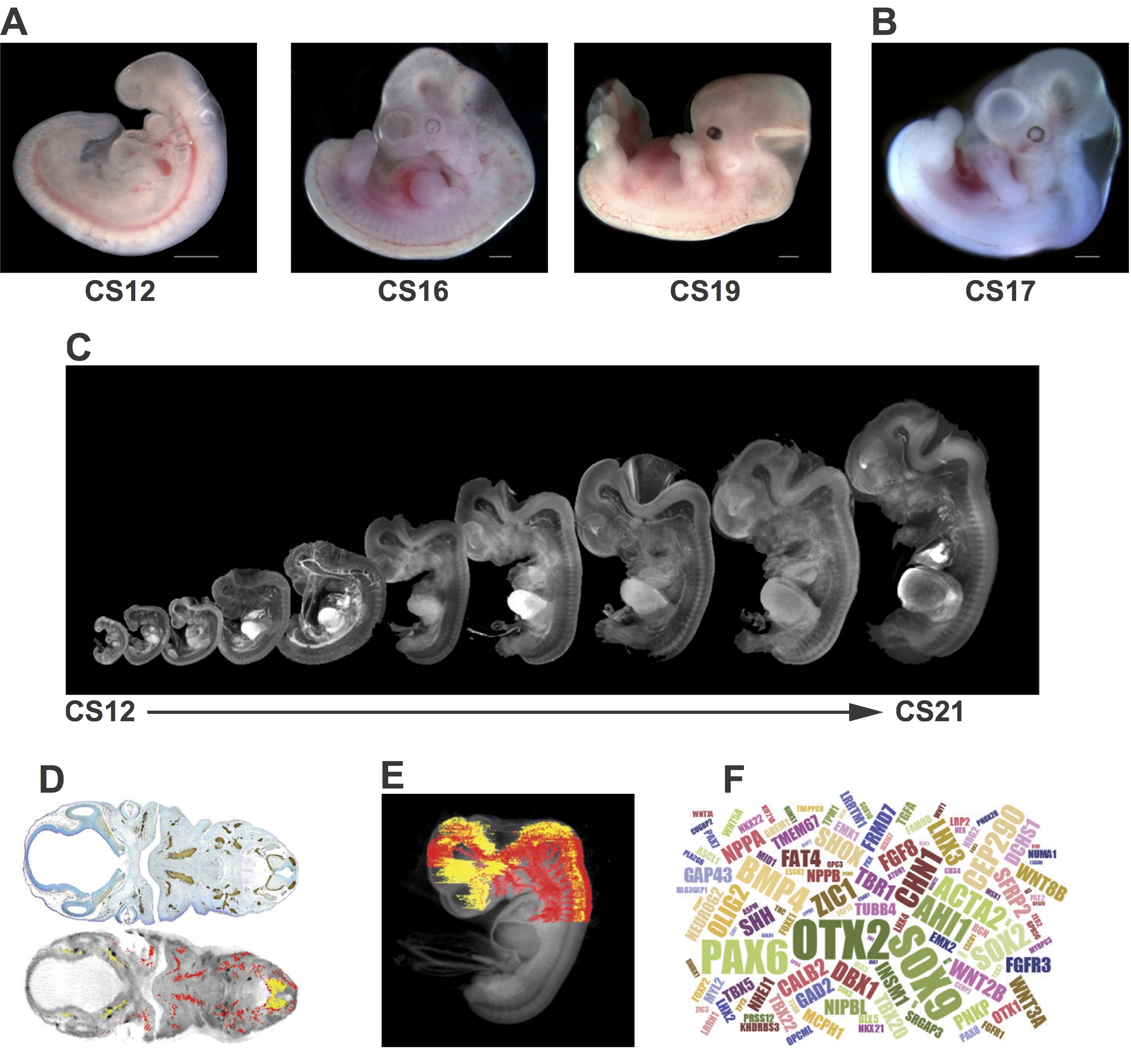
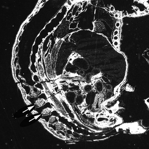
Fig. 1 Examples of resources available from the HDBR. (A) Images of embryos at (from left to right) CS12 [∼26 days post conception (dpc)], CS16 (∼37 dpc) and CS19 (∼47 dpc). (B) A CS17 (∼41 dpc) embryo with trisomy 21. (C) CS12-CS21 series of 3D models, generated using optical projection tomography (OPT) from embryos in the HDBR collection, displayed at their relative sizes. Movies and images of the models can be viewed or the full models requested via the HuDSeN website (www.hudsen.org). (D) The HDBR provides access to curated gene expression data. The upper image shows a section through a CS17 embryo stained with anti-GAP43 antibody (brown); the lower image is of the corresponding section in the CS17 OPT model onto which the GAP43 expression data have been mapped (red, strong expression; yellow, moderate expression). Expression data were mapped from experimental sections to digital sections using MAPaint software (http://www.emouseatlas.org/emap/home.html). (E) 3D expression domain of GAP43 expression in the head and part of the body built up by mapping data from sections as shown in D. Experimental and mapped data are uploaded to a spatial database (www.hudsen.org). (F) Word cloud representation of the expression data available in the HuDSeN human gene expression spatial database (www.hudsen.org). The word cloud represents the number of entries for each gene and there are currently data from 128 genes. Scale bars: 1 mm.
 

The HDBR also provides a gene expression service using RNA in situ hybridisation or protein immunohistochemistry. Sectioned embryonic or fetal tissue is used to identify the temporal and spatial expression of specific genes or proteins. Gene expression data from this service, as well as data generated by external projects, are digitally imaged and made accessible via an open access database (www.hudsen.org). Some stained sections are also available at high resolution (http://nbb-slidepath.ncl.ac.uk/dih; username and password available on request).

Material can be provided to researchers in the UK without the need for project ethics review, as well as to international groups with relevant ethical review body approvals. Over 10,000 slides and tissue samples have been distributed to registered users in the last 5 years, and ∼100 projects are registered per year. The majority of projects are based in the UK, but a growing number of users are in the USA (17%), mainland Europe (11%) and other locations worldwide. Distribution of human embryonic and fetal tissue is prioritised to projects that investigate congenital disorders. Of particular interest are studies that aim to understand the function of genes important for early development, genes linked to human-specific functions (e.g. cognitive function and language) and genes associated with significant anatomical or functional differences between mice and humans.

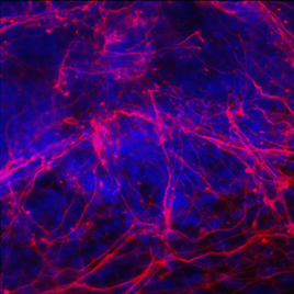
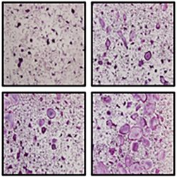
A wide variety of studies have been carried out using HDBR materials (Fig. 2). These range from investigation of single genes underlying genetic disorders (Tischfield et al., 2010;Thomas et al., 2014) to high-throughput studies of transcriptomes (Kang et al., 2011) and regulatory sequences (Necsulea et al., 2014). Advances in high-throughput sequencing technologies have opened the door to large-scale cross-species comparisons that are highlighting differences in transcript sequence, alternative splicing, expression level and regulation between humans and model organisms (Cotney et al., 2013; Bae et al., 2014). Some studies, such as the investigation of tail bud development in chick and human (Olivera-Martinez et al., 2012), dissect key processes of early development. Organ-specific tissues have been used for the derivation of primary cell lines and stem cells (U et al., 2014).

DEV122820F2 copy

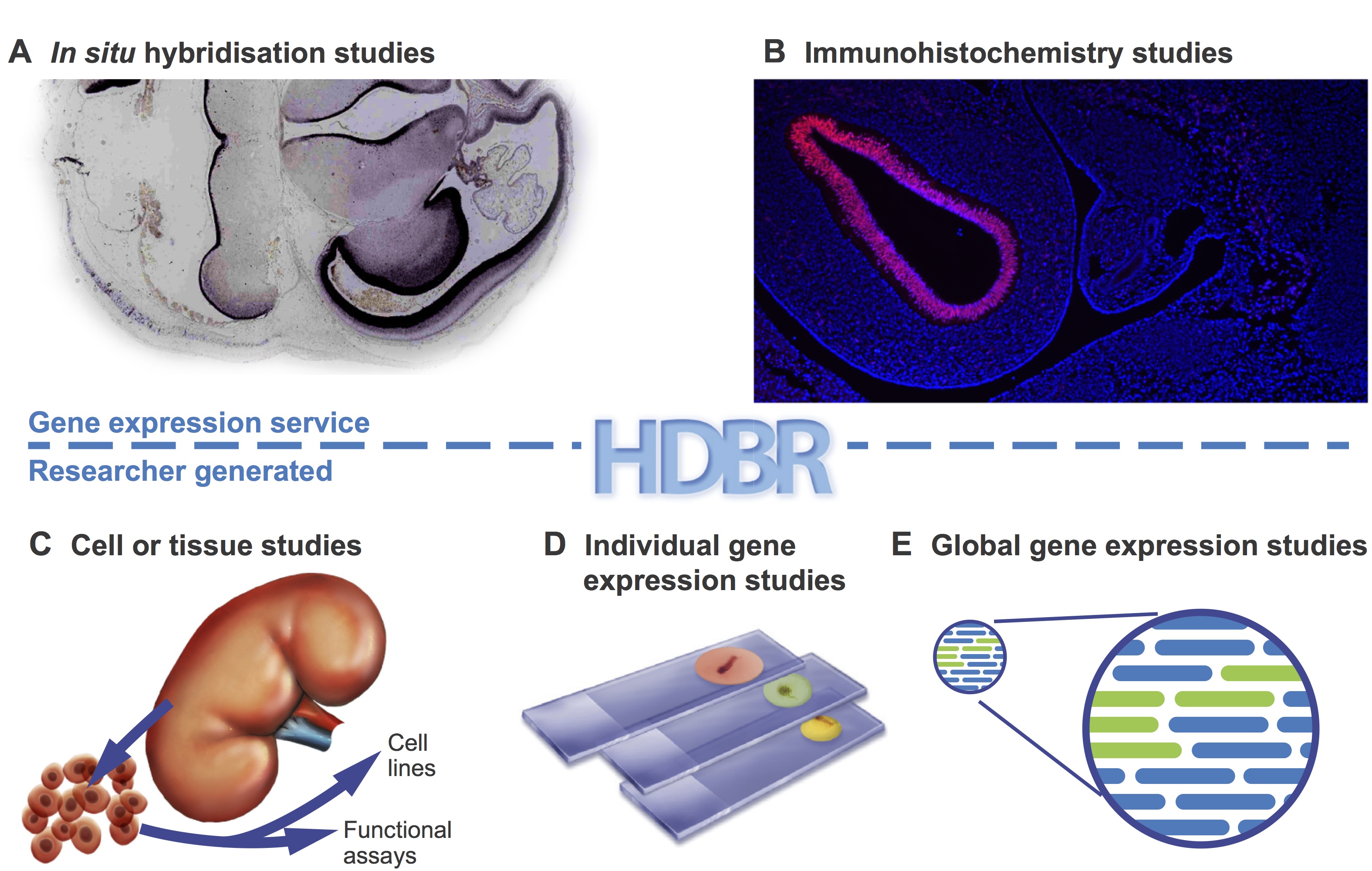
Fig 2. HDBR material has been used in many different types of studies.Examples are shown of the types of study carried out using HDBR material. (A,B) Examples from the gene expression service. HDBR staff perform gene expression analysis on sectioned tissue by in situ hybridisation (A) or immunohistochemistry (B) on behalf of researchers. (C-E) The types of studies that researchers carry out in their own laboratories with material provided by HDBR: to produce cell lines or perform functional analysis (C); gene expression studies in paraffin fixed sections (D); and transcriptomics studies in cells or tissues (E).


Other tissue banks

A number of other groups, in the UK and USA, have set up collections of human fetal material. These are normally organised around defined scientific projects and the samples are not usually available to the wider scientific community. However, there are exceptions where banks have been established to provide material for use in research. For example, the South Wales Initiative for Fetal Tissue (http://www.biobankswales.org.uk/swift-research-tissue-bank/) provides clinical grade fetal tissue (5 and 12 pcw) primarily for human therapeutics, and the University of Maryland Brain and Tissue Bank in the USA (http://medschool.umaryland.edu/btbank/catalog.asp) can provide access to both adult and fetal tissue.


Bioinformatics portals

A number of sites provide valuable information on human development for researchers. For example, the BrainSpan project (www.brainspan.org) has generated transcriptome data and gene expression data from multiple regions of the embryonic, fetal and adult brain. The UNSW Embryology portal is an education and research website that has links to many collections of human embryonic and fetal specimens (https://embryology.med.unsw.edu.au/embryology/index.php/).

In the UK a consortium of UKCRC funders has established The National Tissue Directory and Coordination Centre (www.biobankinguk.org). This directory will enable researchers to find human biobanks within the UK and gain access to their collections through one system. These and other bioinformatics databases are essential resources for researchers, particularly those whose access to primary tissue is limited.


Archive resources

The Carnegie collection (www.ehd.org/virtual-human-embryo/)

Seven thousand human embryos are stored in the Carnegie collection at the National Museum of Health and Medicine, Washington, D.C. This material was used to develop the comprehensive Carnegie staging system based on internal as well as external features. Carnegie staging (CS1-23) is now employed universally in human embryo research. Individual embryos throughout the embryonic period have been sectioned and, in an attempt to make the collection more accessible for research and teaching, digital images from a subset of these have been acquired. The images are labelled with standard anatomy terms to help with interpretation, or used to generate 3D models and 252 movies.


The Kyoto collection (http://bird.cac.med.kyoto-u.ac.jp/index_e.html)

This is the largest human embryo collection in the world, with over 44,000 specimens between CS7 and CS23. Maternal epidemiological data and detailed clinical information on the pregnancies were collected for each specimen. Five hundred normal and 500 abnormal embryos have been serially sectioned, and a further 1300 staged human embryos have been digitally imaged by magnetic resonance (MR) microscopy and 3D reconstructions produced.


Conclusions

The landscape for developmental biology and clinical research has changed within the last few years, with more emphasis being placed on large-scale sequencing projects such as the Genotype-Tissue Expression Project (GTEx Consortium, 2013), Geuvadis (Lappalainen et al., 2013) and the UK 100,000 Genomes Project (Siva, 2015). These projects and numerous others aim to map variations in gene expression in thousands of individual patients and correlate this with disease phenotypes and bioinformatics information on sites such as the Encyclopedia of DNA Elements (ENCODE; https://www.encodeproject.org/;Kellis et al., 2014). Other studies are using novel algorithms to interrogate the sequencing data to investigate gene networks (Liu et al., 2014). These projects are now identifying candidate genes that could play a role in a range of human diseases and syndromes.

The next step in this gene discovery pipeline will be to test candidate genes in model organisms and human tissue. This analysis needs to be performed at the cellular level to determine whether the genes are expressed in tissues and cell types relevant to the disease under study. The HDBR is ideally placed to assist researchers wishing to investigate genes thought to be responsible for congenital abnormalities. In addition, some groups are using material provided by the HDBR to study epigenetic regulation and in functional studies. Other projects include the derivation of stem cells and scaffolds for tissue engineering projects. It is likely in the coming years that the HDBR will not only support projects wishing to understand key developmental processes but will also increasingly provide resources to underpin translational research.


References

Bae, B.-I., Tietjen, I., Atabay, K. D., Evrony, G. D., Johnson, M. B., Asare, E., Wang, P. P., Murayama, A. Y., Im, K., Lisgo, S. N. et al. (2014). Evolutionarily dynamic alternative splicing of GPR56 regulates regional cerebral cortical patterning. Science 343, 764-768. doi:10.1126/science.1244392

Bae, B.-I., Jayaraman, D. and Walsh, C. A. (2015). Genetic changes shaping the human brain. Dev. Cell 32, 423-434. doi:10.1016/j.devcel.2015.01.035

Blaschke, R. J. and Rappold, G. (2006). The pseudoautosomal regions, SHOX and disease. Curr. Opin. Genet. Dev. 16, 233-239. doi:10.1016/j.gde.2006.04.004

Cadman, S. M., Kim, S.-H., Hu, Y., González-Martínez, D. and Bouloux, P.-M. (2007). Molecular pathogenesis of Kallmann’s syndrome. Horm. Res. 67, 231-242. doi:10.1159/000098156

Cotney, J., Leng, J., Yin, J., Reilly, S. K., DeMare, L. E., Emera, D., Ayoub, A. E., Rakic, P. and Noonan, J. P. (2013). The evolution of lineage-specific regulatory activities in the human embryonic limb. Cell 154, 185-196. doi:10.1016/j.cell.2013.05.056

Dolk, H., Loane, M. and Garne, E. (2010). The prevalence of congenital anomalies in Europe. Adv. Exp. Med. Biol. 686, 349-364. doi:10.1007/978-90-481-9485-8_20

Eichholzer, M., Tönz, O. and Zimmermann, R. (2006). Folic acid: a public-health challenge. Lancet 367, 1352-1361.doi:10.1016/S0140-6736(06)68582-6

Gautam, P., Anstey, K. J., Wen, W., Sachdev, P. S. and Cherbuin, N. (2015). Cortical gyrification and its relationships with cortical volume, cortical thickness, and cognitive performance in healthy mid-life adults. Behav. Brain Res. 287, 331-339. doi:10.1016/j.bbr.2015.03.018

GTEx Consortium (2013). The Genotype-Tissue Expression (GTEx) project. Nat. Genet. 45, 580-585. doi:10.1038/ng.2653

Kang, H. J., Kawasawa, Y. I., Cheng, F., Zhu, Y., Xu, X., Li, M., Sousa, A. M. M., Pletikos, M., Meyer, K. A., Sedmak, G. et al. (2011). Spatio-temporal transcriptome of the human brain. Nature 478, 483-489. doi:10.1038/nature10523

Kellis, M., Wold, B., Snyder, M. P., Bernstein, B. E., Kundaje, A., Marinov, G. K., Ward, L. D., Birney, E., Crawford, G. E., Dekker, J. et al. (2014). Defining functional DNA elements in the human genome. Proc. Natl. Acad. Sci. USA 111, 6131-6138. doi:10.1073/pnas.1318948111

Lappalainen, T., Sammeth, M., Friedländer, M. R., t Hoen, P. A. C., Monlong, J., Rivas, M. A., Gonzàlez-Porta, M., Kurbatova, N., Griebel, T., Ferreira, P. G. et al. (2013). Transcriptome and genome sequencing uncovers functional variation in humans. Nature 501, 506-511. doi:10.1038/nature12531

Liu, L., Lei, J., Sanders, S. J., Willsey, A. J., Kou, Y., Cicek, A. E., Klei, L., Lu, C., He, X., Li, M. et al. (2014). DAWN: a framework to identify autism genes and subnetworks using gene expression and genetics. Mol. Autism 5, 22. doi:10.1186/2040-2392-5-22

Necsulea, A., Soumillon, M., Warnefors, M., Liechti, A., Daish, T., Zeller, U., Baker, J. C., Grützner, F. and Kaessmann, H. (2014). The evolution of lncRNA repertoires and expression patterns in tetrapods. Nature 505, 635-640. doi:10.1038/nature12943

Olivera-Martinez, I., Harada, H., Halley, P. A. and Storey, K. G. (2012). Loss of FGF-dependent mesoderm identity and rise of endogenous retinoid signalling determine cessation of body axis elongation. PLoS Biol. 10, e1001415.doi:10.1371/journal.pbio.1001415

Siva, N. (2015). UK gears up to decode 100,000 genomes from NHS patients. Lancet 385, 103-104. doi:10.1016/S0140-6736(14)62453-3

Thomas, M. G., Crosier, M., Lindsay, S., Kumar, A., Araki, M., Leroy, B. P., McLean, R. J., Sheth, V., Maconachie, G., Thomas, S. et al. (2014). Abnormal retinal development associated with FRMD7 mutations. Hum. Mol. Genet. 23, 4086-4093. doi:10.1093/hmg/ddu122

Tischfield, M. A., Baris, H. N., Wu, C., Rudolph, G., Van Maldergem, L., He, W., Chan, W.-M., Andrews, C., Demer, J. L., Robertson, R. L. et al. (2010). Human TUBB3 mutations perturb microtubule dynamics, kinesin interactions, and axon guidance. Cell 140, 74-87. doi:10.1016/j.cell.2009.12.011

Tookey, P. A. and Peckham, C. S. (1999). Surveillance of congenital rubella in Great Britain, 1971-96. BMJ 318, 769-770.doi:10.1136/bmj.318.7186.769

U, K. P., Subramanian, V., Nicholas, A. P., Thompson, P. R. and Ferretti, P. (2014). Modulation of calcium-induced cell death in human neural stem cells by the novel peptidylarginine deiminase–AIF pathway. Biochim. Biophys. Acta 1843, 1162-1171. doi:10.1016/j.bbamcr.2014.02.018

Yi, Y., Lindemann, M., Colligs, A. and Snowball, C. (2011). Economic burden of neural tube defects and impact of prevention with folic acid: a literature review. Eur. J. Pediatr. 170, 1391-1400.doi:10.1007/s00431-011-1492-8

Yue, F., Cheng, Y., Breschi, A., Vierstra, J., Wu, W., Ryba, T., Sandstrom, R., Ma, Z., Davis, C., Pope, B. D. et al. (2014). A comparative encyclopedia of DNA elements in the mouse genome. Nature 515, 355-364. doi:10.1038/nature13992

Thumbs up (No Ratings Yet)
Loading...

Tags: ,
Categories: Resources