This interview was first published in Development.
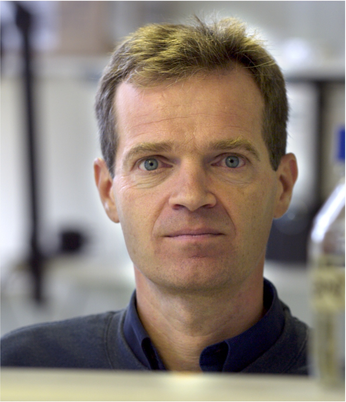
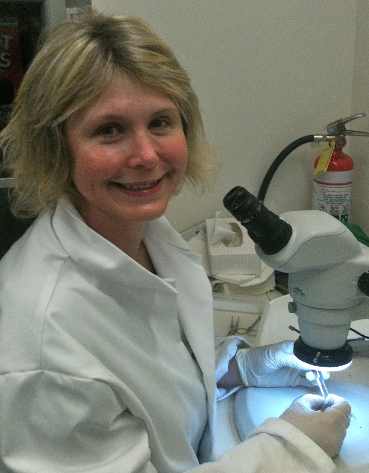
Austin Smith is a stem cell and developmental biologist, who has dedicated his career to the study of pluripotency, stem cell renewal and differentiation. He is currently the Director of the Wellcome Trust MRC Cambridge Stem Cell Institute at the University of Cambridge, UK. We met him there to discuss his research and interests, as well as his role as an editor for Development.
 How did you first become interested in biology? Was there someone who inspired you?
I had a really good biology teacher in school. Back then, he recommended a book by Steven Rose on biochemistry which seemed really interesting, so that is what I chose to do at university, but then I found it mostly boring! Developmental biology was what I found interesting, in particular the concept of pluripotency, which I first heard about in the lectures of Chris Graham. When I finished my degree, I still thought that understanding pluripotency was the most interesting problem, and if I had a chance to do a PhD that is what I would focus on. Embryonic stem cells (ESCs) had just been derived then, so I looked for labs that studied ESCs. And that is what I have worked on ever since!
Why does understanding pluripotency fascinate you?
Pluripotency is interesting in the same way that development is interesting. It is about understanding how tissue complexity develops from a single cell. But the thing that is really enticing, and that I found stunning when I first heard about it, is that these ESCs grow in a dish and retain pluripotency. At that time, it seemed to me that we could use these cells to understand exactly how embryos develop, to dissect development in a way that you couldn’t do in an embryo, and understand how the different cell lineages and cell types were made. Of course, that was a ridiculously naïve idea, but that particular question was for me more interesting than understanding morphogenesis and pattern formation in embryos. Cells in a dish give you the opportunity to control things, or at least that is how it seemed to me then. Actually, we are still fighting to do that properly, as it turns out to be much more complex, and therefore much more interesting, than I thought at that time.
So you are primarily interested in pluripotency from a developmental perspective, rather than its medical applications?
During the first ten years of my research life, while I was a PhD student, a postdoc and then initially running my own lab, everything was in the mouse. No one was talking about human ESCs (hESCs) and it was actually almost taboo. Research was purely curiosity driven, and in general I think that is how it should be for a young scientist. However, as we began to get a better hold on ESCs it became inescapable that we should try to make hESCs. Mice are great because you can do experiments with them, but ultimately we are not interested in understanding a mouse. Of course, we want to know about the diversity of the natural kingdom and of life in general, but fundamentally we want to understand humans, if we can. But you have to have an experimental system! So the motivation for moving into hESCs was directly linked to and emanates from my original interest in pluripotency, but extends it to the most relevant species. Of course, hESCs can have medical applications and, as I become more experienced, I feel some obligation to be involved in promoting this. But it is not what drives my science. The primary driver for me is just a fundamental interest in this remarkable phenomenon of pluripotency.
I am also interested in how pluripotent cells in a dish are related to the cells in the animal. This is why we collaborate with Jenny Nichols, who works on the early embryo, and that dynamic works very well.
What are the next questions that you want your lab to tackle?
At the moment, there is a lot of controversy and uncertainty in the hESC field. I think we are now close to having what I would describe as an authentic, so-called naïve hESC, although this is still a matter of significant debate. I think it is essential for the field to try and clarify this issue over the next year or two, since it has become quite heated and because, from my perspective, it is fundamentally important.
Many people working in the human pluripotency field are not really interested in basic biology, they are interested in applications. This is absolutely fine, but it should not obscure the value in understanding the underlying fundamental biology. A really interesting question that we are trying to answer is the extent to which pluripotency is generic across mammals and whether there are marked species differences. I’m hoping we have the opportunity to study the progression through pluripotency towards lineage specification in humans. It is likely to be quite different from the mouse, but how different is completely unknown at this point. Also, if we really have naïve cells, can we work out how to propagate and differentiate them robustly, and will they be more reliable than the cells currently being used? But the thing I’m most interested in is whether we can explore how these cells begin to become specified. We have done quite a lot of work on this in mouse ESCs, and many people have looked in early mouse embryos, but everything happens so fast that it is really difficult to delineate the causal sequence of events. We speculate that, in humans, it might be easier to pull things apart because the time scale is longer. So, whereas up until now the lab has mostly been doing mouse work with the human work in the background, this is finally going to change. I have been trying to do this for a long time, but until we had the right cell (the naïve hESC) there was no point. Now, I think we have, or very soon will have, the right cell.
Whereas the stem cell field is now a busy field, this was not always the case, and you were one of the few people who originally applied to work with ESCs in the UK. Do you feel like a pioneer, and how has the field changed since the beginning of your career?
The pluripotency field actually shrank, almost disappeared, in the late 1980s, early 1990s. My lab in Edinburgh may have been the only lab in the UK studying pluripotency at the time. When Hitoshi Niwa came to my lab and I asked him why he wanted to work with me, an unknown junior group leader, he said “because you are the only person working on pluripotency”. Whereas now the field has just grown out of all proportion and there are frankly far too many people! We have had ESCs since 1981, but people just used them for gene targeting. The developmental biology community found these cells quite difficult to work with: the dream that you could just put them in factor x and they would all become one cell type, e.g. muscle, did not pan out, so most people just gave up on them. The work that we and Hans Schöler did with Oct4 really demonstrated that through the ESC system you could find a central factor that was relevant in the pluripotent cells of the embryo. I think this awoke interest in the basic biology of pluripotency. Then hESCs came along and that provoked all kinds of controversy and politics, but it also unleashed funding streams. So a lot of people were drawn into the field.
ESCs and stem cell biology used to be considered a marginal branch of developmental biology that most developmental biologists looked down their noses at because it was in vitro. Now, we have an equally unbalanced situation in which there is a huge stem cell biology community that unfairly looks down its nose at developmental biology, not realizing that there are things they need to learn from it. I am surprised at this but my lab is still relatively rare in having a strong foot in both camps. Many people are very firmly in one or the other.
As you say, the stem cell field is moving at a vertiginous speed. Do you feel that this puts particular pressure on people to publish, and if so, what do you think are the potential consequences of this?
The field is not really moving that fast. The publications are coming fast but our understanding of the big questions it is still slow. There is just more noise. More information doesn’t always translate into more knowledge, and certainly not into more understanding. This is a problem that we have in academic research now. People have to publish to survive, never mind to progress, but the real value of each publication unit is diminishing. There is huge pressure and incentives, and not just on young scientists, to publish in the fashion journals and therefore there is temptation to overclaim, to embroider.
The stem cell field has grown so rapidly that there are new developments coming up all the time. It is challenging for the community generally, and particularly for journal editors and reviewers, to really be able to evaluate them, especially if they are cross-disciplinary. It is impossible to be on top of the whole field and this is not helped by seeing completely discordant claims in the literature all the time.
In the past few years, several high-profile ethics issues have brought the stem cell field under scrutiny, including discussion in post-publication peer review forums such as PubPeer, where you have yourself engaged with critiques of your work. Do you think that post-publication discussions are useful for the scientific community?
In contrast to some of my colleagues, who have a rather disdainful approach to PubPeer, I think it is valuable. It is very irritating to have anonymous and non-scientific smears made against your papers, but in the end they are just noise. PubPeer is very useful when issues of falsification and fabrication are identified. Within the community we have a problem of people who cheat, and the incentive and pressure to do so is always there. We need mechanisms of identifying those people and holding them to account. Obviously, there are flaws with PubPeer, but it is one such mechanism that seems actually to be quite effective.
You have also been an editor for Development for the last nine years. How has your experience as an editor been?
Initially, I found it really quite difficult. I was recruited by Jim Smith (Development‘s previous Editor-in-Chief) as a stem cell editor, but actually very few stem cells papers were submitted in those days. Stem cell biologists rightly had the view that developmental biologists looked down their noses at them, and would reject their papers. Sometimes the developmental biologists were right to do that and the papers weren’t of the right standard, but it led to an unfortunate separation between the two communities. That meant that, initially, I was handling many general developmental biology papers, which I wasn’t always the best-equipped to assess or identify good reviewers for! But the reason why I agreed to become an editor was to attract more stem cell papers to the journal, and to make sure that the papers that were published were of good quality. I would still like to see more stem cell papers submitted, but now I get more papers in my area of expertise, and that is quite rewarding because I feel that I can have some positive influence in keeping the standards of the journal high. I hope that we are seen as a venue that publishes quality papers in both stem cell biology and in developmental biology, and particularly papers that are at the interface. What is actually quite nice about being an editor for Development is that generally the reviewers try to be constructive and try to improve the papers. A lot of the papers that I am involved with will go through two rounds of revision but everyone is really working together to make the paper as good as it can be. I do have to reject editorially a lot of papers but I think there is no point wasting authors’ or reviewers’ time by sending a paper out to review that is just not at the right level for the journal.
Is there any particular type of paper, or particular topics, that you would like to see more of in Development?
I would like the journal to publish more papers on adult tissue maintenance and repair. The plasticity of adult stem and progenitor cells in tissues like the intestine, the lung and so on is a really exciting area at the moment. I think Development is missing out on some of those stories, and they are clearly relevant to the developmental biology community. Another related area is regenerative biology, not just in model organisms that are traditionally associated with regeneration research but also in mammals. I think that might be an area that is bubbling under at the moment.
You are also the Director of the Wellcome Trust MRC Stem Cell Institute in Cambridge, and within this role you have been supportive of introducing PhD students at your institute to alternative careers during their training. Why and how have you set about to do this?
Part of my role is to make it clear to students and postdocs that they are licensed to explore alternative careers, but the best way is to let them do it themselves. The important point is to send a clear message from the top that you are not failing, and will not be written off, because you are looking at other careers. We are failing our people if we don’t make sure that they are equipped to be able to contribute to society when they leave the lab. Of course, a measure of a successful lab is that some of your people have progressed within academic research, but it is the simple reality that many of them are going to have to go into other careers. Some of them will go on to be influential in policy, public engagement or industry.
A few years ago, when we were interviewing for PhD studentships, one of the questions we asked the candidates was what their career aspirations were. One of the students was honest enough to say that they wanted to go into industry. One of my colleagues immediately afterwards said “well, we are not taking this person, what are we here for?” I found this quite shocking, but then I had to admit that, five years earlier, it is also the position I would have had. But it shows some clarity for someone to come in and say “ultimately I see myself working in industry” and they shouldn’t be negatively scored for that. There is a culture change and if you are in a leadership position you can either facilitate it or stand against it. Of course, you want to get the best people. But then you have to serve those people, in their interest and not just in your own.
 (6 votes)
 Loading...
 (No Ratings Yet)
 (3 votes)