This article by Göran Hermerén was first published in Development. Also read the companion ethics article here.
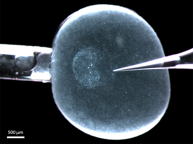
The development of human pluripotent stem cells has opened up the possibility to analyse the function of human cells and tissues in animal hosts, thus generating chimeras. Although such lines of research have great potential for both basic and translational science, they also raise unique ethical issues that must be considered.
A major goal in life science research is to understand human development, physiology and dysfunction, as this will allow better treatment of disease and injury. However, our ability to conduct research using human subjects or samples is obviously limited. In recent years, the isolation of human embryonic stem cells [hESCs; (Thomson et al., 1998)] and the generation of human induced pluripotent stem cells [hIPSCs (Takahashi et al., 2007)] have opened new avenues to study human biology, but there is a need to analyse these cells in an in vivo setting. Consequently, many researchers are now turning to human-animal chimera research.
Lensch et al. (2007) have proposed the following definition of ‘chimera’: “The term chimera […] indicates organisms comprised of cells from two or more individuals of the same or different species. Today, the most common usage describes cellular combinations at the preimplantation blastocyst stage of development, […] also […] other entities created by introducing cells at later stages, including in adult recipients.” This definition makes it clear that there are different kinds of chimeras. This is also obvious from the definition proposed by Behringer (2007): “A chimera is an individual composed of somatic and, in certain cases, germ line tissues derived from more than one zygote. […] If the donor tissue and recipient are of different species, then an interspecific or cross-species chimera is generated.” Different kinds of human-animal chimeras might raise different ethical issues – according, for example, to which tissues the human cells contribute to or how long the chimeric animal survives. Chimeras that include human neural tissue are of particular concern, because the cognitive capacities of the chimeras might be affected, and because of the prevailing special status of humans in our culture.
In this Spotlight article, I will first summarise some of the recent research that uses chimeras, to provide a context for the subsequent discussion on the ethical issues surrounding this kind of research. I then provide a proposal for how such challenges can be addressed – so as to allow research to proceed while respecting these ethical concerns.
Current research avenues using chimeras
Several broad categories of experimental investigation are now making use of human-animal chimeras. Recent work from Jacob Hanna’s lab has used the mouse embryo as an in vivo system to test the potential of human pluripotent cells: creating chimeras by microinjection of hESCs or iPSCs into a mouse morula and analysing the chimeric embryo shortly afterwards (Gafni et al., 2013). Given that these experiments were limited to early embryos (10 days; within the limit allowed for research on human embryos), the ethical concerns here are limited, but it is possible that central nervous system (CNS) tissue containing both mouse and human cells will be found in this chimera.
In a different approach, the EU-funded project ‘Health and the Understanding of Metabolism, Aging and Nutrition’ (HUMAN; http://www.fp7human.eu/) proposes to address the problem of the increased frequency of metabolic diseases in an aging population by creating ‘humanised’ mouse models with cells from the liver and pancreas of human donors. In this way, it will be possible to study the functions of genes in human organs and how, in combination with factors like eating habits and nutrition, they can influence the risk of contracting a metabolic disease. The idea is to study and compare two different groups, namely very old healthy individuals and individuals with metabolic diseases. In contrast to the experiments described above, in which chimeric embryos were destroyed soon after creation, the HUMAN project involves maintaining chimeric animals until old age.
From an ethical standpoint, two avenues of research are particularly interesting: the creation of complete human organs in animals, and the generation of CNS, particularly forebrain, chimeras. The long-term goal of the first approach is to grow organs made exclusively from human cells in a chimeric animal, such as a pig, that could potentially be used for organ transplant. This goal has been pursued most actively by the research group of Hiro Nakauchi, focusing primarily on the pancreas (Kobayashi et al., 2010, 2014; Matsunari et al., 2012; Usui et al., 2012; Rashid et al., 2014), although it still remains at the hypothetical stage.
The second line is exemplified by the research of Steven Goldman and collaborators, who established mice in which the forebrain glial cells were completely replaced by human glia (Han et al., 2013). These animals manifested significantly different cognitive capabilities – showing enhanced plasticity and learning. Repeating such experiments with glia derived from individual patients with neuropsychiatric disorders could allow a better understanding of the pathology of such diseases and might aid in the identification of potential therapeutic targets. The fact that the humanised mice displayed apparently enhanced cognitive capacity raises particular ethical questions, discussed further below.
Ethical issues of chimera research
The examples discussed above suggest that at least two categories of chimera research need to be separated, as they raise partly different ethical issues: in vitro studies using early embryos, and in vivo studies involving sentient animals. The latter raises additional issues of animal health and welfare.
One key ethical question is whether crossing species boundaries is in principle or prima facie ethically wrong. If so, we need to consider whether the generation of stem cell chimeras represents a particular and controversial instance of such boundary crossing. The reason why such issues are raised is that humans and animals are treated differently in our culture and have different ethical and legal status: animals are not legal entities and do not have rights in the way that humans do.
One of the tacit premises in this discussion is that we view ourselves as distinct, and we must consider what defines a human being. Of course, in the history of western civilization, scientific discoveries have challenged and changed our view of ourselves. To begin with, it is necessary then to make some sort of distinction between being human as a biological concept and as a moral concept. In the latter case, the focus is on intentional action, self reflection and self-understanding: on humans as moral and responsible agents.
Concerning the goal of growing human organs in animals such as pigs (as discussed above), a key challenge is the xenogenic barrier – these two species are estimated to have diverged almost 100 million years ago, so is it even feasible to use a pig as an ‘incubator’ for human organs? If this barrier cannot be overcome, could we use primates? Or, taking this to the extreme, might one even conceive of using people in a permanent vegetative state or suffering from senile dementia (who might not display the key characteristics of intentional action, self-reflection and self understanding mentioned above) as incubators? This might
sound like science fiction or a dystopia, but should be discussed before it becomes scientifically feasible.
Two types of particularly problematic research are when cognitive capacities are changed and when germ-line effects are introduced (in which the potential exists for the production of human embryos in animals or vice versa). Thus, focus in the ethics discussion should be on chimeras in which changes have been introduced that might affect their cognitive capacities, and in cases in which the mixture between species is so extensive that confusion might arise as to which species the chimeric individual (and/or its germline) belongs to.
What ethical issues do these research avenues raise, over and above those concerning animal health and welfare? A possible list of concerns (e.g. see Danish Council of Ethics, 2008; and Streiffer, 2010) includes: violation of human dignity; violation of the order of nature; risk and scientific uncertainty; violation of the dignity of the humanised animal; violation of taboo against mixing of species; danger of moral confusion – should resulting chimeras be treated as animals or as humans? Some of these concerns might seem irrelevant: for instance, human dignity is obviously a property of humans, not of human cells, and thus may not apply to chimeric animals. However, it is crucial to examine carefully the arguments for and against particular lines of research, if only to avoid the impression that important issues are swept under the carpet. Important underlying values for those who are pursuing this research include safety and efficacy. Certainly both are valid concerns, but they do not always go together: a particular intervention can be safe but not effective, or effective but not safe.
Subtle ethical questions are raised by how uncertainties and risks in this type of research are to be handled. Proposals have been made as to how germline contribution could be avoided (e.g. the use of progenitor rather than stem cells), but do we know for sure that using progenitors will prevent the introduction of changes in the germline that are inherited? It is always possible to say that no inherited changes have been demonstrated in the experiments conducted so far, but that does not prove that this will never happen. When is ‘safe where to draw a line here is not ethically neutral; it might create opportunities for some and harm and/or difficulties for others.
To make ethically appropriate decisions on whether specific lines of research should be undertaken, we have to try to answer the following four questions: What do we know? What do we want? What are we able to do? What ought to be done? The answers to the first three questions are relevant to, but do not decide, the answer to the crucial fourth question. For instance, we may know something about the attitudes of people towards various aspects of research, but direct inferences from these attitudes cannot be made, as they might be based on incorrect or misleading information, which scientists have a responsibility to correct.
The key focus of ethics is conflicts of values, taken in a wide, non-technical sense, including interests, rights, liberties and obligations. The answer to the question ‘what ought to be done’ has to be informed by the answers to the previous ones, but decided on the basis of the values at stake, ordered in normative importance. However, it should be borne in mind that this analysis might be complicated by the simple fact that ‘we’ might know, want and be able to do different things, depending on the situation and context (discussed in Hermerén, 2014).
A strategy for addressing ethical challenges
What is the best strategy to use in dealing with the ethical challenges raised by chimera research? In addition to the more general strategy developed elsewhere and alluded to above for dealing with ethical concerns, I propose that the following general precepts should be heeded in this particular research area: first, I recommend that one should avoid developing new ethical frameworks or rules for every new type of research. Where possible, it is advisable to use existing frameworks, unless there is something specific about the research that calls for changes in the existing framework. As far as possible, similar cases ought to be treated in similar ways.
Protection of animal welfare will obviously be important for in vivo studies, but what is required in addition to that? And how do we balance the need to limit regulatory hurdles against the importance of ensuring ethical compliance? In contrast to the National Academy of Science panel recommendations (National Research Council, 2005), the International Society for Stem Cell Research (ISSCR) guidelines 2008 (http://www.isscr.org/docs/default-source/clin-trans-guidelines/isscrglclinicaltrans.pdf ) stipulate that the assay of hESCs by teratoma formation should be accepted as routine and be exempt from Stem Cell Research Oversight (SCRO) review.
Why should these assays be exempt? Lensch et al. (2007) have argued that human teratoma formation studies in adult mice are justifiable and should be routinely approved by animal care committees with a minimal need for regulation by the stem cell research oversight process. Their main reason is that “the need for teratoma assays with hESCs is compelling” and that “we believe that the risk of inadvertently creating a rodent chimera with higher, human brain function is negligible.” However, this latter point needs to be discussed against the recent findings by Goldman and collaborators (Han et al., 2013), as discussed above. In particular, this research has shed light on the relative importance of niche and environment versus the origin of the transplanted cells – which defines the final phenotype? This case is also interesting in that the research indicated a cognitive improvement in the transplanted mice. Of course, this result is dependent on the methods of measurement and the criteria used for cognitive improvement, but it is worth discussing in relation to the ISSCR guidelines, which are according to previous studies’ safe enough? The decision about presented and discussed by Insoo Hyun elsewhere in this issue (Hyun, 2015).
Precaution or proportionality?
My personal view is that the precautionary principle has played too important a role in discussions on what should or should not be permitted in terms of chimera research, at least if it is interpreted as saying that, if there is a risk, you should do nothing. Inaction may also be risky and can lead to harm: if we adopt a no-risk scenario, medical research will be stifled and progress will be impossible. Instead, I would advocate some version of the principle of proportionality (Hermerén, 2012). I argue that the risk-benefit analysis can be improved by considering the following questions: Is the research objective important? Are the methods to achieve them feasible and are the facilities adequate? Are there no less risky or controversial methods available? Do the relevant personnel have the training required to deal with the research equipment and the animals?
If the answer to these questions is ‘yes’, then the research should be approved, but with appropriate caveats. Sensible precautions might include using progenitors rather than pluripotent cells, and treating the humanised mice as one would treat genetically modified crop: keep them isolated, make sure they do not mate with wild mice and euthanise them when the research is concluded.
Concluding remarks
Ethical problems arise in a context of beliefs and values. If people have different beliefs about current and future trends, and do not want to achieve and/or avoid the same goals, they will view the problem landscape differently. What is a problem to one person may not be to another. Time is then required for dialogues between the different stakeholders, including researchers, regulators, patients and organizations. In general, issues can not be settled once and for all, for the simple reason that research is developing rapidly, values and preferences change, and so do perceptions of risks and benefits. Top-down approaches must be avoided, as experience shows that they rarely work. Those who are worried must be allowed to express their concerns and in that sense participate in the setting of the agenda.
The advent of pluripotent stem cells and the use of chimera research have unearthed new ethical challenges, but with these approaches to hand, research should be able to proceed without excessive regulation.
References
Behringer, R. R. (2007). Human-animal chimeras in biomedical research. Cell Stem Cell 1, 259-262.
Danish Council of Ethics (2008). Man or Mouse? Ethical aspects of chimaera research. Copenhagen, Denmark.
Gafni, O., Weinberger, L., Mansour, A. A., Manor, Y. S., Chomsky, E., Ben-Yosef, D., Kalma, Y., Viukov, S., Maza, I., Zviran, A. et al. (2013). Derivation of novel human ground state naive pluripotent stem cells. Nature 504, 282-286.
Han, X., Chen, M., Wang, F., Windrem, M., Wang, S., Shanz, S., Xu, Q., Oberheim, N. A., Bekar, L., Betstadt, S. et al. (2013). Forebrain engraftment by human glial progenitor cells enhances synaptic plasticity and learning in adult mice. Cell Stem Cell 12, 342-353.
Hermerén, G. (2012). The principle of proportionality revisited: interpretations and applications. Med. Health Care Philos. 15, 373-382.
Hermerén, G. (2014). Human stem-cell research in gastroenterology: experimental treatment, tourism and biobanking. Best Pract. Res. Clin. Gastroenterol. 28, 257-268.
Hyun, I., Taylor, P., Testa, G., Dickens, B., Jung, K. W., McNab, A., Robertson, J., Skene, L. and Zoloth, L. (2007). Ethical standards for human-to-animal chimera experiments in stem cell research. Cell Stem Cell 1, 159-163.
Hyun, I. (2015). From naïve pluripotency to chimeras: a new ethical challenge. Development 142, 6-8.
Kobayashi, T., Yamaguchi, T., Hamanaka, S., Kato-Itoh, M., Yamazaki, Y., Ibata, M., Sato, H., Lee, Y.-S., Usui, J.-i., Knisely, A. S. et al. (2010). Generation of rat pancreas in mouse by interspecific blastocyst injection of pluripotent stem cells. Cell 142, 787-799.
Kobayashi, T., Kato-Itoh, M. and Nakauchi, H. (2014). Targeted organ generation using MixI1-inducible mouse pluripotent stem cells in blastocyst complementation. Stem Cells Dev. (in press).
Lensch, M. W., Schlaeger, T. M., Zon, L. I. and Daley, G. Q. (2007). Teratoma formation assays with human embryonic stem cells: a rationale for one type of human-animal chimera. Cell Stem Cell 1, 253-258.
Matsunari, H. Nagashima, H.,Watanabe, M., Umeyama, K., Nakano, K., Nagaya, M., Kobayashi, T., Yamaguchi, T., Sumazaki, R., Herzenberg, L. A. et al. (2012). Blastocyst complementation generates exogenic pancreas in vivo in apancreatic cloned pigs. Proc. Natl. Acad. Sci. USA. 110, 4557-4562.
National Research Council (2005). Guidelines for Human Embryonic Stem Cell Research. Washington, DC: National Academies Press.
Rashid, T., Kobayashi, T. and Nakauchi, H. (2014). Revisiting the flight of icarus: making human organs from PSCs with large animal chimeras. Cell Stem Cell 15, 406-409.
Streiffer, R. (2010). Chimeras, moral status, and public policy: implications of the abortion debate for public policy on human/nonhuman chimera research. J. Law Med. Ethics 38, 238-250.
Takahashi, K., Tanabe, K., Ohnuki, M., Narita, M., Ichisaka, T., Tomoda, K. and Yamanaka, S. (2007). Induction of pluripotent stem cells from adult human fibroblasts by defined factors. Cell 131, 861-872.
Thomson, J. A., Itskovitz-Eldor, J., Shapiro, S. S., Waknitz, M. A., Swiergiel, J. J., Marshall, V. S. and Jones, J. M. (1998). Embryonic stem cell lines derived from human blastocysts. Science 282, 1145-1147.
Usui, J.-I., Kobayashi, T., Yamaguchi, T., Knisely, A. S., Nishinakamura, R. and Nakauchi, H. (2012). Generation of kidney from pluripotent stem cells via blastocyst complementation. Am. J. Pathol. 180, 2417-2426.
 (2 votes)
 Loading...
 The vertebrate gastro-intestinal (GI) tract consists of a regionalized epithelial tube surrounded by mesenchyme that later differentiates into smooth muscle. During the early stages of stomach patterning in chick embryos, the primitive GI track is colonized by vagal enteric neural crest cells (vENCCs), which will give rise to the enteric nervous system (ENS). The important role of the ENS in controlling GI function is well understood, but its contribution to the development of the GI tract has never been addressed. On p.
  The apical region of the adult Drosophila testis harbours a stem cell niche that contains germ stem cells, which differentiate into spermatocytes, and somatic cells, which provide nutrients and regulate the proliferation and differentiation of the germline. During spermatogenesis, somatic cells encapsulate the germline cells, isolating them from the environment by providing a permeability barrier. Disruption of either encapsulation or permeability barrier function has catastrophic effects on spermatogenesis, resulting in sterility. Here, Guy Tanentzapf and co-workers investigate the genetic determinants of soma-germline interactions, specifically during germline encapsulation (p.
  Limb and fin morphogenesis start with the formation of the apical ectodermal ridge (AER), an epithelial signalling centre that coordinates appendage development. Wnt signalling is required for AER induction and several extracellular matrix (ECM) components are necessary for proper limb formation. Mahendra Sonawane and colleagues (p.
  The secretory and multiciliated cells of the adult lung are constantly replenished by multipotent epithelial progenitors: the basal cells. Basal cells give rise to parabasal intermediate progenitors, which then terminally differentiate into ciliated or secretory cells. However, the specific molecular mechanisms governing the production of parabasal cells in the lung remain mysterious. Using genetic and pharmacological approaches in air-liquid interface cultures of adult airway progenitors, Wellington Cardoso and colleagues (p.
 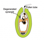 During the development of the plant reproductive lineages – the germlines – typically, single sporophytic (somatic) cells in the flower become committed to undergo meiosis. Here, Grossniklaus and colleagues review recent studies examining the molecular mechanisms underlying cell specification and the acquisition of reproductive fate in sexual and asexual plant species. See the Review on p.
  The neural crest is a cell population that contributes to a variety of derivatives, including sensory and autonomic ganglia, cartilage and bone of the face and pigment cells of the skin. Simões-Costa and Bronner examine neural crest development from a gene regulatory perspective and discuss how the underlying genetic circuitry results in the features that define this unique population. See the Review on p.
  (No Ratings Yet)
