

Outreach activity – Extracting DNA from kiwi fruit
Posted by Katie Howe, on 20 December 2013
DNA extraction from fruit is an easy experiment that makes a great demonstration for kids’ science fairs. I ran a DNA extraction stall at Oxford’s Wow!How? family science fair a few years back. Unfortunately I didn’t take any photos at the time but I had a lot of fun this weekend recreating the experiment in my kitchen!
The experiment is hands on and messy, which kids tend to love, and there’s plenty of opportunity to explain why DNA is important in telling the cells in our body what to do. You could even go into more detail and explain some of the concepts of genetics to older children.
During a busy science fair there might not be time to wait 20 minutes for the extraction solution to work. To avoid having to wait you could set up stations for each of the steps below and every half an hour or so prepare a handful of fruit/extraction buffer solutions and also some alcohol/purified DNA mixtures.
Click here for a downloadable instruction sheet that can be printed off for children/parents to take home.
Here’s what to do:
1) Prepare your equipment
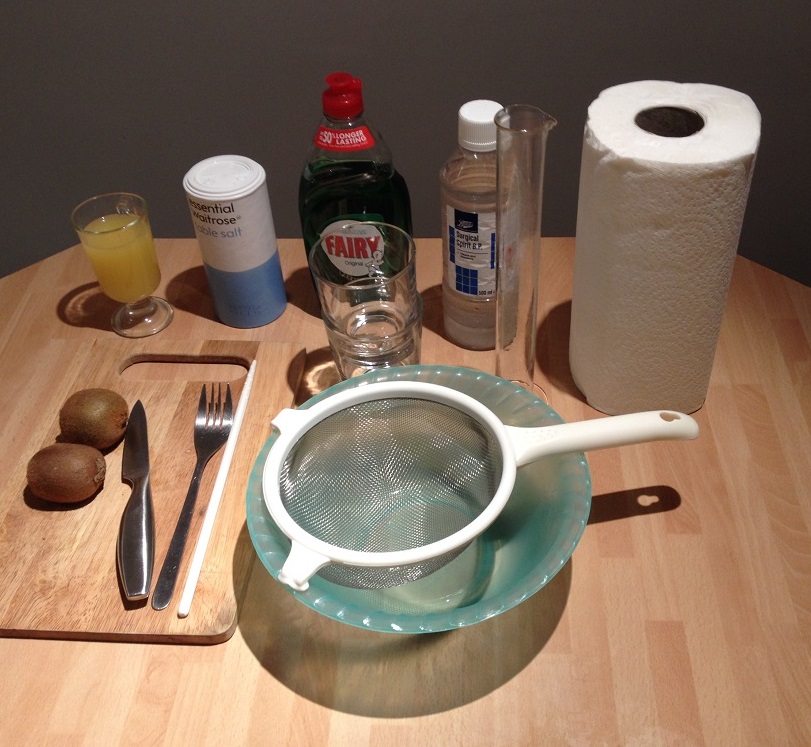
You will need:
– Two kiwis
– Pineapple juice
– Table salt
– Washing up liquid
– Cold alcohol – put in the freezer before you start the experiment (I used surgical spirit but strong rum also works well)
– Two small glass beakers (or plastic cups)
– Sieve
– Bowl
– Tall glass/measuring cylinder
– Kitchen Roll
– Stirring rod (or chopstick)
– Knife
– Fork
– Chopping board
2) Make the extraction solution
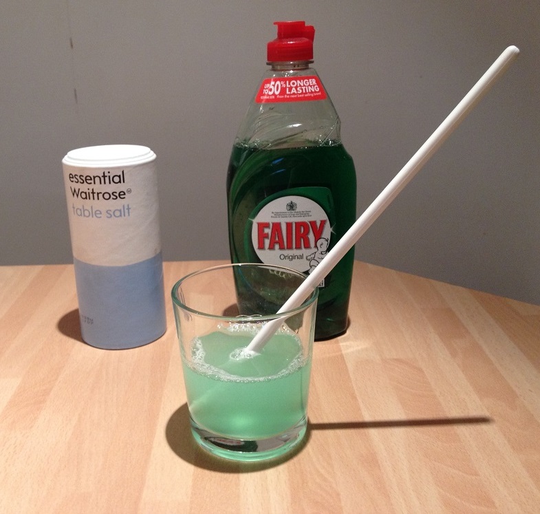
The DNA is tightly packaged inside the nucleus of cells. The membranes of the cell and of the nucleus are rich in fats so we can break them down using a detergent. The salt helps to get rid of the proteins that package the DNA tightly inside the nucleus.
– In one of your beakers measure out about 80mls water
– Add half a teaspoon of salt and stir until dissolved
– Add two teaspoons of washing up liquid and stir gently avoiding making too many bubbles
3) Prepare your fruit mush
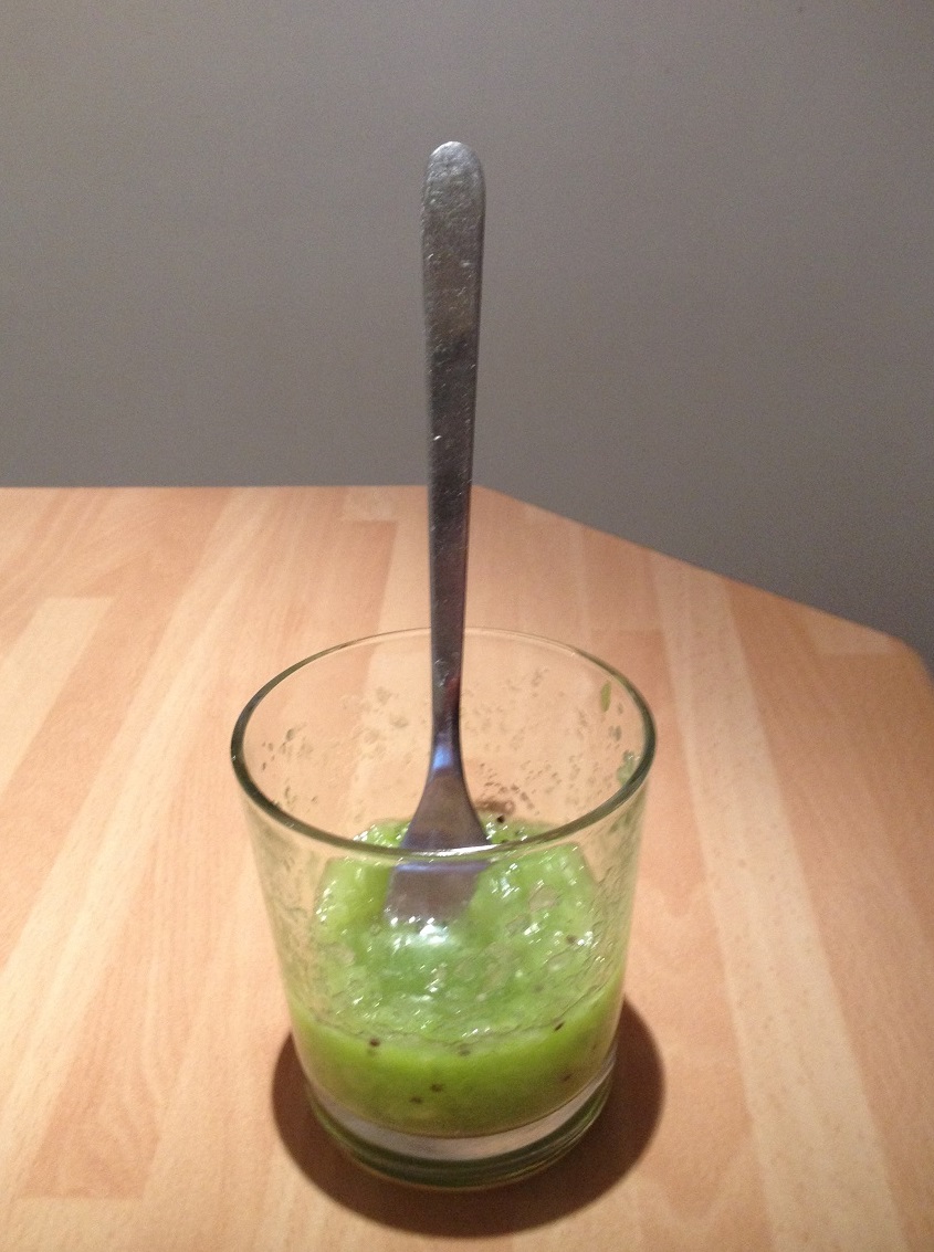
DNA can be extracted from anything living. You could also try this experiment with strawberries or bananas. Make sure you remove the fruit skins as they are mostly dead and don’t contain DNA. The kiwi needs to be broken up so the extraction solution can get to the cells.
– Peel your kiwis and chop into small pieces
– Add the chopped up kiwi to the second small beaker and use the fork to mush it up
4) Add the extraction solution to the fruit mush
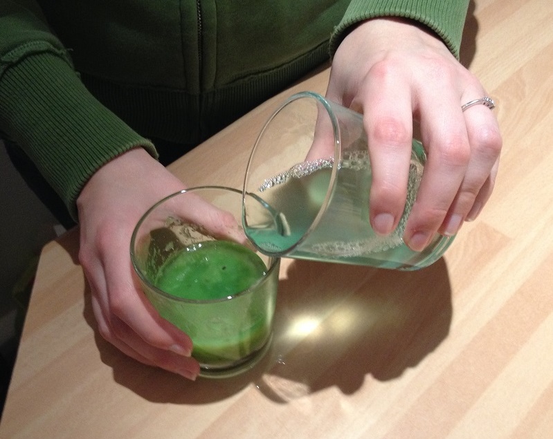
In this step the detergent breaks down the cell membranes so the DNA can be released. The salt removes proteins that are bound to the DNA.
– Add your extraction solution to the kiwi mush
– Leave at room temperature for about 20 minutes
5) Filter the solution
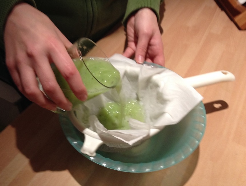
This gets rid of the fruit pulp and seeds and should leave a pure solution of DNA
– Put your sieve over a clean bowl and line the sieve with a few sheets of damp kitchen roll
– Pour your green mush into the sieve carefully, being careful not to break the kitchen roll
– Use a fork to gently push the mixture through the sieve.
– The pulp and seeds should be left in the sieve and there should be a greenish liquid in the bowl. Transfer this to a tall glass or measuring cylinder.
6) Purifying the DNA
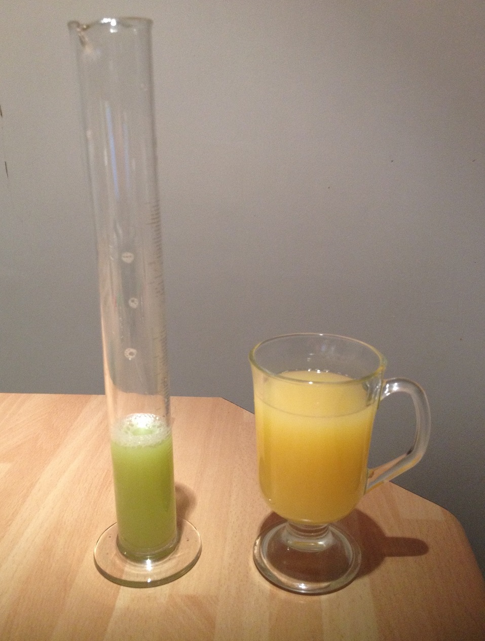
If you want an even purer solution of DNA then we need to remove proteins that are bound to the DNA. Pineapple juice contains an enzyme that breaks down proteins. If you haven’t got any pineapple juice then contact lens cleaning solution can also be used.
– Add pineapple juice to the green liquid. You will need about 1ml of pineapple juice to 5mls of the green DNA solution.
– Leave at room temperature for about 5 minutes
7) Precipitating the DNA
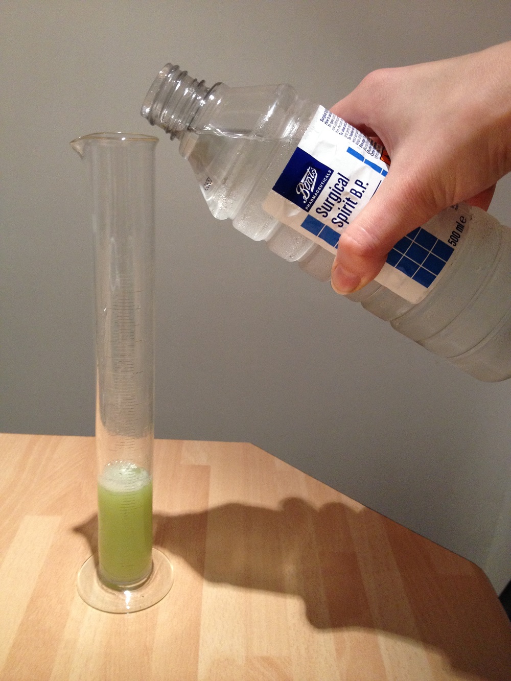
DNA dissolves in water so will not be visible. However, it does not dissolve in alcohol so if we add surgical spirit then the DNA will collect as a white mass at the top of the tube.
– Remove the alcohol from the freezer
– Carefully pour the alcohol down the side of the glass
– You need about equal volumes of DNA solution to alcohol
8) Visualise the DNA sample
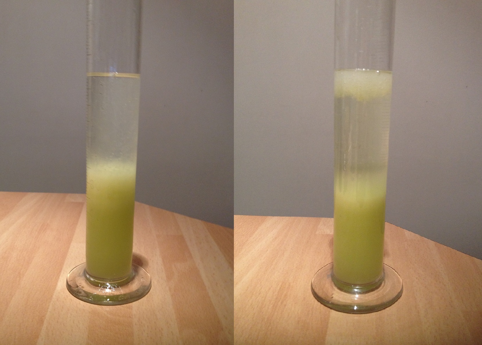
After about 10 minutes you should be able to see a mass of white stringy stuff at the top of the tube (see right hand photo). This is the kiwi DNA! You can fish this out using the chopstick and place it onto a piece of card to take home.
Sources
This protocol is adapted from the following sources:
http://www.funsci.com/fun3_en/dna/dnaen.htm
http://www.nuffieldfoundation.org/practical-biology/extracting-dna-living-things
 This post is part of a series on science outreach. You can read the introduction to the series here and read other posts in this series here.


 Neurotransmitter functions are typically associated with neural function rather than with development, but there is a growing body of evidence suggesting that neurotransmitters may also regulate cell proliferation and differentiation in the nervous system. Recent work has demonstrated that GABAA receptor signalling can control adult neurogenesis in the mouse. Verdon Taylor and co-workers now investigate a role for GABAB receptors in the adult mouse hippocampus (
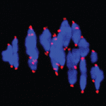 Chromosome mis-segregation during meiosis leads to aneuploidy, and hence to reduced fertility and birth defects. The frequency of aneuploidy increases in older mothers. Various mechanisms have been proposed to account for this correlation between maternal age and chromosome segregation defects, most placing the emphasis on errors occurring in meiosis I (MI). Keith Jones and colleagues (
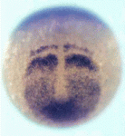 Vertebrate somites form via a sequential process of segmentation, which proceeds from anterior to posterior. In many species, anterior somites form more quickly than posterior ones, but whether this is functionally important, and how the switch in timing might be controlled, is unknown. Now (
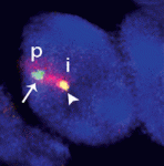 Within the nucleus, active gene loci tend to cluster together in the nuclear interior, while inactive genes are often found at the nuclear lamina. On
 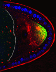 The plus-end directed motor protein Kinesin-1 is a major effector of microtubule-mediated transport, moving a wide range of cargo around cells. How cargo specificity is achieved and how motor transport is regulated are still not fully understood, particularly in in vivo developmental contexts. Isabel Palacios and colleagues (
 The ability to manipulate genomes has been significantly advanced by the development of the CRSIPR/Cas9 technology, in which the Cas9 endonuclease is targeted to specific sites in the genome by single-guide RNAs (sgRNA) – short sequences complementary to the genomic locus of interest. This approach has been further developed to allow regulation of gene expression at particular loci (a system referred to as CRISPRe): a catalytically inactive version of Cas9 (dCas9) can be fused to transcription activation (e.g. VP64) or repression (e.g. KRAB) domains and targeted to the desired genomic locus by sgRNA. Now, René Maehr and colleagues (
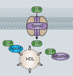 Sphingosine 1-phosphate (S1P) is a lipid mediator formed by the metabolism of sphingomyelin. Recent studies have shown that crucial events during embryogenesis, such as angiogenesis, cardiogenesis, limb development and neurogenesis, are regulated by S1P signalling. Here, Timothy Hla and colleagues provide an overview of S1P signalling in development and in disease.
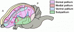 The human cerebral cortex is generally considered the most complex organ, and is the structure that we hold responsible for the repertoire of behavior that distinguishes us from our closest living and extinct relatives. At a recent Company of Biologists Workshop, ‘Evolution of the Human Neocortex: How Unique Are We?’ held in September 2013, researchers considered new information from the fields of developmental biology, genetics, genomics, molecular biology and ethology to understand unique features of the human cerebral cortex and their developmental and evolutionary origin.