The community site for and by
developmental and stem cell biologists

Cycles and cell cycles – EMBO meeting day 1

Posted by , on 23 September 2012

Nice cycle pathThe fourth annual EMBO meeting is currently underway in Nice. I had a few hours to walk around the city before the meeting started, and discovered that Nice is a haven for cyclists, with a network of bicycle paths and a city-wide bike share system. But last night, Nice also became a haven for cell cycle fans, when Paul Nurse gave the opening keynote lecture at the fourth annual EMBO meeting.

Nurse started his talk with a slide of a complicated network of feedback loops of cyclin/CDK regulation, containing all the knowledge we have about which components affect which part of the pathway. “The sole purpose of putting this slide up is to depress you”, he said, but he quickly cheered us up with a more simplified system. His group has studied which parts of the cell cycle pathway are minimally required for particular functions. Using a chimera protein, they were able to show that changing levels of CDK was sufficient for the cell to move from one part of the cell cycle to the next. Nurse emphasized that such studies could also be done for other developmental or cellular systems: when given an extensive network of biological interactions, with multiple feedback loops, it can become very difficult to understand how certain components carry out their role, but by simplifying the system you can find out which processes are relevant for the process that you’re studying.

After the opening keynote, the meeting continued with the first plenary session: Chromatin & chromosomes – the dynamic genome. Three speakers each discussed a different aspect of chromatin dynamics or epigenetic regulation. Steve Henikoff talked about various projects his lab is doing to map nucleosome dynamics and profile epigenetic patterns. Ingrid Grummt focused on epigenetic silencing of rRNA genes. She described a process in which RNA plays a vital role in epigenetic regulation by forming a triplex structure with the two strands of DNA, which then guides a methyltransferase to its target site. The last speaker of the evening, Adrian Bird, illustrated how the study of methyl-CpG interactions is providing insight into Rett Syndrome – an autism spectrum disorder linked to the methyl-CpG-binding protein MeCP2.

opening reception and exhibitThe evening closed with a reception and a visit to the exhibition area, where various companies and organisations have set up a stand for the next few days. There were many vendors and publishers, but also a few institutes: You can find out more about the future Francis Crick Centre in London, or about working at St Jude’s Children’s Hospital in Memphis. The Node is there, too, at the Company of Biologists booth just outside the exhibit hall (next to the job board), so do drop by if you’re attending. I’m there occasionally between talks and I love meeting Node readers.

Thumbs up (1 votes)
Loading...

Tags: ,
Categories: Events

Navigate the archive

Use our Advanced Search tool to search and filter posts by date, category, tags and authors.

Post-Doctoral Research Associate in Developmental Neurobiology

Posted by , on 19 September 2012

Closing Date: 15 March 2021

Applications are invited to join our research group  at UCL Institute of Child Health in London. We are investigating the molecular and developmental mechanisms, genetics and cell biology underlying neural tube closure and neural tube defects (NTDs), mainly using mouse models. Recent research has identified key roles for Grainyhead-like transcription factors in regulating neural tube closure. The purpose of the current project is to further define the role of Grhl genes and their targets in neural tube closure. This project will complement ongoing studies that address the cell and developmental biology of neurulation, analysis of metabolic factors and testing of novel therapies.

This is an MRC-funded position in Dr Nick Greene and Prof Andy Copp’s lab and is available immediately. Applicants should have, or shortly expect to obtain, a PhD in developmental biology and/or molecular biology, preferably with some experience in mammalian systems.  Informal enquiries can be made to Nick Greene (n.greene@ucl.ac.uk). Please apply at  http://www.ucl.ac.uk/hr/jobs/ Ref:1281142 (closing date 4th October 2012)

Thumbs up (No Ratings Yet)
Loading...

Categories: Jobs

In Development this week (Vol. 139, Issue 20)

Posted by , on 18 September 2012

Here are the highlights from the current issue of Development:


S1P1 halts vascular remodelling

During organogenesis, vascular remodelling ensures that developing organs receive sufficient oxygen and nutrients. Factors that induce sprouting angiogenesis, which is essential for vascular remodelling, have been identified, but the mechanisms that terminate sprouting angiogenesis, thereby stabilising the vasculature, remain unclear. Here (p. 3859), Adi Ben Shoham, Elazar Zelzer and colleagues report that sphingosine-1-phosphate receptor 1 (S1P1) inhibits sprouting angiogenesis during vascular development. S1P1 is known to mediate interactions between endothelial cells and mural cells during vascular maturation. Now, though, the researchers report that vessel size aberrations and excessive sprouting occur in the limbs of S1P1-null mice before vessel maturation, which suggests that S1P1 acts as an anti-angiogenic factor independently of mural cells. The effect of S1P1 on sprouting is endothelial cell-autonomous, they report, and similar vascular abnormalities develop in S1p1 knockdown zebrafish embryos. Because the S1P ligand is blood-borne, these findings suggest that blood flow completes a negative feedback loop that inhibits sprouting angiogenesis during development once the vascular bed is established and functional.


Fruitful endoreduplication

Endopolyploidy (increased cell ploidy) occurs during normal development in many eukaryotes. In higher plants, endopolyploidy is usually the result of endoreduplication – endonuclear DNA replication that produces chromosomes with multivalent chromatids. According to the ‘karyoplasmic ratio’ theory, a cell’s cytoplasmic volume is proportional to its nuclear DNA content. On p. 3817, Christian Chevalier and co-workers test this theory by analysing the structure of endoreduplicated nuclei in tomato fruit, which reach very high ploidy levels during their development. The researchers show that endopolyploidy in tomato pericarp (the fleshy part of the fruit) leads to the formation of polytene chromosomes. Pericarp nuclei, they report, have a complex structure in which numerous deep grooves are filled with mitochondria and in which there is a fairly constant ratio between nuclear surface area and the nuclear volume. Finally, they provide the first direct evidence that endoreduplication triggers enhanced transcription. Together, these results support the karyoplasmic theory and suggest that endoreduplication is associated with the complex cellular organisation that is required for tomato fruit development.


Cell-cell interactions set blastomere fate

The inside-outside model of cell-fate specification in pre-implantation mammalian embryos proposes that blastomeres on the inside of the 16-cell stage embryo adopt an inner cell mass (ICM) fate, whereas those on the outside adopt a trophectoderm (TE) fate. Cell-cell contact should therefore be a key factor in this cell-fate specification event. On p. 3722, Chanchao Lorthongpanich and colleagues test this prediction by analysing the gene expression patterns of individual blastomeres separated from two-cell stage mouse embryos and re-separated after every subsequent cell division. Each singled blastomere has a unique gene expression pattern that is not characteristic of either ICM or TE, but that leans towards that of TE. Notably, embryos reconstructed from singled blastomeres are incapable of gastrulation but singled blastomeres preferentially contribute to the TE lineage when aggregated with intact embryos. Thus, the authors propose that a developmental clock drives the random expression of lineage-specific genes in pre-implantation embryos, but correct patterning of lineage-specific gene expression and proper embryonic development requires positional signals and cell-cell interactions.


Epigenetic brain building

During brain development, neural progenitor cells (NPCs) give rise to various types of neurons and finally differentiate into astrocytes via switches in their differentiation competency. These switches involve changes in gene expression profiles that are thought to be governed partly by epigenetic control mechanisms, such as histone modification. Ryoichiro Kageyama and co-workers now report that the histone H3 Lys9 (H3K9) methyltransferase ESET (also known as Setdb1 or KMT1E) plays an essential role during mouse brain development (see p. 3806). ESET, they report, is highly expressed by mouse NPCs at early stages of brain development but is downregulated over time. Conditional ablation of ESET leads to reduced H3K9 trimethylation, misregulation of gene expression (including downregulation of neural gene expression, activation of non-neural gene expression and derepression of endogenous retroposons), severe brain defects and early lethality. Notably, loss of ESET impairs early neurogenesis but enhances astrocyte formation. Thus, the researchers suggest, ESET helps to regulate mouse brain development by epigenetically controlling temporal and tissue-specific gene expression.


Replication origins wear many developmental HATs

The genomic location and S-phase timing of origins of DNA replication change during multicellular development. Chromatin modifications influence differences in origin location and timing among different cells, but how is DNA replication coordinated with development programmes? Brian Calvi and colleagues have been examining developmental gene amplification in Drosophila ovarian follicle cells (p. 3880) and now report that the histone acetyltransferase (HAT) Chameau binds to amplicon origins and is partially required for their function. Unlike its human orthologue HBO1, however, Chameau is not absolutely required for gene amplification or genomic replication. The HAT CBP (Nejire) also binds to amplicon origins and is partially required for amplification, report the researchers, and Chameau and CBP collaborate in origin replication. Finally, Chameau and CBP globally regulate the developmental transition of follicle cells from endocycling to gene amplification. Thus, multiple HATs coordinate amplicon origin activity with follicle cell differentiation, and the researchers propose that origin regulation by multiple chromatin modifiers may be a general theme in development.


Developmentally, β-catenin levels matter

β-Catenin is a central component of adherens junctions, which are required for cell sorting and migration during development, and of canonical Wnt signalling, which controls numerous developmental processes. Mice that lack β-catenin die before gastrulation but mice that express 50% of wild-type levels develop normally. Now, on p. 3711, Stefan Rudloff and Rolf Kemler examine the developmental consequences of other levels of β-catenin expression in mouse embryonic stem (ES) cells and embryos. Expression at ~12.5% of the wild-type level, they report, maintains the morphology and pluripotent characteristics of ES cells but cannot activate canonical target genes upon Wnt stimulation. Expression of β-catenin at ~25% of wild-type levels only partially restores Wnt signalling in vitro or in vivo. In embryos, they find that although both levels of expression partially rescue the knockout phenotype, neither results in proper gastrulation. Moreover, different Wnt targets require different β-catenin levels for expression at their wild-type levels. Thus, in mice, the level of β-catenin expression determines pluripotency, gastrulation and subsequent development.


Plus…

An excitingly predictable ‘omic future

Read the winning entry (written by Joanna Asprer) in the ‘Developments in development’ essay competition, which was run on the Node earlier this year.


Plant developmental biologists meet on stairways in Matera

The third EMBO Conference on Plant Molecular Biology, which focused on ‘Plant development and environmental interactions’, was held in May 2012 in Matera, Italy. In this issue, Beeckman and Friml review some of the topics and themes that emerged from the meeting. See the Meeting Review on p. 3677


Stomatal development: a plant’s perspective on cell polarity, cell fate transitions and intercellular communication

As part of our “Development: The Big Picture” series, Lau and Bergmann review how stomata can provide a conceptual and technical framework for the study of cell fate, stem cells, and cell polarity in plants. See the Primer on p. 3683


Ectodomain shedding and ADAMs in development

Weber and Saftig summarize the fascinating roles of A Disintegin And Metalloproteinases (ADAMs) in embryonic and adult tissue development in both vertebrates and invertebrates. See the Review on p. 3693


Thumbs up (No Ratings Yet)
Loading...

Categories: Research

Developmental Biology in the Wellcome Image Awards

Posted by , on 14 September 2012

Given that most readers of this post will be developmental biologists, it seems slightly unnecessary to point out that development is an amazing, beautiful process.  But it’s true!  The expansion and rearrangement of a little ball of cells that gradually resolves into the shape of an animal is awe-inspiring when you see it for the first time, and still pretty cool when you watch it happen several times a week.  And with the imaging technologies now available, there are plenty of opportunities to capture development as it happens.

Images are vital for communicating science at every level, from illustrating a concept to other researchers, to engaging the general public.  The Wellcome Trust takes an active role in public engagement with science, and they recognise the great ability of pictures to capture an audience’s attention.  They encourage researchers to share their work in this format through a high-profile competition: the Wellcome Image Awards.  This year, two of the sixteen winning images came from developmental biology research, and both were submitted by Vincent Pasque of the University of Cambridge.

His first image shows a collection of Xenopus oocytes, which he has used extensively in his research on somatic cell nuclear reprogramming in the lab of Xenopus pioneer, John Gurdon.  The large oocytes are still within the ovary, surrounded by their support network of tiny follicle cells and blood vessels.  Hoescht staining shows the nuclei of the follicle cells as miniscule blue dots, with the pale vegetal poles of the underlying oocytes glowing in cyan.  This image is exciting because it will appear quite abstract to most people; it begs the question, “What is that?”

In contrast, his other image shows something that is instantly recognisable as alive.  It is a whole chick embryo, with fluorescent dye running through the complex network of blood vessels between it and its yolk.  This image was created by carefully injecting dye into the largest blood vessels at the posterior end of the vascular network.  The action of the beating heart then gradually pumped the dye around the circulatory system, and within a few minutes, the entire network of vessels was alight.

All sixteen winning images from the Wellcome Image Awards are available to view on their website and are on display at the Wellcome Collection in London until 31st December.

Thumbs up (8 votes)
Loading...

Tags: , ,
Categories: Images

Stem cells—not just for the young!

Posted by , on 12 September 2012

Stem cells are not just for the (super) young!  As I get older, I’ve come to grips that my body needs some help…some more yoga, some more salads, some more brain-stretching Sudoku.  Thankfully, my adult stem cells are pulling their weight too, by replacing old or damaged cells.  A recent paper in Development describes the importance of p57kip2 in regulating the cell fate decisions in adult neural stem cells.

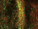
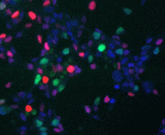
The adult brain has two stem cell niches—one in the subventricular zone (SVZ) and one in the subgranular zone (SGZ).  Within these niches are neural stem cells (NSCs) that can self-renew and differentiate into neurons and glia.  The nervous system has limited regeneration abilities, as seen in patients with traumatic injuries or degenerating diseases like multiple sclerosis (MS).  MS patients experience myelin loss and axon degeneration, and can experience some phases of recovery due to remyelination.  Understanding the regulation that drives NSCs towards different glial cell fates should help guide the development of treatment for MS patients.  A recent Development paper describes the importance of p57kip2 (Cdkn1) in regulating NSC cell fate decisions.  Jadasz and colleagues found that suppression of p57kip2 in cultured adult NSCs skewed the resulting cell fates away from astroglial fates, and toward oligodendroglial fates.  Oligodendrocytes are the glial cells that produce myelin.  The resulting cells also showed an induction of BMP antagonists, in line with the knowledge that the BMP pathway normally promotes astroglial differentiation.  When p57kip2-suppressed NSCs were transplanted into adult spinal cords, markers for oligodendroglial cells increased.  The images above show the stem cell niches in the SVZ and SGZ.  Sox2 (red) and GFAP (blue) double-positive cells have been proposed to be NSCs in these niches.  These cells also show p57kip2 (green) immunostaining.

For a more general description of this image, see my imaging blog within EuroStemCell, the European stem cell portal.

Jadasz JJ, Rivera FJ, Taubert A, Kandasamy M, Sandner B, Weidner N, Aktas O, Hartung HP, Aigner L, & Küry P ResearchBlogging.org(2012). p57kip2 regulates glial fate decision in adult neural stem cells. Development (Cambridge, England), 139 (18), 3306-15 PMID: 22874918

Thumbs up (4 votes)
Loading...

Tags: ,
Categories: Images, Research

Research Technician – Cell and Developmental Biology – MRC-NIMR

Posted by , on 12 September 2012

Closing Date: 15 March 2021

Applications are invited for a Research Technician position in the research group of James Briscoe at the MRC National Institute for Medical Research, London. The lab studies the embryonic development of the vertebrate central nervous system. We combine cutting edge transgenic and genomic approaches with the latest imaging and cell biology techniques to investigate how morphogens and transcriptional networks generate spatial pattern.

http://www.nimr.mrc.ac.uk/research/james-briscoe/

Experience in molecular biology is required and experience of cell culture, mouse genetics and embryological techniques desirable. Enthusiasm, reliability and organisational skills are essential. The successful applicant will be expected to support and become engaged in specific projects aimed at elucidating the mechanisms of neural patterning. The group, which will move to the Francis Crick Institute, London, in 2015, currently comprises 11 scientists and is well supported by both MRC and external funds. The Institute provides excellent training for researchers in a multi-disciplinary environment and is equipped with state of the art facilities. Specialist training to support the development of skills will be given where necessary.

The applicant is expected to have a Degree or equivalent in a relevant subject.

This position is for 5 years in the first instance. Salary range is from £21,565 per annum inclusive of location allowance. MRC final salary pension scheme is also available.

Situated in Mill Hill, North West London, the MRC National Institute for Medical Research is the largest MRC institute, supporting 70 research groups and 500 bench scientists. Facilities include genetic modification of mice, imaging, histology, FACS and high throughput sequencing.

Applications are handled by the RCUK Shared Services Centre; to apply please visit our job board at https://ext.ssc.rcuk.ac.uk and complete an online application form.  Applicants who would like to receive this advert in an alternative format (e.g. large print, Braille, audio or hard copy), or who are unable to apply online should contact us by telephone on 01793 867003, please quote reference number

Closing date: 8th October 2012

The MRC is an Equal Opportunities Employer

Final appointments will be subject to a pre employment screening.

Thumbs up (No Ratings Yet)
Loading...

Categories: Careers, Jobs

Update about the Node updates

Posted by , on 12 September 2012

Have you updated your profile on the Node yet, as suggested in this post? If not, you have a few more weeks: we’re delaying the public profiles (and other updates) while we fix some existing issues with the site.

The site updates are ready to go, and we’re really excited about them, but we want to make sure everything is working perfectly without them first.

On that note, if you’re having any technical issues with the Node, please do let us know right away. You can use the email form or email us directly at thenode [at] biologists.com, or use Twitter (@the_Node).

Thanks for your understanding, and sorry to keep you waiting!

Thumbs up (1 votes)
Loading...

Categories: News

World Cell Race

Posted by , on 12 September 2012

The Olympics and the Paralympics may be over, but there’s still time for sporting success this year, albeit on a rather smaller scale. The 2012 World Cell Race is on. Over the last year or so, I’ve heard both Matthieu Piel and Manuel Thery talk about the 2011 version of the race, and this morning I noticed that the results have just been published in Current Biology, officially* revealing that the fastest cell is a human mesenchymal stem cell.

Think you can beat that? They’re still accepting submissions for this year’s race, so if you have the single-celled equivalent of a Usain Bolt or an Oscar Pistorius in your freezer, send it in!

*Well, officially out of the 54 cell types they tested under the specific conditions they used…

Thumbs up (2 votes)
Loading...

Tags: ,
Categories: Events, Research

Grand Challenge PhD in Rihel Lab at UCL

Posted by , on 11 September 2012

Closing Date: 15 March 2021

PhD Position
University College London
Department of Cell and Developmental Biology
PhD Supervisor: Dr. Jason Rihel
Application Deadline: Oct. 15, 2012

Funding Availability:  Funding PhD Project (Grand Challenge; European/UK Students; including a stipend of £18,000 a year, Home/EU fees and £5000 per year research funds).  Non-UK/EU citizens are eligible but must have the ability to pay the difference between the Home/EU fee and the International fee for the 3 years.

Sleep is a fundamental biological process that has a major impact on human health, cognitive performance, and quality of life, yet the genetic and neural mechanisms that regulate sleep/wake behaviour are largely unknown. In the Rihel lab, we use zebrafish as a model system to study the regulation of sleep because it combines the powerful genetics of invertebrate models with the basic brain structures that regulate sleep in humans. We use high throughput behavioural assays to measure sleep behaviours in the fish and exploit genetic tools to manipulate critical regulators of sleep, such as the functionally conserved hypocretin/orexin (Hcrt) signalling system.  Recently, we have performed both small molecule and genetic screens to identify potential novel regulators of sleep in zebrafish. 

The successful PhD student will combine cutting edge techniques in molecular biology and behavioural neuroscience to explore the function of novel sleep genes and drugs.  In particular, the research will aim to map the neural circuits that are altered by small molecule and neuropeptide manipulations.       

Applicants should have a degree in molecular biology, neurobiology, or a similar field.  A 2:1 or better is normally required according to UCL eligibility criteria.

Send all enquires to Dr Jason Rihel (j.rihel@ucl.ac.uk).  Applicants should send a CV and names and contact details of two referees.

Thumbs up (No Ratings Yet)
Loading...

Categories: Careers, Jobs

Vote for a Development cover – Woods Hole – Round 4

Posted by , on 5 September 2012

This is the final round of images from the 2011 Woods Hole embryology course. The students of the 2012 course have also taken some beautiful images, and you’ll see those on the Node later this year.
But for now, vote in the poll below the images for the one you would like to see on the cover of Development. (Click any of the images to see a bigger version.) Poll closes on September 26, noon GMT.

1. Alcian blue staining of a Stage 17 bat (Carollia perspicillata) embryo. This image was taken by Lingyu Wang and Ketty Lee.

2. 3D reconstruction (face on view) showing the head vasculature of a zebrafish embryo. Visualization of gata1: dsRed (red; blood cells) and flk1: EGFP (blue, endothelial cells including blood vessels). This image was taken by Meghan Morrissey and Lynn Kee.

3. Day 10.5 mouse embryo immunostained for PECAM in green, Phospho-Histone H3 in red, and DAPI (nuclei) in blue. This image was taken by Juliette Petersen and Rachel K. Miller.

4. Skeletal preparation of a turtle, the red-eared slider (Trachemys scripta elegans). This image was taken by Megan Martik, Jane Yu, John Young and Eric Brooks.


Thumbs up (20 votes)
Loading...

Tags: , , , , ,
Categories: Images