The community site for and by
developmental and stem cell biologists

Development: New Executive Editor

Posted by , on 30 November 2011

I’m the new Executive Editor at Development, taking over after Jane Alfred’s eight years at the journal, and I’d like to take this opportunity to introduce myself. I’m starting here fresh off the plane from Heidelberg, Germany, where I have been working as a scientific editor at The EMBO Journal for the last three years, handling manuscripts in the fields of developmental and cell biology. Before then, my research life is probably best described as “trying to understand how to make an eye”: firstly during my PhD with Matthew Freeman at the Laboratory of Molecular Biology in Cambridge working with Drosophila (where I published my first ever paper in Development!), and subsequently studying morphogenesis of the fish retina in Jochen Wittbrodt’s lab at EMBL Heidelberg.

While I’m no longer in the lab, I’m still fascinated by the subject, and am excited to be getting back to my developmental biologist roots here at the journal. To me, Development is all about publishing by and for the community, and The Node is a big part of that: I’ve been reading it since its inception last year, and I look forward to playing a more active role from now on – I’m sure you’ll be hearing more from me in the future. I also hope to be meeting many of you in person over the coming months and years. For now, though, all that remains is for me to thank Jane for the fantastic job she’s done here: I have big boots to fill, but I hope I’m up to the challenge!

Thumbs up (8 votes)
Loading...

Tags: ,
Categories: News

Navigate the archive

Use our Advanced Search tool to search and filter posts by date, category, tags and authors.

This month on the Node – November 2011

Posted by , on 30 November 2011

What was new on the Node this month? Here are a few of the highlights from November:

New Research:
Several exciting new research papers were discussed on the Node this month. In one post, Stas Shvartsman introduces a recent Development paper from his own lab that describes a method to quantify the spatial range of morphogen gradients.

“Our paper provides a practical definition of the range of a morphogen gradient, a statistical procedure for estimating this range, a demonstration of this procedure in practice, and several independent experimental tests of derived estimates. From the biological standpoint, the range of a gradient can be viewed as the distance over which it acts as a spatial regulator of cell responses.”

This method from the Shvartsman lab can be applied to other systems. Find out more in the post.

Elsewhere, Erin Campbell highlights an image from a paper by Andrei Mardaryev et al., showing that Lhx2 in hair follicle stem cells regulates epidermal regeneration after injury.

Paul O’Neill writes about a new Nature paper from Yoshiki Sasai’s lab at RIKEN CDB, in which the authors describe how they generated functional pituitary gland tissue from mouse ES cells in vitro.

Graduate students
The Node also addressed graduate student issues this month, both the fictional and the factual.

For the past 14 years, the web comic Piled Higher and Deeper has looked specifically at the ups and downs of graduate student life. The comic is now a movie, and the Node had a chance to catch up with creator Jorge Cham at a screening of the film in London.

If you’d rather watch a more serious film involving graduate students, take a look at Stand With Science, in which MIT students urge US Congress not to cut science funding.

Also on the Node:
– Over the next few weeks, we’ll re-post this year’s batch of book reviews for Development, starting with this Star Trek-themed review of “Imaging in Developmental Biology”. We’ll also have some reviews unique to the Node, so keep an eye on the site!

– Finally, Elena Kardash explains how to find a place to practice piano when you’re in Barcelona for a brief research stint…

Thumbs up (No Ratings Yet)
Loading...

Categories: Highlights

December desktop calendar

Posted by , on 29 November 2011

And here it is: the last of the desktop wallpaper calendars. In June we celebrated our first birthday, and decided to give all our readers a virtual gift. It ended up being six gifts: one desktop calendar wallpaper for each remaining month of 2011. If you want to see all the images, or download the latest one, visit the calendar page. All images were chosen from either the intersection image contest or from the images we’ve featured from the Woods Hole Embryology 2010 course.

december_thumbnailOn the december calendar wallpaper, a dorsal view of the central nervous system of a Drosophila embryo.
This image, taken by Joshua Clanton of Vanderbilt University, was one of the candidates in the third Development cover image voting round of images taken at the 2010 Woods Hole Embryology course.

Visit the calendar page to select the resolution you need for your screen.

Thumbs up (No Ratings Yet)
Loading...

Tags:
Categories: Images

Postdoctoral positions to study the Hippo pathway

Posted by , on 25 November 2011

Closing Date: 15 March 2021

Peter MacCallum Cancer Centre is the largest cancer research group in Australia, uniquely integrating basic, translational and clinical research with patient care in the setting of a specialist cancer hospital.

You will be part of the Cell Growth and Proliferation Laboratory, led by Dr Kieran Harvey, which is investigating organ size-control and tumorigenesis controlled by the Salvador-Warts-Hippo (SWH) pathway. The focus of your research will involve determining the mechanism by which activity of the SWH pathway is controlled, and how this pathway controls tissue growth and tumorigenesis.

Skills required include a PhD, with a broad base of molecular, genetic and cell biology techniques. You will have the ability to teach and supervise undergraduate and postgraduate students, and technical staff. You have a strong background in cell signalling with an emphasis on signal transduction pathways and molecular mechanisms regulating cell proliferation and growth. Experience with Drosophila will be advantageous but is not essential.

Selected References:

CLC Poon, JI Lin, X Zhang and KF Harvey (2011). The sterile 20-like kinase Tao-1 controls tissue growth by regulating the Salvador-Warts-Hippo pathway. Dev Cell. 21: 896-90

X Zhang, J George, S Deb, JL Degoutin, EA Takano, SB Fox, AOCS Study Group, DDL Bowtell and KF Harvey (2011). The Hippo pathway transcriptional co-activator, YAP, is an ovarian cancer oncogene. Oncogene. 30: 2810-2822.

X Zhang, CC Milton, CLC Poon, W Hong and KF Harvey (2011). Wbp2 cooperates with Yorkie to drive tissue growth downstream of the Salvador-Warts-Hippo pathway. Cell Death Diff. 18: 1346-1355.

FC Bennett and KF Harvey (2006). Fat Cadherin Modulates Organ Size in Drosophila via the Salvador/Warts/Hippo Signaling Pathway. Curr Biol. 16, 2101-2110.
Enquiries to: Dr Kieran Harvey: Kieran.Harvey[at]petermac.org

Thumbs up (No Ratings Yet)
Loading...

Tags: , , , ,
Categories: Jobs

Book review: Fast-forward: the fourth dimension in development

Posted by , on 23 November 2011

Development issue 24 features several book reviews. Over the next few weeks, these book reviews will also appear here on the Node. In this first one, Elaine Dzierzak and Catherine Robin compare developmental biology to Star Trek in their review of “Imaging in Developmental Biology: A Laboratory Manual” (Edited by James Sharp and Rachel O. Wong)
(Originally in Development.)

Book info:
Imaging in Developmental Biology: A Laboratory Manual Edited by James Sharp, Rachel O. Wong Series Editor, Rafael Yuste Cold Spring Harbor Laboratory Press (2011) 883 Pages ISBN 978-0-879699-40-6 (paperback), 978-0-879699-39-0 (hardback) $165 (paperback), $246 (hardback)

Development is a bit like Star Trek, the long-running television series in which ‘space’ is the final frontier. For development, the final frontier is the fourth dimension, ‘time’. Time travel through the embryo, from the zygote to gastrulation, to organogenesis, and birth, has been a subject of fascination and science (fiction?) for centuries. This fascination is reflected in the many historical drawings of developing embryos and by advances in the field of embryology that came with the invention of the microscope. With the aid of microscopy, the field advanced from drawings of embryos to static images of fixed sections, which could be rendered, with some mental effort, into three-dimensional (3D) structures. However, comparisons of embryos at different formative stages could only hint at the patterns of dynamic cell growth and morphological change that occur during development, which recent molecular and genetic analyses have begun to uncover. Importantly, the current advances being made in innovative, real-time imaging technologies and in the computational processing of images have now fast-forwarded the field boldly into the dynamic fourth dimension. These advances are now summarized and explained in a newly published book on imaging, Imaging in Developmental Biology, edited by James Sharp and Rachel O. Wong, both experts in this field.

Imaging in Developmental Biology is an excellent resource from which both novices and experienced researchers can obtain current state-of-the-art embryo-imaging protocols for studying key developmental events, such as cell-fate determination, morphogen gradient formation, cell-cell interactions, cell migration and morphogenesis. The eye-catching cover immediately attracted passing lab members, encouraging them to browse the book, which they did with increasing interest. The first comment often expressed was: “I did not know that we could do so much!” Upon first perusal, this comprehensive book seems almost overwhelming with an impressive 57 chapters and seven appendices. But it does contain just about everything known about imaging embryos. This is not surprising as the volume is based, in part, on the popular and excellent Cold Spring Harbor imaging course. The editors have organized the book into four large sections, which contain chapters that are frequently and conveniently cross-referenced. A particularly helpful table is provided in Chapter 1 that guides the reader to specific protocols of interest in different animal models.

(more…)

Thumbs up (6 votes)
Loading...

Tags:
Categories: Book Reviews

Spatial range of a morphogen gradient

Posted by , on 23 November 2011

Estimating the range of a morphogen gradient

Morphogen gradient, defined as a concentration field of a chemical factor that acts as a dose-dependent regulator of cell differentiation, is an established concept in developmental biology. However, morphogen gradients in real systems are difficult to measure and mechanisms by which they control patterns of cell fates are heavily debated in the literature. In order to resolve some of the outstanding questions, it is essential to measure morphogen gradients in large numbers of embryos, at multiple developmental time points, and across multiple genetic backgrounds. We have developed a high throughput experimental approach that greatly facilitates these tasks for morphogens in the Drosophila embryo, a model system at the forefront of quantitative studies of development (Chung, Kim, et. al., Nature Methods 2011). In our recent Development paper, we used this technique to quantify the spatial range of the graded distribution of nuclear Dorsal, a transcription factor that subdivides the early fly embryo into presumptive muscle, skin, and nerve tissues (Kanodia et al, Development, 2011).

Our paper provides a practical definition of the range of a morphogen gradient, a statistical procedure for estimating this range, a demonstration of this procedure in practice, and several independent experimental tests of derived estimates. From the biological standpoint, the range of a gradient can be viewed as the distance over which it acts as a spatial regulator of cell responses.

Consider a common scenario in which the level of a morphogen decays from the maximal level at the point of morphogen production to the basal level at the position most distant from the peak of the gradient. As a practical definition of the spatial range of a gradient, we propose to use the distance from the peak value at which the level of the measured signal first becomes indistinguishable from the baseline.

This position can be estimated as follows. First, by measuring morphogens gradients in a collection of embryos, one can construct an empirical distribution function for the morphogen levels at multiple positions within the tissue. Second, based on these distribution functions, one can compare the mean of the morphogen level at a specific location to the baseline value. The larger the distance between from the position of the peak of the gradient, the smaller is the difference from the baseline value. The range is defined is the largest distance at which the means of the two distributions (at a current position and at the position most distant from the peak) are different from each other.

Clearly, this definition of the range leads to an estimate that is affected by the variability in the analyzed dataset. The larger the variability, the smaller is the estimated range. Since some sources of the observed variability come from the experimental procedure, such as uncertainty associated with determining the developmental stage, our estimate for the range is conservative. In other words, the true range of the gradient is actually larger than that predicted by our analysis.

Following this procedure, the range of the nuclear Dorsal gradient is estimated around 2/3 of the dorsoventral axis. As an independent estimate for the range of this gradient, we characterized the spatial extent of its transcriptional effects. For this, we use short gastrulation (sog), a well-studied transcriptional target of Dorsal and quantified the spatial pattern of its expression within the tissue. Remarkably, the spatial extent of sog expression came out to be very close to our estimate of the spatial range of Dorsal, which acts as a direct regulator of sog.

One of the main outcomes of our studies is the conclusion about the size of the dataset needed to estimate of the range of a spatially distributed signal. An accurate estimate can be obtained based on a dataset from ~40 embryos, which is within the reach for a large number of experimental systems. When combined with the fact that our computational procedures are easy to implement and require only the basic knowledge of statistics, we expect that our approach should be applicable in multiple developmental events controlled by morphogen gradients.

ResearchBlogging.orgKanodia, J., Kim, Y., Tomer, R., Khan, Z., Chung, K., Storey, J., Lu, H., Keller, P., & Shvartsman, S. (2011). A computational statistics approach for estimating the spatial range of morphogen gradients Development, 138 (22), 4867-4874 DOI: 10.1242/dev.071571

Thumbs up (11 votes)
Loading...

Tags:
Categories: Research

In Development this week (Vol. 138, Issue 24)

Posted by , on 22 November 2011

Here are the research highlights from the current issue of Development:

Getting to the heart of Flk1 expression

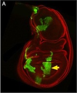
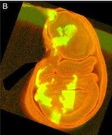
The Flk1 gene, which encodes a VEGF-A receptor, is expressed in the multipotent mesodermal progenitor cells of mouse embryos that give rise to various haemato-cardiovascular cell lineages. FLK1 expression also marks haemato-cardiovascular cell lineages in differentiating human embryonic stem (ES) cells, lineages that could be useful for the treatment of human cardiovascular diseases if the molecular regulation of Flk1 expression can be unravelled. Masatsugu Ema and colleagues now identify a novel enhancer in the mouse Flk1 gene that is required for the mesodermal expression of Flk1 in the early embryo and in differentiating ES cells (see p. 5357). This enhancer region, they report, is activated by Bmp, Wnt and Fgf, and contains binding sites for the transcription factors Gata, Cdx, Tcf/Lef, ER71/Etv2 and Fox, all of which are controlled by Bmp, Wnt and Fgf signalling. The researchers suggest, therefore, that early Flk1 expression might be induced by cooperative interactions between this set of transcription factors under the control of Bmp, Wnt and Fgf signalling.

Eph/ephrin signals guide muscle rebuilding

Skeletal muscle regeneration after injury is dependent on satellite cells (skeletal muscle stem cells) that, in response to local myofibre damage, proliferate to build up a supply of adult myoblasts that repair the damage. But do satellite cells relocate within the muscle to respond to distant myofibre damage? If so, how do they find their way? On p. 5279, D. D. W. Cornelison and co-workers investigate whether Ephs and ephrins – molecules that are usually associated with axon guidance but that are expressed by activated satellite cells – modulate satellite cell motility and patterning. Using an ephrin ‘stripe’ assay, they show that multiple ephrins elicit a repulsive migratory response in activated satellite cells and affect the patterning of differentiating satellite cells. Importantly, the same ephrins are present on the surface of healthy myofibres and increase during regeneration, which suggests that muscle regeneration could involve ephrin-mediated guidance. Given their results, the researchers propose that Eph/ephrin signalling might regulate multiple aspects of satellite cell behaviour during muscle regeneration.

Noncanonical Wnts and PAR-1 drive neural crest fate

Neural crest (NC) cells are multipotent progenitors that form at the neural plate border, undergo epithelial-mesenchymal transition, and then migrate to give rise to numerous cell types in vertebrate embryos. Noncanonical Wnt signalling is known to be involved in NC migration, but is it, like canonical Wnt signalling, required for NC specification? On p. 5441, Olga Ossipova and Sergei Sokol implicate noncanonical Wnt11-like proteins in NC specification in Xenopus embryos. They show that Wnt11R, which is expressed in the neuroectoderm next to the NC territory, is required for NC formation. The authors also show that Wnt11-like signals regulate the localisation and activity of the cell polarity determinant PAR-1. Importantly, PAR-1 itself is required for NC specification, they report, and PAR-1 RNA rescues NC markers in embryos in which noncanonical Wnt signalling has been blocked. Together, these results identify roles for noncanonical Wnt signalling and PAR-1 in NC specification and reveal an unexpected connection between cell polarisation and cell fate.

Dual embryonic origin for the inner ear

It is widely accepted that the inner ear labyrinth and the neurons of the cochleovestibular ganglion (CVG), which innervates the inner ear’s sensory epithelia, derive entirely from the otic placode, an ectodermal region that invaginates during embryogenesis to form the otic vesicle (OV) and the CVG. Here (p. 5403), by genetically labelling cranial neuroepithelial cell (NEC) lineages, including neural crest cells, in mice, Bernice Morrow and colleagues show that cells from the neural tube invade the otic epithelium in vivo and that NEC descendants constitute a significant proportion of the OV. NEC descendants, they report, are localised within the sensory epithelia of the saccule and utricle (the inner ear structures that are sensitive to movement) and the cochlea (the auditory portion of the inner ear) throughout development and into adulthood, and differentiate into neurons, hair cells and supporting cells. By revealing the inner ear’s dual embryonic origin, these results challenge the current model for the neurosensory development of the inner ear.

Heads up for new Noggin functions

The secreted protein Noggin1 antagonises the BMP family of TGFβ ligands and, as a consequence, plays a key role in many processes during embryogenesis. Here (p. 5345), Andrey Zaraisky and colleagues unexpectedly reveal that Noggin1 and its homologue Noggin2 also antagonise, albeit less effectively, the non-BMP TGFβ ligands ActivinB, Xnr2 and Xnr4 (Nodal homologues), and XWnt8 during early Xenopus embryogenesis. Inactivation of these ligands is essential for head induction, and the researchers show that both Noggin proteins can induce a secondary head, including a forebrain, if ectopically produced at high concentrations in Xenopus embryos. During normal development, they report, the Noggin1 concentration in the presumptive forebrain is only sufficient for its BMP-antagonizing function whereas the higher concentration of Noggin2 produced in the anterior margin of the neural plate protects the developing forebrain from inhibition by ActivinB and XWnt8 signalling. Thus, the researchers conclude, forebrain specification in Xenopus requires the inhibition of Activin/Nodal, BMP and Wnt signalling not only during gastrulation but also at post-gastrulation stages.

Seven up works double time in neuroblasts

Neural progenitor cells generate different cell types at different times during nervous system development. In Drosophila neuroblasts, the sequential expression of Hunchback (Hb), Kruppel (Kr) and several other transcription factors controls temporal competence changes. The transcription factors in this temporal cascade regulate each other’s expression but, in addition, Seven up (Svp) acts as a switching factor to ensure the Hb to Kr transition. Now, Stefan Thor and co-workers uncover a second role for Svp during the development of the Drosophila embryonic thoracic neuroblast 5-6 (NB5-6T) lineage (see p. 5311). The researchers show that svp is expressed in two distinct pulses in this lineage. In the early pulse, they report, svp acts as a switching factor by suppressing hb expression. However, in the second pulse, which occurs later in the NB5-6T lineage, svp acts as a sub-temporal gene to establish the alternative fates of four interneurons expressing the transcription factor Apterous. Thus, one gene can play two temporal roles in the development of one neural lineage.

Plus…

This issue, the last of the 2011 volume, contains Development’s annual Book Review section, which covers a broad range of topics that are becoming increasingly important to developmental biologists. The titles reviewed include:

– Mathematical Models of Biological Systems
(reviewed by Lance Davidson)

– Principles of Development
(reviewed by Richard Harland)

– Imaging in Developmental Biology: A Laboratory Manual
(reviewed by Elaine Dzierzak and Catherine Robin)

– Molecular Biology of RNA
(reviewed by Ilan Davis)

– Epigenetics Linking Genotype and Phenotype in Development and Evolution
(reviewed by Mellissa R. W. Mann)

– The Nucleus
(reviewed by Wendy A. Bickmore)

– Human Stem Cell Technology and Biology: A Research Guide and Laboratory Manual
(reviewed by Neil Singh and Ludovic Vallier)


Thumbs up (No Ratings Yet)
Loading...

Categories: Research

Doing Science in Barcelona

Posted by , on 22 November 2011

Blog entry

When thinking Barcelona, what first comes to mind is probably the football and Olympic games, the beaches, relaxed Mediterranean lifestyle, siesta and long nights out. All of it is accompanied with the Gaudi’s whimsical architecture at the background as an extra bonus. People usually go to Barcelona for a vacation or … for a science meeting. I was in this colorful city already several times because of science and always loved the place. And then a perfect chance came to spend a little bit more time in Barcelona when I was awarded the Development Travelling Fellowship supporting my visit for collaborative purposes. I was hosted by the lab of Enrique Martin-Blanco at IBMB at the University of Barcelona and the main goal of my stay was to work out the conditions for measuring protein activity during early zebrafish development. I embraced this opportunity to learn more about the place and its local customs by living there. What follows, are some of the experiences I have made in those two months in Barcelona.

Science reflection

Despite the economical situation and the ever-tightening budget, people do amazing science in Barcelona. I was exposed to a rich spectrum of different projects during the lab group meetings and also during department seminars. The microscopy facilities were impressive and I was happy to have more that one choice for a particular setup to use in my experiments. I could test different excitation and emission combinations as well as various optical variants to find the best conditions for imaging. Although the facilities were very busy and shared by many labs, I could get access to all the necessary equipment to conduct experiments and collect enough data that kept me busy with the analysis when the microscopes were occupied by other data hunters. Here, I would like to express my deep gratitude to the members of Enruque’s lab and to the heads of the facilities for making my time there very smooth in terms of organization and planning. The first two weeks were made very easy for me, because the girls who were working with zebrafish, made all the necessary arrangements such as booking injection stations and the microscope slots, preparing certain reagents and also making spare time to show me around. This was an enormous help during the short visit when every second counts.

As other travelers here had already commented, a good thing about going to a different place is that it allows refreshing ones thinking and the general attitude to the lab routines. For example, it became clear very quickly how spoiled we are back home in Germany. While it is good to focus on the work without thinking much about the costs involved, one can get easily carried away and waste unduly amount of reagents and materials. While I never felt limited in any reagent or equipment I had to use during my visit, it became apparent how important is to be economical in the way one works. For example, it is perfectly possible reusing the plastic Petri dishes in which the embryos are kept, which is often for half a day only. There is no good reason for discarding the dishes. It is not about the cost of the dishes, which is rather low, bur more about the plastic waste that is generated that way. Certainly, it made me think more carefully of the amount of waste produced in a lab and I keep applying my new working habits.

Cultural differences

When in Spain, do like Spanish do, or at least try to. I was very curious about the local customs and the differences between the Spain and Germany.

People in Barcelona are very friendly and one can easily get around with speaking English, but knowing a few words in a local language would definitely be helpful. Barcelona is the capital of Catalonia, therefore there are two official languages spoken in Barcelona: the Catalan and Castilian Spanish, which could be very confusing at the beginning, especially when trying to read the signs. It always makes a nice impression if you could say “bom dia” instead of “bueans dias” and some other simple phrases in Catalan.

Strangers at the street and in a public transport stare at one another much more directly than they do in Germany. That was a striking difference from the way the strangers interact in Northern Europe, where people mind their own businesses. The personal distance is much shorter and in the metro it would be very normal to lean over to see better the book a fellow passenger is reading.

For someone who is used to have a lunch break at 11:30, which is typical in Germany, it might be a bit unusual to shift this time for an hour or more. People in Spain normally go for lunch around 1 pm or later. It is very typical to conclude the lunch with a coffee and maybe a cigarette.

It was relatively easy to find a place to stay. I got a room in a shared apartment with four other people in a very nice neighborhood with large streets and beautifully designed houses. Every day going to the metro, I would fight my way to get in because of the tourists crowding in front of casa Battlo, one of the famous Gaudy houses turned into museum, right next to the station. My flatmates were on various kinds of the study exchange programs or other types of visits and all from different backgrounds, which was a very enriching experience. I feel very lucky with my choice because I had the nicest flatmates, easygoing and very friendly.

Although it is acceptable, I would not advice drinking the tab water. It is full of chlorine, which gives a peculiar taste to it. The bottled water is relatively cheap and the 5 to 8 liter cans are available everywhere. Barcelona is rather expensive city but good deals are possible. There are plenty of grocery stores and little private kiosks that run till late but the price range could be huge and I spent some time comparing prices before deciding where to shop.

The sunlight is very intense in Barcelona and I was constantly using sunglasses. My room had no curtain and the effect was amazing: I did not need an alarm clock anymore getting up with the sun every day. If only the sun could get up a bit later on the weekends!

Barcelona is generally a very safe city but one must be wary of the theft, which is apparently a very popular business in the tourist areas. It is well known and has been discussed thoroughly in many blogs giving travel advices. The thieves will try to steal the stuff from you while you are admiring the architecture and other street wonders. Being vigilant and keeping an eye on your belonging is enough to stay away form the trouble.

How to find a piano

While this would not be an issue for the most visiting scientists, I felt desperate facing with the idea of spending two months without a piano. That’s why during the first week I kept annoying my colleagues asking them to call different piano stores and enquire about a possibility of renting one. Soon it became clear that renting a piano for two months is not an option. I found a place nearby where I could play for a small fee, but I could never make it there during the opening hours. At the end I was quite lucky to find a cheap digital piano on ebay, which I could later re-sell to one of the colleagues who got inspired and decided she wanted to perfect her musical skills.

Thumbs up (3 votes)
Loading...

Tags: ,
Categories: Funding, Lab Life, Research

Stand With Science

Posted by , on 21 November 2011

A group of graduate students at MIT have written a letter on behalf of American graduate students, urging the United States Congress not to cut science funding. The Congress Joint Select ‘super’ Committee on Deficit Reduction will make their decision later this week, and today is the last day to sign the letter.

If you’re in the US, have a look at the Stand With Science website for more info.

Thumbs up (1 votes)
Loading...

Categories: Funding, News

Post-doctoral scientist –Peter MacCallum Cancer Centre, Melbourne, Australia

Posted by , on 21 November 2011

Closing Date: 15 March 2021

Applications are invited for a post-doctoral research scientist post in Melbourne, to join a newly funded research group within the Cancer Cell Biology Program. The research focuses on understanding the mechanisms regulating the growth of neural stem cell derived tumours in Drosophila. We are part of a vibrant fly community working alongside 400 cancer biologists located in the centre of Melbourne.

Funding is available for 3 years, starting July 2012. Candidates should have a PhD in developmental biology or cell biology, proven experience in molecular biology, immunohistochemistry and/or biochemistry, experience in fly genetics is also desired. For more information and application, please write to Dr Louise Cheng at lcheng@nimr.mrc.ac.uk

References:

1. Cheng et al. Anaplastic Lymphoma Kinase Spares Organ Growth during Nutrient Restriction in Drosophila. Cell (2011) vol. 146 (3) pp. 435-47

2. Sousa-Nunes et al. Regulating neural proliferation in the Drosophila CNS. Curr Opin Neurobiol (2010) vol. 20 (1) pp. 50-7

3. Maurange et al. Temporal transcription factors and their targets schedule the end of neural proliferation in Drosophila. Cell (2008) vol. 133 (5) pp. 891-902

Thumbs up (No Ratings Yet)
Loading...

Tags: , , ,
Categories: Jobs